In coronary arteries, final stent area is an important predictor for subsequent clinical events and restenosis. Acute stent recoil has been identified as a contributor for inadequate stent expansion. However, assessment of acute recoil is complex. We describe a method for real time imaging of acute recoil with optical coherence tomography (OCT) in a patient treated for a significant lesion in the left circumflex artery.
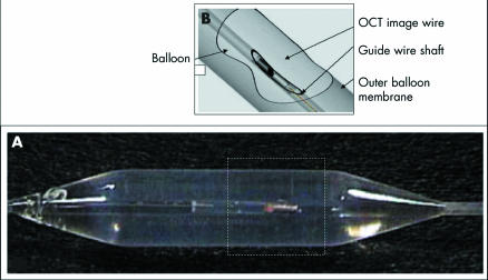
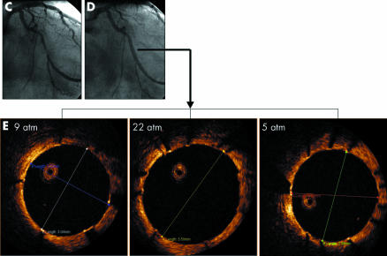
Panels A and B illustrate the principle. The dedicated OCT imaging wire (diameter 0.014 inch; light source 1300 nm; LightLab Imaging LLC, Westford, Massachusetts, USA) is introduced into the guidewire shaft of an over‐the‐wire balloon. Cross sectional images of the artery are obtained by imaging through the inflated balloon (mixture of contrast medium and saline). Panels C, D, and E demonstrate the OCT findings during postdilatation of the stent (Cypher 3.0 mm diameter/18 mm length; Cordis, Johnson & Johnson, Warren, New York, USA). The balloon (3.0 mm diameter, Open sail, Guidant, Diegem, Belgium) was stepwise inflated at 9 atm and 22 atm and then deflated to a pressure of 5 atm with the OCT imaging wire in place. At each pressure level, the maximum stent diameter was measured online by OCT. Stent diameter was 3.04 mm at 9 atm, increased to 3.50 mm at 22 atm, and decreased to 2.94 mm immediately after deflating the balloon to 5 atm. This corresponds to acute stent recoil of 16%.
Our case illustrates a new approach with OCT to image stent expansion directly during balloon inflation. This might be useful to study stent behaviour in the clinical setting and to guide improvements of stent characteristics based on in vivo findings.


