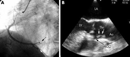
A 52 year old man was admitted for acute inferior myocardial infarction evolving for two hours. He was a mild active smoker, and had no history of heart disease. Coronary angiography revealed occlusions of both the posterior interventricular and retroventricular branches of the right coronary artery (RCA) (panel A). Because of the angiographic appearance, coronary embolism was suspected and a transoesophageal echocardiographic (TOE) examination was performed in search of a cardiac source of the embolism. TOE revealed the presence of an 18 × 7 mm pedunculated floating mass in the right coronary sinus of Valsalva. The mass was implanted on a small, minimally calcified atheromatous plaque, and had a highly mobile extremity prolapsing into the RCA ostium during diastole (panel B). These features were suggestive of an ascending aortic thrombus complicated with right coronary embolisation. After five days of medical treatment, including unfractionated heparin, aspirin (75 mg/day), clopidogrel (75 mg/day), and high dose atorvastatin (80 mg/day), a further TOE showed complete disappearance of the thrombus. The patient was discharged from hospital on β blocker, antiplatelet agents, and statin treatment. He is being followed up on an outpatient basis.
(A) Angiographic data. Right coronary artery selective angiogram (left anterior view) showing TIMI grade 2 flow caused by occlusion of the retroventricular and interventricular branches (arrows). (B) Transoesophageal echocardiographic study. Long axis view showing a mobile pedunculated mass (white arrows) of the ascending aorta. The highly mobile free floating extremity of the thrombus prolapses into the right coronary ostium (RCO, black arrows) during diastole. Ao, aorta; LA, left atrium; LV, left ventricle; RCO, right coronary artery ostium; Thr, thrombus.


