A 50‐year‐old woman presented with a six‐month history of progressive dyspnoea and signs of right heart failure. Armoured heart was suspected by clinical examination, chest x‐ray and two‐dimensional echocardiography.
Tissue Doppler imaging (TDI) revealed an E′‐velocity above the cut‐off value (8 cm/s) indicating cardiac constriction.
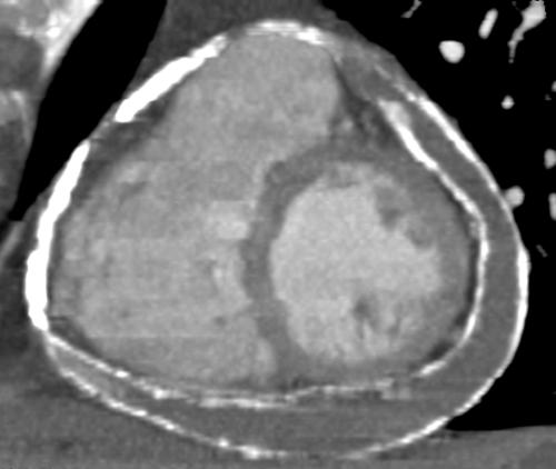
ECG‐synchronised contrast enhanced multislice computed tomography (MSCT) demonstrated severe calcifications expanding over nearly the entire heart in an inhomogeneous pattern. An inner and an outer shell representing epicardium and pericardium were differentiated. Encapsulated pericardial effusion was located above extended parts of the left and right ventricle including the atrioventricular groove (see panel).
Haemodynamics demonstrated elevated end‐diastolic filling pressures and a dip‐and‐plateau phenomenon supporting the suspected diagnosis. The patient underwent pericardectomy which confirmed “pericarditis constrictiva calcarea”.
Modern cardiac imaging techniques are helpful to differentiate constrictive from restrictive cardiac disease. MSCT demonstrated a large pericardial effusion whereas TDI showed an increased velocity of the mitral annulus. We propose the large pericardial effusion as a possible pathophysiologic mechanism responsible for the late onset of symptoms despite extensive calcification.
Multislice computed tomography (MSCT) based maximum intensity projection demonstrating the inhomogeneous pattern of epi‐ and pericardial calcification encapsulating a large pericardial effusion.


