

A 64‐year‐old man with a prior history of anterior wall myocardial infarction treated with thrombolysis in 1992, followed 10 years later by coronary artery bypass surgery, underwent cardiovascular magnetic resonance examination (CMR) for quantification of residual left ventricular function. Steady state free precession cine‐CMR demonstrated thinned myocardium with dyskinesis of the distal anteroseptal and apical left ventricular regions consistent with an old myocardial infarction. Areas of unusual signal intensity compared to that of the surrounding myocardium were noted on cine‐CMR within the infarcted region. These areas were confirmed to be composed of adipose tissue due to their characteristic high signal intensity on T1‐weighted turbo spin echo images and complete disappearance of signal using fat suppression. The adipose tissue also had high signal intensity on two‐dimensional inversion recovery images obtained before administration of gadolinium‐based contrast (see panel) and was surrounded by areas of myocardial fibrosis demonstrated on late contrast‐enhanced (LCE) images. These findings are consistent with lipomatous metaplasia in the region of an old myocardial infarction. This case demonstrates that a high signal intensity on LCE images may not always represent fibrosis alone in regions of old myocardial infarction.
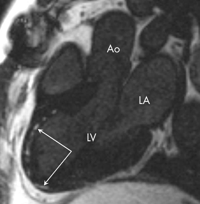
Left ventricular outflow tract view; two‐dimensional inversion recovery image before administration of contrast, arrows indicating high signal intensity in infarcted area. Ao, aorta; LA, left atrium; LV, left ventricle.