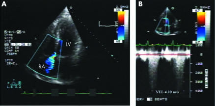
A 76‐year‐old man with a history of renal cell carcinoma was referred for evaluation of chest pain and progressive dyspnoea on exertion. Physical examination showed distended neck veins up to 10 cm above the sternal angle with large V waves and on auscultation a loud 3/6 systolic murmur was heard along the left parasternal border radiating toward the right side of the upper sternum. The patient was evaluated by spiral computed tomography (CT) to rule out the presence of pulmonary embolus. A right retrocrural metastatic adenopathy was observed but there was no evidence of pulmonary emboli. A subsequent transthoracic echocardiogram (TTE) demonstrated enlarged right sided chambers. Colour Doppler interrogation revealed a left‐to‐right shunt from the left ventricle (LV) to the right atrium (RA) across the ventriculo‐atrial septum immediately above the tricuspid valve (panel A). The estimated gradient between LV to RA was 70.2 mm Hg (panel B). This finding was consistent with Gerbode defect, a rare defect of the ventriculo‐atrial septum.
To further define the anatomy of the shunt, a live three dimensional echocardiogram (L3D, Philips 7500) was performed. Full volume images were cropped to localise the right atrial, tricuspid, and septal structures in three dimensions. Three dimensional colour Doppler interrogation of the defect demonstrated acceleration of flow on the ventricular side of the septal leaflet of the tricuspid valve. The LV to RA communication involved the base of the septal leaflet of the tricuspid valve as it extended across the ventriculo‐atrial septum into the right atrium (video files 1 and 2; to view video footage visit the Heart website—http://www.heartjnl.com/supplemental).
LV to RA communications could be either congenital or acquired defects from endocarditis, myocardial infarction, aortic or mitral valve replacement and trauma. The cause of our patient's defect remains unclear. The patient did not demonstrate clinical or echocardiographic evidence of endocarditis. Myocardial infarction was ruled out by serial myocardial enzymes and ECGs. After aggressive diuresis the patient clinically improved. Given the advanced stage of his malignancy, he requested hospice care.
(A) Transthoracic two dimensional echocardiogram with colour Doppler from apical four chamber view shows colour flow jet directed from left ventricle (LV) to right atrium (RA) across the ventriculoatrial septum. (B) Continuous wave Doppler through the ventriculoatrial septum demonstrates the left to right shunt with a maximum velocity of 4.19 m/s. This calculates to a LV to RA gradient of 70.2 mm Hg.
Live three dimensional echocardiography delineated the origin and the path of the left to right shunt involving the base of the septal leaflet of the tricuspid valve and the ventriculoatrial septum. To our knowledge, this is the first reported case of a Gerbode defect demonstrating the involvement of the tricuspid valve in the shunt. These findings have clinical implications when surgical repair is planned.
Supplementary Material
Associated Data
This section collects any data citations, data availability statements, or supplementary materials included in this article.


