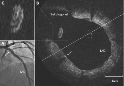
Although drug‐eluting stents have dramatically reduced restenosis rates after percutaneous coronary intervention, concerns remain with regard to the potential risk of stent thrombosis. For bifurcation lesions, a stent thrombosis rate up to 3.6% has been reported, and incomplete stent apposition has been proposed as a possible mechanism. We report the case of a 52‐year‐old man with diabetes and three‐vessel disease. Initially, three overlapping sirolimus‐eluting stents (Cypher, Cordis) were deployed to the left anterior descending artery. No further kissing post‐dilatation was performed because of TIMI‐3 flow in the diagonal branches. Angiography at 4 months of follow‐up showed widely patent stents with TIMI‐3 flow to both diagonals (panel A). Optimal coherence tomography (LightLab Imaging Inc, Westford, Massachusetts, USA) at the bifurcation of the left anterior descending artery to the first diagonal (arrow) showed good stent apposition, with minimal (<0.07 mm) intimal layer (panel B, arrow) on almost all struts. One strut was protruding into the lumen at the origin of the diagonal branch circumferentially covered by a thick tissue layer (0.25 mm, panel C) extending to its connection with the vessel wall proximally and distally. On the basis of tissue density and uniform growth on the free strut with minimal growth on other apposed struts, the case strongly suggests that this tissue consists of thrombus, likely fully organised and endothelialised. These findings lead to two thoughts on stent thrombosis in the presence of malapposed drug‐eluting stents. Firstly, unapposed struts can nestle thrombus triggering stent occlusion and secondly, the process can be self‐limiting, eventually allowing growth of tissue on the stent strut and possibly promoting its complete endothelialisation.
. 2007 Mar;93(3):378. doi: 10.1136/hrt.2006.091876
Do unapposed stent struts endothelialise? In vivo demonstration with optical coherence tomography
1J Tanigawa, P Barlis, C Di Mario, c.dimario@rbht.nhs.uk
Copyright © 2007 BMJ Publishing Group and British Cardiovascular Society
PMCID: PMC1861443 PMID: 17322520


