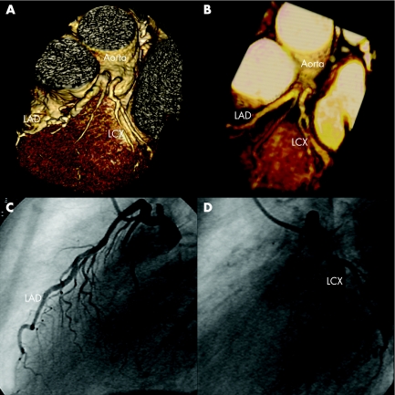
A 62‐year‐old man with onset of atypical angina pectoris was referred to our institution to rule out coronary artery disease. He had no cardiovascular risk factors other than smoking and arterial hypertension. Electrocardiogram and stress electrocardiogram were both negative. We performed non‐invasive coronary angiography using both multislice computed tomography and magnetic resonance imaging. Neither examination showed any major coronary stenoses in the left coronary artery system, but a split origin of this coronary artery (fig 1A, B). Both the left anterior descending artery and the left circumflex artery originated from separate but adjacent ostia in the left sinus of Valsalva. This anomaly is the most common coronary artery anomaly, with a prevalence of approximately 0.4%, which causes no haemodynamic impairment and thus, should be considered benign. The absence of major stenoses and the presence of a split left coronary artery were confirmed on conventional coronary angiography, with injections of selective contrast agents into the left anterior descending artery (fig 1C) and the left circumflex artery (fig 1D). Both multislice computed tomography and magnetic resonance imaging have recently been shown to allow non‐invasive detection of coronary anomalies. As these images show, both non‐invasive methods are of clinical value for assessing proximal coronary anomalies.
. 2007 Apr;93(4):538. doi: 10.1136/hrt.2006.094227


