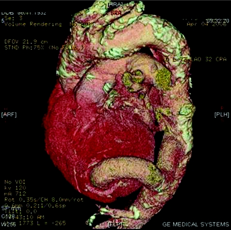
Three‐dimensional reconstruction; anterior view showing the correct position of the conduit and the extensive calcification of the aortic wall.
A 74‐year‐old male patient previously operated on for coronary artery bypass grafting was admitted to our institute because of recurrent episodes of pulmonary oedema. A coronary angiogram showed a patent internal mammary graft to the left anterior descending artery and a severe stenosis of a venous bypass to the right coronary artery. A concomitant severe aortic stenosis was diagnosed. Because of extensive aortic wall calcification, the standard approach of valve replacement was considered to be of prohibitively high risk. In view of haemodynamic instability, it was decided to implant a valve conduit from the left ventricular apex to the descending thoracic aorta. Postoperative multidetector computed tomography (MDCT) showed an extensive degree of aortic wall calcification and the correct position of the conduit. The patient had an excellent outcome and was discharged home on the 10th day after surgery, following percutaneous transluminal coronary angioplasty (PTCA) of the right coronary graft.


