

Abstract
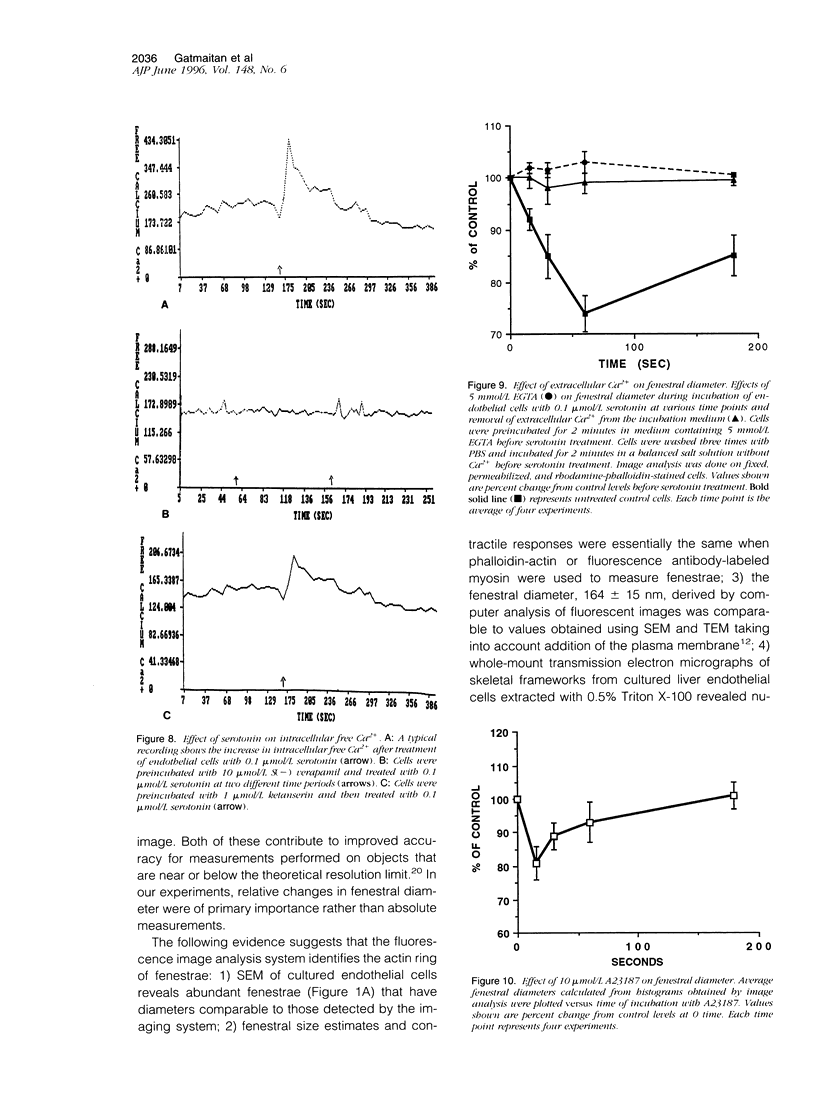
Liver endothelial cells possess fenestrae, which are pores supported by a cytoskeleton ring composed of actin and myosin. Fenestrae are dynamic structures that can contract or dilate, although the mechanism for this phenomenon remains to be elucidated. Staining of actin and/or of myosin permitted measurement of fenestral diameter and area in cultured rat liver endothelial cells using digitized video-intensified fluorescence microscopy with image analysis. Within 1 minute of incubation with 0.1 micromol/L serotonin, fenestral diameter and area decreased by 24 +/- 5% and 56 +/- 7%, respectively. Contraction of fenestrae by serotonin was inhibited by chelation of extracellular Ca2+ with EGTA and by addition of Ca2+ channel blockers, such as dilthiazem and verapamil. The response of fenestrae to serotonin was mimicked by addition of a Ca2+ ionophore, A23187. Serotonin inhibited cAMP production, had no effect on inositol phosphate production, and activated phospholipase A2, causing release of arachidonic acid. These results suggest that contraction of fenestrae is associated with Ca2+ influx. In response to 0.1 micromol/serotonin, intracellular Ca2+ levels increased within 3 to 5 seconds from 150 nmol/L to >400 nmol/l followed by rapid phosphorylation of the 20-kd subunit of myosin light chain; both events dependent on extracellular Ca2+.
Full text
PDF


Images in this article


Selected References
These references are in PubMed. This may not be the complete list of references from this article.
- Adelstein R. S., Conti M. A., Hathaway D. R., Klee C. B. Phosphorylation of smooth muscle myosin light chain kinase by the catalytic subunit of adenosine 3': 5'-monophosphate-dependent protein kinase. J Biol Chem. 1978 Dec 10;253(23):8347–8350. [PubMed] [Google Scholar]
- Affolter H., Erne P., Bürgisser E., Pletscher A. Ca2+ as messenger of 5HT2-receptor stimulation in human blood platelets. Naunyn Schmiedebergs Arch Pharmacol. 1984 Apr;325(4):337–342. doi: 10.1007/BF00504378. [DOI] [PubMed] [Google Scholar]
- Arias I. M. The biology of hepatic endothelial cell fenestrae. Prog Liver Dis. 1990;9:11–26. [PubMed] [Google Scholar]
- Barak L. S., Yocum R. R., Nothnagel E. A., Webb W. W. Fluorescence staining of the actin cytoskeleton in living cells with 7-nitrobenz-2-oxa-1,3-diazole-phallacidin. Proc Natl Acad Sci U S A. 1980 Feb;77(2):980–984. doi: 10.1073/pnas.77.2.980. [DOI] [PMC free article] [PubMed] [Google Scholar]
- Berridge M. J., Dawson R. M., Downes C. P., Heslop J. P., Irvine R. F. Changes in the levels of inositol phosphates after agonist-dependent hydrolysis of membrane phosphoinositides. Biochem J. 1983 May 15;212(2):473–482. doi: 10.1042/bj2120473. [DOI] [PMC free article] [PubMed] [Google Scholar]
- Berridge M. J. Rapid accumulation of inositol trisphosphate reveals that agonists hydrolyse polyphosphoinositides instead of phosphatidylinositol. Biochem J. 1983 Jun 15;212(3):849–858. doi: 10.1042/bj2120849. [DOI] [PMC free article] [PubMed] [Google Scholar]
- Braet F., De Zanger R., Baekeland M., Crabbé E., Van Der Smissen P., Wisse E. Structure and dynamics of the fenestrae-associated cytoskeleton of rat liver sinusoidal endothelial cells. Hepatology. 1995 Jan;21(1):180–189. [PubMed] [Google Scholar]
- Brauneis U., Gatmaitan Z., Arias I. M. Serotonin stimulates a Ca2+ permeant nonspecific cation channel in hepatic endothelial cells. Biochem Biophys Res Commun. 1992 Aug 14;186(3):1560–1566. doi: 10.1016/s0006-291x(05)81585-2. [DOI] [PubMed] [Google Scholar]
- Burch R. M., Luini A., Axelrod J. Phospholipase A2 and phospholipase C are activated by distinct GTP-binding proteins in response to alpha 1-adrenergic stimulation in FRTL5 thyroid cells. Proc Natl Acad Sci U S A. 1986 Oct;83(19):7201–7205. doi: 10.1073/pnas.83.19.7201. [DOI] [PMC free article] [PubMed] [Google Scholar]
- Burch R. M., Luini A., Mais D. E., Corda D., Vanderhoek J. Y., Kohn L. D., Axelrod J. Alpha 1-adrenergic stimulation of arachidonic acid release and metabolism in a rat thyroid cell line. Mediation of cell replication by prostaglandin E2. J Biol Chem. 1986 Aug 25;261(24):11236–11241. [PubMed] [Google Scholar]
- Crouch M. F., Lapetina E. G. Dual mechanisms of platelet hormone receptor desensitization. Differential importance between agonists of protein kinase C-dependent and -independent pathways. J Biol Chem. 1989 Jan 5;264(1):584–588. [PubMed] [Google Scholar]
- Doyle V. M., Creba J. A., Rüegg U. T., Hoyer D. Serotonin increases the production of inositol phosphates and mobilises calcium via the 5-HT2 receptor in A7r5 smooth muscle cells. Naunyn Schmiedebergs Arch Pharmacol. 1986 Jun;333(2):98–103. doi: 10.1007/BF00506510. [DOI] [PubMed] [Google Scholar]
- Fey E. G., Wan K. M., Penman S. Epithelial cytoskeletal framework and nuclear matrix-intermediate filament scaffold: three-dimensional organization and protein composition. J Cell Biol. 1984 Jun;98(6):1973–1984. doi: 10.1083/jcb.98.6.1973. [DOI] [PMC free article] [PubMed] [Google Scholar]
- Frandsen E. K., Krishna G. A simple ultrasensitive method for the assay of cyclic AMP and cyclic GMP in tissues. Life Sci. 1976 Mar 1;18(5):529–541. doi: 10.1016/0024-3205(76)90331-3. [DOI] [PubMed] [Google Scholar]
- Frazer A., Maayani S., Wolfe B. B. Subtypes of receptors for serotonin. Annu Rev Pharmacol Toxicol. 1990;30:307–348. doi: 10.1146/annurev.pa.30.040190.001515. [DOI] [PubMed] [Google Scholar]
- Grynkiewicz G., Poenie M., Tsien R. Y. A new generation of Ca2+ indicators with greatly improved fluorescence properties. J Biol Chem. 1985 Mar 25;260(6):3440–3450. [PubMed] [Google Scholar]
- Hathaway D. R., Haeberle J. R. A radioimmunoblotting method for measuring myosin light chain phosphorylation levels in smooth muscle. Am J Physiol. 1985 Sep;249(3 Pt 1):C345–C351. doi: 10.1152/ajpcell.1985.249.3.C345. [DOI] [PubMed] [Google Scholar]
- Horn T., Christoffersen P., Henriksen J. H. Alcoholic liver injury: defenestration in noncirrhotic livers--a scanning electron microscopic study. Hepatology. 1987 Jan-Feb;7(1):77–82. doi: 10.1002/hep.1840070117. [DOI] [PubMed] [Google Scholar]
- Hoyer L. W., De los Santos R. P., Hoyer J. R. Antihemophilic factor antigen. Localization in endothelial cells by immunofluorescent microscopy. J Clin Invest. 1973 Nov;52(11):2737–2744. doi: 10.1172/JCI107469. [DOI] [PMC free article] [PubMed] [Google Scholar]
- Inoué S. Imaging of unresolved objects, superresolution, and precision of distance measurement with video microscopy. Methods Cell Biol. 1989;30:85–112. doi: 10.1016/s0091-679x(08)60976-0. [DOI] [PubMed] [Google Scholar]
- Itoh T., Izumi H., Kuriyama H. Mechanisms of relaxation induced by activation of beta-adrenoceptors in smooth muscle cells of the guinea-pig mesenteric artery. J Physiol. 1982 May;326:475–493. doi: 10.1113/jphysiol.1982.sp014207. [DOI] [PMC free article] [PubMed] [Google Scholar]
- Kamm K. E., Stull J. T. The function of myosin and myosin light chain kinase phosphorylation in smooth muscle. Annu Rev Pharmacol Toxicol. 1985;25:593–620. doi: 10.1146/annurev.pa.25.040185.003113. [DOI] [PubMed] [Google Scholar]
- LOWRY O. H., ROSEBROUGH N. J., FARR A. L., RANDALL R. J. Protein measurement with the Folin phenol reagent. J Biol Chem. 1951 Nov;193(1):265–275. [PubMed] [Google Scholar]
- McGuire R. F., Bissell D. M., Boyles J., Roll F. J. Role of extracellular matrix in regulating fenestrations of sinusoidal endothelial cells isolated from normal rat liver. Hepatology. 1992 Jun;15(6):989–997. doi: 10.1002/hep.1840150603. [DOI] [PubMed] [Google Scholar]
- Murayama T., Ui M. Receptor-mediated inhibition of adenylate cyclase and stimulation of arachidonic acid release in 3T3 fibroblasts. Selective susceptibility to islet-activating protein, pertussis toxin. J Biol Chem. 1985 Jun 25;260(12):7226–7233. [PubMed] [Google Scholar]
- Persechini A., Kamm K. E., Stull J. T. Different phosphorylated forms of myosin in contracting tracheal smooth muscle. J Biol Chem. 1986 May 15;261(14):6293–6299. [PubMed] [Google Scholar]
- Rogers G. W., Dobbs B. R., Fraser R. Decreased hepatic uptake of cholesterol and retinol in the dimethylnitrosamine rat model of cirrhosis. Liver. 1992 Oct;12(5):326–329. doi: 10.1111/j.1600-0676.1992.tb00581.x. [DOI] [PubMed] [Google Scholar]
- Silver P. J., DiSalvo J. Adenosine 3':5'-monophosphate-mediated inhibition of myosin light chain phosphorylation in bovine aortic actomyosin. J Biol Chem. 1979 Oct 25;254(20):9951–9954. [PubMed] [Google Scholar]
- Sladeczek F., Homburger V., Mauger J. P., Gozlan H., Lucas M., Bouhelal R., Pantaloni C., Bockaert J. Turnover of adrenergic receptors under normal and desensitized conditions. J Recept Res. 1984;4(1-6):69–89. doi: 10.3109/10799898409042540. [DOI] [PubMed] [Google Scholar]
- Steffan A. M., Gendrault J. L., Kirn A. Increase in the number of fenestrae in mouse endothelial liver cells by altering the cytoskeleton with cytochalasin B. Hepatology. 1987 Nov-Dec;7(6):1230–1238. doi: 10.1002/hep.1840070610. [DOI] [PubMed] [Google Scholar]
- Voyta J. C., Via D. P., Butterfield C. E., Zetter B. R. Identification and isolation of endothelial cells based on their increased uptake of acetylated-low density lipoprotein. J Cell Biol. 1984 Dec;99(6):2034–2040. doi: 10.1083/jcb.99.6.2034. [DOI] [PMC free article] [PubMed] [Google Scholar]
- Wisse E. An ultrastructural characterization of the endothelial cell in the rat liver sinusoid under normal and various experimental conditions, as a contribution to the distinction between endothelial and Kupffer cells. J Ultrastruct Res. 1972 Mar;38(5):528–562. doi: 10.1016/0022-5320(72)90089-5. [DOI] [PubMed] [Google Scholar]
- Yasuura S., Ueno T., Watanabe S., Hirose M., Namihisa T. Immunocytochemical localization of myosin in normal and phalloidin-treated rat hepatocytes. Gastroenterology. 1989 Oct;97(4):982–989. doi: 10.1016/0016-5085(89)91507-2. [DOI] [PubMed] [Google Scholar]
- Zahlten R. N., Hagler H. K., Nejtek M. E., Day C. J. Morphological characterization of Kupffer and endothelial cells of rat liver isolated by counterflow elutriation. Gastroenterology. 1978 Jul;75(1):80–87. [PubMed] [Google Scholar]
- de Leeuw A. M., Barelds R. J., de Zanger R., Knook D. L. Primary cultures of endothelial cells of the rat liver: a model for ultrastructural and functional studies. Cell Tissue Res. 1982;223(1):201–215. doi: 10.1007/BF00221510. [DOI] [PubMed] [Google Scholar]