Abstract
The antiphospholipid syndrome (APS), characterized by thrombosis and pregnancy loss that occur in the presence of antiphospholipid (aPL) antibodies, is a leading cause of miscarriage and maternal and fetal morbidity. Using a mouse model of APS induced by passive transfer of human aPL antibodies, we have shown that complement activation plays an essential and causative role in pregnancy loss and fetal growth restriction, and that blocking activation of the complement cascade rescues pregnancies. Given that the primary treatment for APS patients is anticoagulation throughout pregnancy, usually with sub-anticoagulant doses of heparin, we considered the possibility that heparin prevents pregnancy loss by inhibiting complement. We found that heparin inhibits activation of complement on trophoblasts in vivo and in vitro and that anticoagulation, in and of itself, is not sufficient to prevent pregnancy complications in our experimental model of APS. Our studies underscore the importance of inflammation in fetal injury associated with aPL antibodies and emphasize the importance of developing and testing targeted complement inhibitory therapy for patients with APS.
Introduction
The antiphospholipid antibody syndrome (APS) is characterized by arterial and venous thrombosis and pregnancy complications, including fetal death and growth restriction, in association with antiphospholipid (aPL) antibodies. The APS is a leading cause of miscarriage and maternal and fetal morbidity (1–3). In addition to recurrent miscarriage (including fetal death), pregnancy complications in women with APS include preeclampsia, placental insufficiency, and intrauterine growth restriction (IUGR). APL antibodies are a family of autoantibodies that exhibit a broad range of target specificities and affinities, all recognizing various combinations of phospholipids, phospholipid-binding proteins, or both. Although the specific antigenic reactivity of aPL antibodies is critical to their effect, the pathogenic mechanisms that lead to injury in vivo are incompletely understood and the therapy for pregnant women with APS, currently aimed at preventing thrombosis (3,4), is only partially successful in averting pregnancy loss.
Recent experimental observations suggest that altered regulation of complement, an ancient component of the innate immune system, can cause and may perpetuate complications of pregnancy (5,6). We have found that aPL antibodies mediate pregnancy complications by initiating activation of the complement cascade, and that the local increase in complement activation fragments is highly deleterious to the developing fetus (6,7). Thus, the identification of this new mechanism for pregnancy loss in women with aPL antibodies holds the promise of new, safer and better treatments.
Complement activation and tissue injury
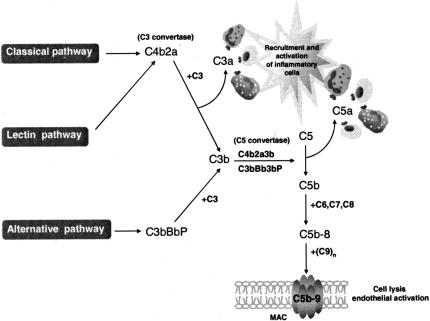
The complement system, composed of over 30 proteins that act in concert to protect the host against invading organisms, initiates inflammation and tissue injury (Figure 1) (8,9). Complement activation promotes chemotaxis of inflammatory cells and generates proteolytic fragments that enhance phagocytosis by neutrophils and monocytes. The classical pathway is activated when natural or elicited antibodies (Ab) bind to antigen and unleash potent effectors associated with humoral responses in immune-mediated tissue damage. Activation of the classical pathway by natural Ab plays a major role in the response to neoepitopes unmasked on ischemic endothelium, and thus may be involved in reperfusion injury (10). The mannose-binding lectin (MBL) pathway is activated by MBL recognition of carbohydrates (often on infectious agents) and MBL-associated serine protease-2, which autoactivates and cleaves complement component 2 (C2) and C4. Alternative pathway activation differs from classical and MBL activation because it is initiated directly by spontaneous deposition of complement on cell surfaces. Under normal physiologic conditions, C3 undergoes low-grade spontaneous hydrolysis and deposits on target surfaces, allowing binding and activation of factor B, formation of the alternative pathway C3 convertase, and further amplification of C3 cleavage. This pathway is antibody-independent and is triggered by the activity of factor B, factor D and properdin. Properdin enhances complement activation by binding to and stabilizing the C3 and C5 convertases. Properdin, the only regulator of complement that amplifies its activation, is produced by T cells, monocytes/macrophages, and polymorphonuclear leukocytes (PMN). Thus, a proinflammatory amplification loop may result from alternative pathway activation of anaphylatoxin-responsive, properdin-secreting inflammatory cells. In addition, recent data show that oxidative stress initiates complement activation by all three pathways (11–13). By means of these recognition and activation mechanisms the complement system identifies and responds to “dangerous” situations presented by foreign antigens, pathogens, tissue injury, ischemia, apoptosis and necrosis (14). This capacity places the complement system at the center of many clinically important responses to pathogens, as well as, to fetal injury mediated by cellular or humoral immune mechanisms.
Fig. 1.
Complement cascade. Schematic diagram of the three complement activation pathways and the products they generate. From Hughes Syndrome, 2nd Edition, Khamashta, MA (Ed.), 2006, page 396, chapter 31, by Girardi, G and Salmon, J, Figure 31.1. With kind permission of Springer Science and Business Media.
The convergence of three complement activation pathways on the C3 protein results in a common pathway of effector functions (Figure 1). The initial step is generation of the fragments C3a and C3b. C3a, an anaphylatoxin that binds to receptors on leukocytes and other cells, causes activation and release of inflammatory mediators (15). C3b and its further sequential cleavage fragments, iC3b and C3d, are ligands for complement receptors 1 and 2 (CR1 and CR2) and the β2 integrins, CD11b/CD18 and CD11c/CD18, present on a variety of inflammatory and immune accessory cells (16,17). C3b attaches covalently to targets, followed by the assembly of C5 convertase with subsequent cleavage of C5 to C5a and C5b. C5a is a potent soluble inflammatory, anaphylatoxic and chemotactic molecule that promotes recruitment and activation of neutrophils and monocytes and mediates endothelial cell activation through its receptor. Expression of C5aR, initially thought to be limited to neutrophils, monocytes and eosinophils, has recently been shown to be more widespread and to include endothelial cells, liver parenchymal cells, vascular smooth muscle cells, bronchial epithelium, alveolar cells, mast cells, astrocytes and human mesangial cells (18).
Binding of C5b to the target initiates the non-enzymatic assembly of the C5b-9 membrane attack complex (MAC). Insertion of C5b-9 MAC into cell membranes causes erythrocyte lysis through changes in intracellular osmolarity, while C5b-9 MAC damages nucleated cells primarily by activating specific signaling pathways through the interaction of the membrane-associated MAC proteins with heterotrimeric G proteins (19,20). Pro-inflammatory and tissue damaging phenotypic outcomes that follow these signaling events include the proliferation and release of reactive oxygen intermediates, leukotrienes, thromboxane, platelet-derived growth factor and procoagulant activity (21–24). That MAC plays a major role in human disease is further supported by the finding that MAC is present at sites of tissue damage in rheumatoid arthritis, and glomerulonephritis (22). Indeed, deficiency or inactivation of CD59, the complement regulatory protein that restricts MAC formation, seen in paroxysmal nocturnal hemoglobinuria and diabetes, respectively, is associated with thrombosis and vasculopathy (25).
Role of complement in fetal tolerance
Because activated complement fragments have the capacity to bind and damage self-tissues, it is imperative that autologous bystander cells be protected from the deleterious effects of complement. To this end, most human and murine cells express soluble and membrane-bound molecules that limit the activation of various complement components (8,26). Studies in mice show that complement regulatory proteins critically determine the sensitivity of host tissues to complement injury in autoimmune, ischemic and inflammatory disorders (26,27). The importance of complement regulatory proteins in humans is underscored by recent reports of a strong association between mutations in membrane cofactor protein (MCP) and factor H (causing ineffective C3 inactivation) with the hemolytic uremic syndrome and glomerular endothelial injury (28,29). Though activated complement components are present in normal placentas (30,31), it appears that in successful pregnancy uncontrolled complement activation is prevented by three regulatory proteins present on the trophoblast membrane: decay accelerating factor (DAF), MCP and CD59 (32–34). All three proteins are strategically positioned on the trophoblast and provide a mechanism to protect the fetus from damage due to complement pathway activation by alloantibodies.
Studies in mouse experimental models underscore the importance of complement regulation in fetal control of maternal processes that mediate tissue damage. In mice, Crry, a membrane-bound intrinsic complement regulatory protein (like DAF and MCP) blocks C3 and C4 activation on self-membranes (35) and thereby inhibits classical and alternative pathway C3 convertases and blocks C3, C5, and subsequent MAC activation. That appropriate complement inhibition is an absolute requirement for normal pregnancy has been demonstrated by the finding that Crry deficiency in utero leads to progressive embryonic lethality (5). Crry−/− embryos are surrounded by activated C3 fragments and PMN, primarily around the ectoplacental cone and surrounding trophoectoderm. Importantly, Crry−/− embryos are completely rescued from 100% lethality and live pups are born at a normal Mendelian frequency when C3 deficiency or factor B deficiency is introduced to Crry−/− embryos (5,36). Defects in placental formation were associated with pregnancy loss and were solely dependent on alternative pathway activation and required maternal C3 (36). These observations provide genetic proof that Crry−/− embryos die in utero due to their inability to suppress complement activation mediated by C3. Based on these findings, we proposed that aPL antibodies activate complement within decidual tissue, overwhelm the normally adequate inhibitory mechanisms described above and induce inflammation and fetal damage.
Role of complement in fetal damage induced by aPL antibodies in a mouse model
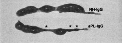
During trophoblast differentiation, phosphatidylserine is externalized on the trophoblast outer leaflet where it provides a target for aPL antibodies (37,38). Indeed, human and mouse studies have shown that aPL antibodies are specifically targeted to trophoblasts. Our in vivo studies show that aPL antibodies are selectively removed from the circulation in pregnant mice and specifically bound to decidual tissues (39). We hypothesized that aPL antibodies bound to trophoblasts activate complement via the classical pathway generating split products that mediate placental injury and cause fetal loss and growth restriction. Using a murine model of APS induced by passive transfer of human aPL antibodies, we have shown that complement activation plays an essential and causative role in pregnancy loss and fetal growth restriction (39,40). Passive transfer of IgG from women with recurrent miscarriage and aPL antibodies (aPL-IgG) results in a 40% frequency of fetal resorption compared to <10% in mice treated with IgG from healthy individuals (NH-IgG), and a 35% reduction in the average weight of surviving fetuses (40). We observed a similar rate of pregnancy failure using IgG from 5 different patients or using monoclonal human aPL antibodies. These in vivo pathogenic effects require both recognition of relevant target antigens by aPL antibodies and Fc domain-mediated complement activation that initiates effector functions. Figure 2 shows representative uteri from mice treated with NH-IgG (6 fetuses, no resorptions) and aPL-IgG (4 fetuses, 3 resorptions).
Fig. 2.
aPL antibodies cause pregnancy loss. Representative uteri from day 15 of pregnancy mice are shown. The upper panel from a mouse treated with normal human IgG (NH-IgG) shows 6 fetuses and no resorptions. The lower panel from a mouse treated with aPL antibodies (aPL-IgG) shows 4 fetuses and 3 resorptions (asterisks).
In our initial studies to determine whether complement activation is necessary for aPL-IgG induced fetal loss, we focused on C3 because the three complement activation pathways converge at C3 and C3 is a nexus for complement effector pathways. We used Crry-Ig, an inhibitor of classical and alternative pathway complement C3 convertases, to block complement activation in vivo (41). BALB/c mice were treated with aPL-IgG (10 mg), control human IgG (10 mg), or saline on days 8 and 12 of pregnancy. Half the mice received Crry-Ig (3 mg) every other day from days 8–12. On day 15, pregnant mice were sacrificed and the uterine contents evaluated. Treatment with aPL-IgG caused a nearly 4-fold increase in the frequency of fetal resorption, while simultaneous treatment with Crry-Ig prevented aPL antibody-induced pregnancy losses. Crry-Ig also prevented aPL antibody-mediated growth restriction in surviving fetuses. As an alternate test of the hypothesis that complement activation is required for aPL-induced pregnancy complications, we studied mice deficient in complement component C3. As predicted by experiments with Crry-Ig, aPL-IgG did not increase fetal resorption in C3−/− mice. The protective effects of a complete lack of C3 were also evident in fetal weights.
To determine whether excessive complement activation occurs within the placenta in aPL-treated mice we conducted immunohistological analyses of decidua after the first treatment with aPL-IgG or control IgG. In aPL-treated mice, the decidua was abnormal morphologically, showing focal necrosis and apoptosis with PMN infiltrates and loss of fetal membrane elements. These findings contrasted with relatively normal appearing deciduas of mice treated with control human IgG or aPL-IgG and Crry-Ig. Staining with antibodies against murine C3 revealed extensive complement deposition within the decidua and on the extraembryonic membranes in mice treated with aPL-IgG. In contrast, in mice treated with control human IgG, only small amounts of C3 staining were detectable, mostly in extraembryonic tissues; the decidua was not inflamed and had normal cellular elements. We infer that membrane bound complement inhibitors, such as Crry, are unable to prevent complement deposition when aPL-IgG within the decidua triggers the activation of large amounts of complement.
We have performed studies to identify initiating pathways of complement activation. That C4-deficient pregnant mice treated with aPL-IgG were protected from fetal loss and growth restriction suggests that aPL antibodies trigger the complement cascade through either the classical or lectin pathways. Activation of the alternative pathway also contributes to aPL-associated fetal loss, because mice deficient in Factor B were protected, as were mice treated with an anti-factor B mAb. We hypothesize that the classical (or lectin) pathway(s) generates C3 at sites of injury, initiating a positive feedback loop through the alternative pathway to amplify complement activation and generate further complement fragments.
Complement C5 activation and C5a-C5aR interactions are required for aPL antibody-induced fetal loss
To distinguish the importance of complement activation at the level of C3 from that at C5, we blocked C5 activation with an anti-C5 monoclonal antibody (mAb). Inhibition at the level of C5 prevents the formation of MAC and generation of C5a, while preserving complement-derived immunoprotective functions such as opsonization and immune complex clearance. Treatment with anti-C5 mAb prevented aPL-induced pregnancy losses (fetal resorption frequency: aPL-IgG vs aPL-IgG + anti-C5 mAb: 44 ± 11% vs 5 ± 3%, p < 0.001) and growth restriction. To confirm that C5 activation is required for fetal loss, we studied mice deficient in C5. In C5+/+ mice, both aPL-IgG and human monoclonal aPL antibodies caused a 4-fold increase in fetal resorption frequency and a significant decrease in embryo weight and development compared to control IgG. In contrast, C5−/− mice were protected from aPL antibody-induced pregnancy complications (6).
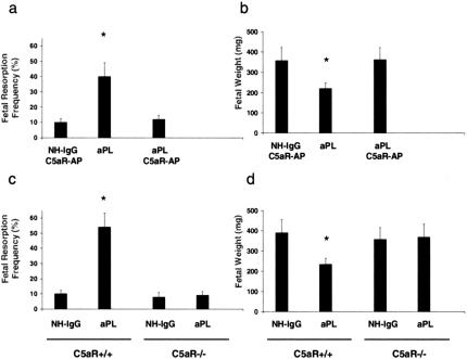
Two complement effector pathways are initiated by cleavage of C5: C5a, a potent anaphylatoxin and cell activator, and C5b, which leads to formation of the C5b-9 membrane attack complex. We used two methods to distinguish the role of C5a and the C5a receptor (C5aR) from that of MAC seeded by C5b. First, we treated pregnant mice that had received aPL-IgG with a highly specific peptide antagonist of C5aR (C5aR-AP), AcPhe[L-ornithine-Pro-D-cyclohexylalanine-Trp-Arg], which possesses potent in vivo anti-inflammatory activities in murine models of endotoxic shock, renal ischemia-reperfusion injury and the Arthus reaction (42–45). Administration of C5aR-AP prevented aPL antibody-induced pregnancy loss and growth restriction, but had no effect on either frequency of fetal resorption or fetal size in the absence of aPL antibodies (Figure 3a and 3b). As a second approach to test the hypothesis that C5a-C5aR interactions mediate pregnancy complications, we performed studies in mice deficient in C5aR. In the background strain, C5aR+/+, there was a 5-fold increase in fetal resorption after treatment with aPL-IgG (Figure 3b), while, as predicted by experiments with the C5aR-AP, aPL-IgG did not increase the frequency of fetal resorption in C5aR−/− mice (Figure 3c). The protective effects of the total absence of C5aR were also observed when fetal weights were examined (Figure 3d). Taken together, our experiments with C5aR−/− mice and C5aR antagonist peptide identify the C5a-C5aR interaction as a critical effector of aPL antibody-induced injury.
Fig. 3.
Blockade of C5a-C5aR interaction protects pregnancies from aPL antibody-associated injury. a and b, Pregnant BALB/c mice were given aPL-IgG or NH-IgG on days 8 and 12, and some also received C5aR antagonist peptide (C5aR-AP) on day 8, 30 minutes before administration of aPL-IgG. Mice were sacrificed on day 15 of pregnancy, uteri were dissected, fetuses were weighed, and frequency of fetal resorption calculated (number of resorptions/number of fetuses + number of resorptions). Treatment with C5aR-AP prevented fetal loss and growth inhibition (*P < 0.01, aPL vs. aPL + C5aR-AP). c and d, The effects of aPL-IgG on pregnancy outcomes in C5aR−/− and C5aR+/+ mice were compared. Pregnant mice were treated with aPL-IgG or NH-IgG as described above. C5aR−/− mice were protected from aPL-IgG-induced fetal resorption and growth inhibition (*P < 0.05, aPL-IgG vs. NH-IgG). From Girardi et al., Complement C5a receptors and neutrophils mediate fetal injury in the antiphospholipid syndrome. J Clin Invest 2003;112:1644–1654. Republished with permission.
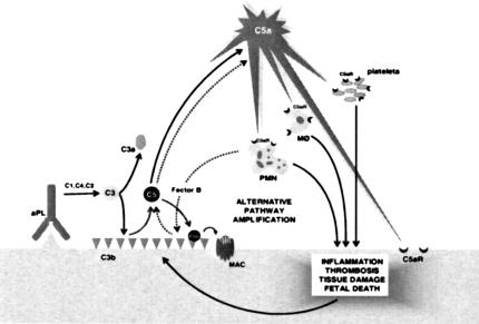
Based on the results of our murine studies, we proposed a mechanism for pregnancy complications in APS (Figure 4): aPL antibodies preferentially targeted at decidua and placenta activate complement via the classical and, perhaps, lectin pathways, (Fc- and C4-dependent), leading to the generation of potent anaphylatoxins (C3a and C5a) and mediators of effector cell activation. Recruitment of inflammatory cells accelerates local alternative pathway activation and creates a proinflammatory amplification loop that enhances C3 activation and deposition, generates additional C3a and C5a, and results in further influx of inflammatory cells into the placenta (39). Depending on the extent of damage, either death in utero or fetal growth restriction ensues.
Fig. 4.
Proposed mechanism for the pathogenic effects of aPL antibodies on tissue injury. APL antibodies are preferentially targeted to the placenta where they activate complement via the classical pathway. The complement cascade is initiated; C3 and subsequently C5 are activated. C5a is generated and attracts and activates neutrophils, monocytes and platelets, and stimulates the release of inflammatory mediators, including reactive oxidants, proteolytic enzymes, chemokines, cytokines and complement factors. Complement activation is amplified by the alternative pathway. This results in further influx of inflammatory cells and ultimately fetal injury. Depending on the extent of damage, either death in utero or fetal growth restriction ensues. (PMN, neutrophil; Mφ, monocyte-macrophage) From Girardi et al., Complement C5a receptors and neutrophils mediate fetal injury in the antiphospholipid syndrome. J Clin Invest 2003; 112:1644–1654. Republished with permission.
Inhibition of complement activation: A novel mechanism for the protective effects of heparin in aPL antibody-induced pregnancy loss.
The studies described above underscore the importance of inflammation, rather than thrombosis, in fetal injury associated with aPL antibodies, yet therapy for pregnant women with APS is focused on preventing thrombosis, and anticoagulation is only partially successful in averting miscarriage. Histopathologic findings in placentas from women with APS also argue that pro-inflammatory factors may contribute to tissue injury (46,47). Interestingly, as early as 1929, heparin, the standard anticoagulant treatment used to prevent obstetric complications in patients with APS, was shown to have “anti-complementary effects” (48). Given the importance of complement split products as mediators of aPL antibody-induced fetal injury, we considered the possibility that heparin prevents pregnancy loss by inhibiting complement activation on trophoblasts and that anticoagulation, in and of itself, is not sufficient to prevent pregnancy complications in APS. In experiments in our model of APS, treatment with unfractionated or low molecular weight heparins (LMWH) protected pregnancies from aPL antibody-induced damage, whereas treatment with hirudin or fondaparinux (anticoagulants without anti-complement effects) was not protective. Mice that received unfractionated heparin (UFH) 20U, LMWH, fondaparinux, or hirudin were anticoagulated, as demonstrated by increased partial thromboplastin time (PTT) or decreased factor Xa activity, while PTTs in mice receiving UFH 10U were not different from control. Importantly, heparins did not alter binding of aPL-IgG to decidual tissue. These findings indicate that even in the absence of a detectable anticoagulant effect, as in mice treated with UFH 10U, heparins protected mice from aPL-induced pregnancy loss (Table 1).
TABLE 1.
Heparins prevent pregnancy loss and inhibit complement activation
| Anticoagulation | Prevention of pregnancy loss | Complement inhibition | |
|---|---|---|---|
| UFH (10 U) | − | + | + |
| UFH (20 U) | + | + | + |
| LMWH | + | + | + |
| Fondaparinux | + | − | − |
| Hirudin | + | − | − |
LMWH, low molecular weight heparin; UFH, unfractionated heparin.
To assess the effect of heparin on complement activation in vivo, we focused on the products of aPL-IgG-initiated C3 cleavage: C3a in plasma (measured as C3a desArg) and C3 activation fragments bound to trophoblasts. Mice treated with aPL-IgG showed a marked increase in plasma C3a desArg levels (compared to mice treated with NH-IgG) and this increase in C3a desArg levels was completely abolished in the presence of heparins. Neither fondaparinux nor hirudin affected C3 activation as detected by plasma C3a desArg (Table 1).
In addition, we developed an in vitro model to directly examine the effect of heparin on aPL-IgG-initiated cleavage of C3 by measuring C3a in supernatants and cell-bound C3b using trophoblast-like BeWo cells. Phosphatidylserine, externalized on BeWo cells as on human villous cytotrophoblasts, is recognized by aPL antibodies (38). BeWo cells opsonized with aPL-IgG were incubated with mouse or human serum, as a source of complement, in the presence or absence of UFH, LMWH, hirudin, or fondaparinux, at concentrations comparable to those administered in the in vivo experiments. In the presence of UFH or LMWH, however, activation of complement was completely inhibited. In contrast to the effects of the heparins, neither hirudin nor fondaparinux blocked the generation of complement split products. The results of these in vitro experiments confirmed the findings of our in vivo studies.
Our data indicate that heparins prevent obstetrical complications in women with APS by limiting aPL antibody-triggered complement activation and the ensuing inflammatory response at the maternal-fetal interface, rather than by inhibiting thrombosis. This work provides a framework for understanding how sub-anticoagulant doses of heparin exert beneficial effects in antibody-mediated tissue injury.
Conclusion
We have identified an important and previously unrecognized role for complement as an early effector in pregnancy loss associated with aPL antibodies and placental inflammation. Our experiments indicate that aPL antibodies targeted to decidual tissues damage pregnancies by engagement of the classical pathway of complement activation, followed by amplification through the alternative pathway, and it appears the heparins prevent obstetrical complications caused by aPL antibodies because they block activation of complement. Because treatment with heparin injections throughout pregnancy is neither safe, well tolerated, nor completely effective, our findings emphasize the importance of developing and testing targeted complement inhibitory therapy for patients with APS.
We have initiated the The PROMISSE Study (Predictors of pRegnancy Outcome: bioMarkers In antiphospholipid antibody Syndrome and Systemic lupus Erythematosus), a prospective, multi-center observational study to translate our findings in mice to humans and evaluate the role of complement in aPL antibody-induced pregnancy loss in women. The PROMISSE Study will test the hypothesis that classical, alternative and terminal complement pathway activation will be detected in the circulation and placentas of patients with aPL antibodies and will be associated with poor pregnancy outcomes. Characterization of surrogate markers that predict poor pregnancy outcome will identify clinically applicable indicators that will enable us to initiate an interventional trial of complement inhibition in patients at risk for aPL antibody-associated fetal loss.
ACKNOWLEDGMENTS
This research was supported in part by grants from NIH, the Mary Kirkland Center for Lupus Research at Hospital for Special Surgery, the Alliance for Lupus Research, and the S.L.E. Foundation, Inc.
DISCUSSION
Blaser: New York: Jane, that was beautiful progressive work. I wonder if you can extend this very specific model of tissue injury in lupus and determine whether this is a prototype for injury in other organ systems in lupus, and that C5a inhibitors might be useful for renal disease or other manifestations.
Salmon: New York: My belief is that it will be the case. Dr. Girardi has set up a mouse model of thrombotic microangiopathy. Again, this is an antiphospholipid antibody-induced injury model, but importantly, complement inhibitors effectively protect mice from renal damage. There is extensive experience using a C5 monoclonal antibody to treat mouse models of lupus and rheumatoid arthritis, and this intervention can limit renal disease and joint disease. Unfortunately, the initial human trials were not effective in rheumatoid arthritis, and the lupus studies were discontinued early. I believe there is still hope for targeting C5 in antibody-mediated injury. There are new drugs, including anti-C5 antibodies, anti-C5a receptor antibodies and C5a receptor antagonist peptides that are already, or nearly, in clinical trials.
Sacher: Cincinnati: Very nice presentation. One of the strategies that is being used in recurrent fetal loss, at least historically, is high dose polyvalent immunoglobulin therapy. Have you thought of exploring this in your model, because, of course, we know that this compound has pleiotrophic multiple effects, and it is beneficial in many diseases. Could there be an anticomplementary effect, or could it be anti-idiotype, or could it be all of those?
Salmon: My sense is that meta-analysis of all of studies with IVIG for recurrent pregnancy loss do not support its use. On the other hand, studies in mice show that IVIG is “anti-inflammatory”. IVIG changes the balance of stimulatory and inhibitory Fc receptors to favor inhibition; it binds complement split products, and it blocks FcRn resulting in increased catabolism of pathogenic antibodies. We have shown that IVIG treatment lowers the titer of antiphospholipid antibodies in mice. There is rationale for using IVIG, but it hasn't been overwhelmingly effective in patients, and I'm not sure why.
Coggins: Boston: Very nice paper. Of course, in situations other than pregnancy, people often benefit from long-term anticoagulation with warfarin, presumably with very different mechanisms, but I wonder if you had any speculations about the possibility of complement approaches to those people as well.
Salmon: I think complement inhibition could be an effective way to prevent thrombophilia associated with aPL antibodies. In a mouse model of injury-induced venous thrombosis, a standard pinch injury to the femoral vein results in a small local clot, but in aPL antibody-treated mice, a gigantic clot is formed. In this example of aPL antibody-induced thombophilia, complement inhibitors prevent enhanced clot formation. To move the field forward, we must create an inhibitor of complement for safe chronic use, one that will not predispose patients to encapsulated microbial infection.
Key: Chapel Hill: It was a very nice presentation. Of course antiphospholipid syndrome is characterized by its heterogeneity. Some people get arterial thrombosis, others venous, other women get these recurrent miscarriages. Do you have an assay to predict this, I mean prospectively? You may have said this, do samples from all comers with antiphospholipid antibodies have this activity, or did you select your patient samples and consider these clinical differences?
Salmon: The samples of immunoglobulin we used in the mouse studies were from many individuals with the antiphosphopholipid syndrome (with or without lupus). They were very sick patients who were treated with plasmapheresis. We selected these patients because we needed very large quantities of purified immunoglobulins with high titers of anticardiolipin antibodies to perfume multiple mouse experiments with the same aPL antibodies. The immunoglobulins we injected into mice were from patients with different phenotypes. I think we've studied eight or nine patients, and we never found a preparation that didn't cause fetal loss. In addition, we've done studies with a range of monoclonal human and mouse antiphospholipid antibodies. If the monoclonal antibody is of a subclass that activates complement, we see fetal loss, but if the monoclonal antibody does not activate, we do not see injury.
Falk: Chapel Hill: Jane, that was beautiful as usual. Two quick questions. Can you accelerate your model by using an FcRIIb knockout? Secondly, how do you know this is not the mannose lectin-binding pathway, because there can be direct activation of C4 and, if Mike Holers is right, additional activation through C3 that would have therapeutic effects downstream for the inhibitors of the mannose lectin-binding pathway that may be different than those that you are suggesting now?
Salmon: We desperately tried to find animals with more severe pregnancy outcomes. We studied FcRIIb knockout mice and we studied FcRIIa transgenic mice looking for a worse phenotype, but neither had a worse phenotype. I was surprised, and can't explain these results. In response to your question about MBL, we've looked at mice that are doubly deficient in factor D and C1q, and they are protected, arguing that MBL pathway alone cannot mediate injury in our model. Nonetheless, we are trying to get the double MBL knockouts to directly address the question, because I think the MBL pathway is important, particularly in hypoxic states.
Falk: Can you rescue by adding factor B?
Salmon: We haven't done those experiments yet, but we've studied factor B-deficient mice and treated mice with anti-factor B monoclonal antibody from the Holer's laboratory. These strategies rescue pregnancies.
REFERENCES
- 1.Wilson WA, Gharavi AE, Koike T, Lockshin MD, Branch DW, Piette JC, Brey R, Derksen R, Harris EN, Hughes GR, Triplett DA, Khamashta MA. International consensus statement on preliminary classification criteria for definite antiphospholipid syndrome: report of an international workshop. Arthritis Rheum. 1999;42:1309–1311. doi: 10.1002/1529-0131(199907)42:7<1309::AID-ANR1>3.0.CO;2-F. [DOI] [PubMed] [Google Scholar]
- 2.Lockshin MD, Sammaritano LR, Schwartzman S. Validation of the Sapporo criteria for antiphospholipid syndrome. Arthritis Rheum. 2000;43:440–443. doi: 10.1002/1529-0131(200002)43:2<440::AID-ANR26>3.0.CO;2-N. [DOI] [PubMed] [Google Scholar]
- 3.Levine JS, Branch DW, Rauch J. The antiphospholipid syndrome. N Engl J Med. 2002;346:752–763. doi: 10.1056/NEJMra002974. [DOI] [PubMed] [Google Scholar]
- 4.Derksen RH, Khamashta MA, Branch DW. Management of the obstetric antiphospholipid syndrome. Arthritis Rheum. 2004;50:1028–1039. doi: 10.1002/art.20105. [DOI] [PubMed] [Google Scholar]
- 5.Xu C, Mao D, Holers VM, Palanca B, Cheng AM, Molina H. A critical role for murine complement regulator crry in fetomaternal tolerance. Science. 2000;287:498–501. doi: 10.1126/science.287.5452.498. [DOI] [PubMed] [Google Scholar]
- 6.Girardi G, Berman J, Redecha P, Spruce L, Thurman JM, Kraus D, Hollmann TJ, Casali P, Caroll MC, Wetsel RA, Lambris JD, Holers VM, Salmon JE. Complement C5a receptors and neutrophils mediate fetal injury in the antiphospholipid syndrome. J Clin Invest. 2003;112:1644–1654. doi: 10.1172/JCI18817. [DOI] [PMC free article] [PubMed] [Google Scholar]
- 7.Holers VM, Girardi G, Mo L, Guthridge JM, Molina H, Pierangeli SS, Espinola R, Xiaowei LE, Mao D, Vialpando CG, Salmon JE. Complement C3 activation is required for antiphospholipid antibody-induced fetal loss. J Exp Med. 2002;195:211–220. doi: 10.1084/jem.200116116. [DOI] [PMC free article] [PubMed] [Google Scholar]
- 8.Abbas AK, Lichtman AH, Pober JS. In Cellular and molecular immunology. Philadelphia, PA: W.B. Saunders Company; 2000. Effector mechanisms of humoral immunity; pp. 316–334. [Google Scholar]
- 9.Schmidt BZ, Colten HR. Complement: a critical test of its biological importance. Immunol Rev. 2000;178:166–176. doi: 10.1034/j.1600-065x.2000.17801.x. [DOI] [PubMed] [Google Scholar]
- 10.Weiser MR, Williams JP, Moore FD, Jr, Kobzik L, Ma M, Hechtman HB, Carroll MC. Reperfusion injury of ischemic skeletal muscle is mediated by natural antibody and complement. J Exp Med. 1996;183:2343–2348. doi: 10.1084/jem.183.5.2343. [DOI] [PMC free article] [PubMed] [Google Scholar]
- 11.Thurman JM, Ljubanovic D, Edelstein CL, Gilkeson GS, Holers VM. Lack of a functional alternative complement pathway ameliorates ischemic acute renal failure in mice. J Immunol. 2003;170:1517–1523. doi: 10.4049/jimmunol.170.3.1517. [DOI] [PubMed] [Google Scholar]
- 12.Hart ML, Walsh MC, Stahl GL. Initiation of complement activation following oxidative stress. In vitro and in vivo observations. Mol Immunol. 2004;41:165–171. doi: 10.1016/j.molimm.2004.03.013. [DOI] [PubMed] [Google Scholar]
- 13.Gadjeva M, Takahashi K, Thiel S. Mannan-binding lectin—a soluble pattern recognition molecule. Mol Immunol. 2004;41:113–121. doi: 10.1016/j.molimm.2004.03.015. [DOI] [PubMed] [Google Scholar]
- 14.Fearon DT. Seeking wisdom in innate immunity. Nature. 1997;388:323–324. doi: 10.1038/40967. [DOI] [PubMed] [Google Scholar]
- 15.Hugli TE. Structure and function of C3a anaphylatoxin. Curr Top Microbiol Immunol. 1990;153:181–208. doi: 10.1007/978-3-642-74977-3_10. [DOI] [PubMed] [Google Scholar]
- 16.Brown EJ. Complement receptors and phagocytosis. Curr Opin Immunol. 1991;3:76–82. doi: 10.1016/0952-7915(91)90081-b. [DOI] [PubMed] [Google Scholar]
- 17.Holers VM. In Complement, principles and practices of clinical immunology. In: Rich R, editor. Mosby, St. Louis, MO: 1995. p. 363. [Google Scholar]
- 18.Wetsel RA. Structure, function and cellular expression of complement anaphylatoxin receptors. Curr Opin Immunol. 1995;7:48–53. doi: 10.1016/0952-7915(95)80028-x. [DOI] [PubMed] [Google Scholar]
- 19.Shin ML, Rus HG, Nicolescu FI. Membrane attack by complement: assembly and biology of terminal complement complexes. Biomembranes. 1996;4:123–149. doi: 10.1007/s12026-011-8239-5. [DOI] [PMC free article] [PubMed] [Google Scholar]
- 20.Morgan BP, Meri S. Membrane proteins that protect against complement lysis. Springer Semin Immunopathol. 1994;15:369–396. doi: 10.1007/BF01837366. [DOI] [PubMed] [Google Scholar]
- 21.Devin DV. The effects of complement activation on platelets. Curr Top Microbiol Immunol. 1992;178:101–113. doi: 10.1007/978-3-642-77014-2_7. [DOI] [PubMed] [Google Scholar]
- 22.Morgan BP. Effects of the membrane attack complex of complement on nucleated cells. Curr Top Microbiol Immunol. 1992;178:115–140. doi: 10.1007/978-3-642-77014-2_8. [DOI] [PubMed] [Google Scholar]
- 23.Benzaquen LR, Nicholson-Weller A, Halperin JA. Terminal complement proteins C5b-9 release basic fibroblast growth factor and platelet-derived growth factor from endothelial cells. J Exp Med. 1994;179:985–992. doi: 10.1084/jem.179.3.985. [DOI] [PMC free article] [PubMed] [Google Scholar]
- 24.Hattori R, Hamilton KK, McEver RP, Sims PJ. Complement proteins C5b-9 induce secretion of high molecular weight multimers of endothelial von Willebrand factor and translocation of granule membrane protein GMP-140 to the cell surface. J Biol Chem. 1989;264:9053–9060. [PubMed] [Google Scholar]
- 25.Manzi S, Rairie JE, Carpenter AB, Kelly RH, Jagarlapudi SP, Sereika SM, Medsger TA, Jr, Ramsey-Goldman R. Sensitivity and specificity of plasma and urine complement split products as indicators of lupus disease activity. Arthritis Rheum. 1996;39:1178–1188. doi: 10.1002/art.1780390716. [DOI] [PubMed] [Google Scholar]
- 26.Song WC. Membrane complement regulatory proteins in autoimmune and inflammatory tissue injury. Curr Dir Autoimmun. 2004;7:181–199. doi: 10.1159/000075693. [DOI] [PubMed] [Google Scholar]
- 27.Yamada K, Miwa T, Liu J, Nangaku M, Song WC. Critical protection from renal ischemia reperfusion injury by CD55 and CD59. J Immunol. 2004;172:3869–3875. doi: 10.4049/jimmunol.172.6.3869. [DOI] [PubMed] [Google Scholar]
- 28.Noris M, Brioschi S, Caprioli J, Todeschni M, Bresin E, Porrati F, Gamba S, Remuzzi G. Familial haemolytic uraemic syndrome and an MCP mutation. Lancet. 2003;362:1542–1547. doi: 10.1016/S0140-6736(03)14742-3. [DOI] [PubMed] [Google Scholar]
- 29.Goodship TH, Liszewski MK, Kemp EJ, Richards A, Atkinson JP. Mutations in CD46, a complement regulatory protein, predispose to atypical HUS. Trends Mol Med. 2004;10:226–231. doi: 10.1016/j.molmed.2004.03.006. [DOI] [PubMed] [Google Scholar]
- 30.Weir PE. Immunofluorescent studies of the uteroplacental arteries in normal pregnancy. Br J Obstet Gynaecol. 1981;88:301–307. doi: 10.1111/j.1471-0528.1981.tb00985.x. [DOI] [PubMed] [Google Scholar]
- 31.Wells M, Bennett J, Bulmer JN, Jackson P, Holgate CS. Complement component deposition in uteroplacental (spiral) arteries in normal human pregnancy. J Reprod Immunol. 1987;12:125–135. doi: 10.1016/0165-0378(87)90040-4. [DOI] [PubMed] [Google Scholar]
- 32.Cunningham DS, Tichenor JR., Jr Decay-accelerating factor protects human trophoblast from complement-mediated attack. Clin Immunol Immunopathol. 1995;74:156–161. doi: 10.1006/clin.1995.1023. [DOI] [PubMed] [Google Scholar]
- 33.Tedesco F, Narchi G, Radillo O, Meri S, Ferrone S, Betterle C. Susceptibility of human trophoblast to killing by human complement and the role of the complement regulatory proteins. J Immunol. 1993;151:1562–1570. [PubMed] [Google Scholar]
- 34.Liszewski MK, Farries TC, Lublin DM, Rooney IA, Atkinson JP. Control of the complement system. Adv Immunol. 1996;61:201–283. doi: 10.1016/s0065-2776(08)60868-8. [DOI] [PubMed] [Google Scholar]
- 35.Kim YU, Kinoshita T, Molina H, Hourcade D, Seya T, Wagner LM, Holers VM. Mouse complement regulatory protein Crry/p65 uses the specific mechanisms of both human decay-accelerating factor and membrane cofactor protein. J Exp Med. 1995;181:151–159. doi: 10.1084/jem.181.1.151. [DOI] [PMC free article] [PubMed] [Google Scholar]
- 36.Mao D, Xiaobo W, Molina H. A dispensable role for neutrophils, C5, and the classical pathway of complement activation in the abnormal fetomaternal tolerance found in crry-deficient mice. International Immunopharmacology. 2002;2:1233. [Google Scholar]
- 37.Rote NS, Stetzer BP. Autoimmune disease as a cause of reproductive failure. Clin Lab Med. 2003;23:265–293. doi: 10.1016/s0272-2712(03)00022-2. [DOI] [PubMed] [Google Scholar]
- 38.Rote NS, Vogt E, DeVere G, Obringer AR, Ng AK. The role of placental trophoblast in the pathophysiology of the antiphospholipid antibody syndrome. Am J Reprod Immunol. 1998;39:125–136. doi: 10.1111/j.1600-0897.1998.tb00344.x. [DOI] [PubMed] [Google Scholar]
- 39.Girardi G, Berman J, Redecha P, Spruce L, Thurman JM, Kraus D, Hollmann TJ, Casali P, Caroll MC, Wetsel RA, Lambris JD, Holers VM, Salmon JE. Complement C5a receptors and neutrophils mediate fetal injury in the antiphospholipid syndrome. J Ciln Invest. 2003;112:1644–1654. doi: 10.1172/JCI18817. [DOI] [PMC free article] [PubMed] [Google Scholar]
- 40.Holers VM, Girardi G, Mo L, Guthridge JM, Molina H, Pierangeli SS, Espinola R, Xiaowel LE, Mao D, Vialpando CG, Salmon JE. Complement C3 activation is required for antiphospholipid antibody-induced fetal loss. J Exp Med. 2002;195:211–220. doi: 10.1084/jem.200116116. [DOI] [PMC free article] [PubMed] [Google Scholar]
- 41.Quigg RJ, Kozono Y, Berthiaume D, Lim A, Salant DJ, Weinfeld A, Griffin P, Kremmer E, Holers VM. Blockade of antibody-induced glomerulonephritis with Crry-Ig, a soluble murine complement inhibitor. J Immunol. 1998;160:4553–4560. [PubMed] [Google Scholar]
- 42.Finch AM, Wong AK, Paczkowski NJ, Wadi SK, Craik DJ, Fairlie DP, Taylor SM. Low-molecular-weight peptidic and cyclic antagonists of the receptor for the complement factor C5a. J Med Chem. 1999;42:1965–1974. doi: 10.1021/jm9806594. [DOI] [PubMed] [Google Scholar]
- 43.Mastellos D, Papadimitriou JC, Franchini S, Tsonis PA, Lambris JD. A novel role of complement: mice deficient in the fifth component of complement (C5) exhibit impaird liver regeneration. J Immunol. 2001;166:2479–2486. doi: 10.4049/jimmunol.166.4.2479. [DOI] [PubMed] [Google Scholar]
- 44.Strachan AJ, Woodruff TM, Haaima G, Fairlie DP, Taylor SM. A new small molecule C5a receptor antagonist inhibits the reverse-passive Arthur reaction and endotoxic shock in rats. J Immunol. 2000;164:6560–6565. doi: 10.4049/jimmunol.164.12.6560. [DOI] [PubMed] [Google Scholar]
- 45.Arumugam TV, Shiels IA, Strachan AJ, Abbenante G, Fairlie DP, Taylor SM. A small molecule C5a receptor antagonist protects kidneys from ischemia/reperfusion injury in rats. Kidney Int. 2003;63:134–142. doi: 10.1046/j.1523-1755.2003.00737.x. [DOI] [PubMed] [Google Scholar]
- 46.Out HJ, Koolijman CD, Bruinse HW, Derksen RH. Histopathological findings in placentae from patients with intra-uterine fetal death and anti-phospholipid antibodies. Eur J Obstet Gynecol Reprod Biol. 1991;41:179–186. doi: 10.1016/0028-2243(91)90021-c. [DOI] [PubMed] [Google Scholar]
- 47.Magid MS, Kaplan C, Sammaritano LR, Peterson M, Druzin ML, Lockshin MD. Placental pathology in systemic lupus erythematosus: a prospective study. Am J Obstet Gynecol. 1998;179:226–234. doi: 10.1016/s0002-9378(98)70277-7. [DOI] [PubMed] [Google Scholar]
- 48.Ecker E, Gross P. Anticomplementary Power of Heparin. J Infect Dis. 1929;44:250. [Google Scholar]


