Abstract
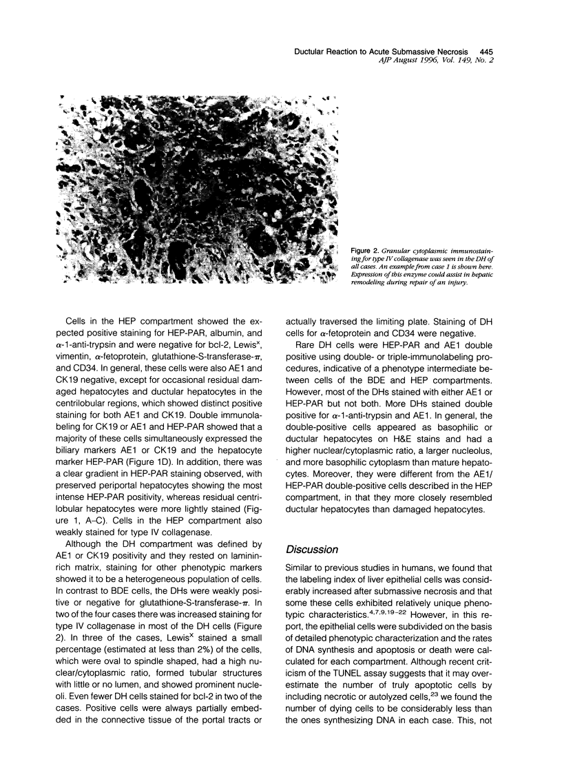
The ductular reaction to acute submassive necrosis was studied in human livers removed at the time of orthotopic liver transplantation. Single, double, and triple immunohistochemical labeling in combination with morphometry was used to analyze the phenotype and proliferative and apoptotic rates of various epithelial cell compartments. These were divided on the basis of immunohistochemistry and morphology into three subtypes: 1) CK19+/AE1+ mature bile duct epithelium, 2) HEP-PAR+ mature hepatocytes (HEPs), and 3) CK19+/AE1+ ductular hepatocyte (DH) cells lying at the interface between the portal tract connective tissue and the hepatic lobules. Cycling cells were defined as those showing Ki-67+ (MIB-1) nuclear labeling. Apoptotic cells were identified with in situ labeling using the terminal deoxynucleotidyl transferase-mediated dUTP-digoxigenin nick end labeling assay. Special emphasis was placed on DHs that appeared at the interface between the portal tracts and hepatic lobules. During the recovery phase from submassive hepatic necrosis, subtraction of the rate of cell death from the proliferative index shows that all of the epithelial compartments experience a net increase in the number of cells. The highest proliferation rate occurs in the DHs, which is significantly (P < 0.0001) higher than the proliferation rate seen in either the HEP or bile duct epithelium compartments. Immunohistochemical analysis of the highly proliferative DH compartment shows it to be a heterogeneous population with unique phenotypic features. Like epithelial cells in the ductal plate of fetal liver and cholangiocarcinomas, DHs are positioned on a laminin-rich matrix and focally express vimentin and Lewis(x) and show up-regulation of bcl-2 and type IV collagenase. However, unlike ductal plate cells, DHs are CD34 and alpha-fetoprotein negative. Although a subpopulation of DHs share phenotypic features with mature bile duct epithelium (AE1/cytokeratin 19 and type IV collagenase positive) or HEP (HEP-PAR, albumin, and alpha-1-antitrypsin positive), they are also clearly separate from both populations; DHs are negative or only weakly stain for glutathione-S-transferase-pi and are type IV collagenase positive. Moreover, occasional DHs also co-expressed HEP-PAR or alpha-1-antitrypsin and AE1, indicative of both hepatocyte and ductular differentiation. These findings suggest that DHs seen in human livers after submassive necrosis may represent a transient amplifying population arising from a progenitor population located in or near the canals of Herring. In addition, injured hepatocytes can express cytokeratin 19 and AE1, which normally are biliary intermediate filaments.
Full text
PDF


Images in this article
Selected References
These references are in PubMed. This may not be the complete list of references from this article.
- Blakolmer K., Jaskiewicz K., Dunsford H. A., Robson S. C. Hematopoietic stem cell markers are expressed by ductal plate and bile duct cells in developing human liver. Hepatology. 1995 Jun;21(6):1510–1516. [PubMed] [Google Scholar]
- Brill S., Holst P., Sigal S., Zvibel I., Fiorino A., Ochs A., Somasundaran U., Reid L. M. Hepatic progenitor populations in embryonic, neonatal, and adult liver. Proc Soc Exp Biol Med. 1993 Dec;204(3):261–269. doi: 10.3181/00379727-204-43662. [DOI] [PubMed] [Google Scholar]
- Burt A. D., MacSween R. N. Bile duct proliferation--its true significance? Histopathology. 1993 Dec;23(6):599–602. doi: 10.1111/j.1365-2559.1993.tb01258.x. [DOI] [PubMed] [Google Scholar]
- Callea F., Fevery J., Massi G., de Groote J., Desmet V. J. Storage of alpha-1-antitrypsin in intrahepatic bile duct cells in alpha-1-antitrypsin deficiency (Pi Z phenotype). Histopathology. 1985 Jan;9(1):99–108. doi: 10.1111/j.1365-2559.1985.tb02973.x. [DOI] [PubMed] [Google Scholar]
- Charlotte F., L'Herminé A., Martin N., Geleyn Y., Nollet M., Gaulard P., Zafrani E. S. Immunohistochemical detection of bcl-2 protein in normal and pathological human liver. Am J Pathol. 1994 Mar;144(3):460–465. [PMC free article] [PubMed] [Google Scholar]
- Dabeva M. D., Shafritz D. A. Activation, proliferation, and differentiation of progenitor cells into hepatocytes in the D-galactosamine model of liver regeneration. Am J Pathol. 1993 Dec;143(6):1606–1620. [PMC free article] [PubMed] [Google Scholar]
- Delladetsima J. K., Kyriakou V., Vafiadis I., Karakitsos P., Smyrnoff T., Tassopoulos N. C. Ductular structures in acute hepatitis with panacinar necrosis. J Pathol. 1995 Jan;175(1):69–76. doi: 10.1002/path.1711750111. [DOI] [PubMed] [Google Scholar]
- Evarts R. P., Nagy P., Marsden E., Thorgeirsson S. S. A precursor-product relationship exists between oval cells and hepatocytes in rat liver. Carcinogenesis. 1987 Nov;8(11):1737–1740. doi: 10.1093/carcin/8.11.1737. [DOI] [PubMed] [Google Scholar]
- Evarts R. P., Nagy P., Nakatsukasa H., Marsden E., Thorgeirsson S. S. In vivo differentiation of rat liver oval cells into hepatocytes. Cancer Res. 1989 Mar 15;49(6):1541–1547. [PubMed] [Google Scholar]
- Fausto N. The american journal of pathology-are we there? Am J Pathol. 1993 Jul;143(1):1–2. [PMC free article] [PubMed] [Google Scholar]
- Fucich L. F., Cheles M. K., Thung S. N., Gerber M. A., Marrogi A. J. Primary vs metastatic hepatic carcinoma. An immunohistochemical study of 34 cases. Arch Pathol Lab Med. 1994 Sep;118(9):927–930. [PubMed] [Google Scholar]
- Fukuda Y., Imoto M., Koyama Y., Miyazawa Y., Nakano I., Hattori M., Urano F., Kodama S., Iwata K., Hayakawa T. Immunohistochemical study on tissue inhibitors of metalloproteinases in normal and pathological human livers. Gastroenterol Jpn. 1991 Feb;26(1):37–41. doi: 10.1007/BF02779506. [DOI] [PubMed] [Google Scholar]
- Gerber M. A., Thung S. N., Shen S., Stromeyer F. W., Ishak K. G. Phenotypic characterization of hepatic proliferation. Antigenic expression by proliferating epithelial cells in fetal liver, massive hepatic necrosis, and nodular transformation of the liver. Am J Pathol. 1983 Jan;110(1):70–74. [PMC free article] [PubMed] [Google Scholar]
- Grasl-Kraupp B., Ruttkay-Nedecky B., Koudelka H., Bukowska K., Bursch W., Schulte-Hermann R. In situ detection of fragmented DNA (TUNEL assay) fails to discriminate among apoptosis, necrosis, and autolytic cell death: a cautionary note. Hepatology. 1995 May;21(5):1465–1468. doi: 10.1002/hep.1840210534. [DOI] [PubMed] [Google Scholar]
- Grisham J. W., Coleman W. B., Smith G. J. Isolation, culture, and transplantation of rat hepatocytic precursor (stem-like) cells. Proc Soc Exp Biol Med. 1993 Dec;204(3):270–279. doi: 10.3181/00379727-204-43663. [DOI] [PubMed] [Google Scholar]
- Hamanoue M., Kawaida K., Takao S., Shimazu H., Noji S., Matsumoto K., Nakamura T. Rapid and marked induction of hepatocyte growth factor during liver regeneration after ischemic or crush injury. Hepatology. 1992 Dec;16(6):1485–1492. doi: 10.1002/hep.1840160626. [DOI] [PubMed] [Google Scholar]
- Hine C. H., Pasi A., Stephens B. G. Fatalities following exposure to 2-nitropropane. J Occup Med. 1978 May;20(5):333–337. [PubMed] [Google Scholar]
- Inoue M., Kratz G., Haegerstrand A., Ståhle-Bäckdahl M. Collagenase expression is rapidly induced in wound-edge keratinocytes after acute injury in human skin, persists during healing, and stops at re-epithelialization. J Invest Dermatol. 1995 Apr;104(4):479–483. doi: 10.1111/1523-1747.ep12605917. [DOI] [PubMed] [Google Scholar]
- Kayano K., Yasunaga M., Kubota M., Takenaka K., Mori K., Yamashita A., Kubo Y., Sakaida I., Okita K., Sanuki K. Detection of proliferating hepatocytes by immunohistochemical staining for proliferating cell nuclear antigen (PCNA) in patients with acute hepatic failure. Liver. 1992 Jun;12(3):132–136. doi: 10.1111/j.1600-0676.1992.tb00571.x. [DOI] [PubMed] [Google Scholar]
- Kleiner D. E., Jr, Stetler-Stevenson W. G. Structural biochemistry and activation of matrix metalloproteases. Curr Opin Cell Biol. 1993 Oct;5(5):891–897. doi: 10.1016/0955-0674(93)90040-w. [DOI] [PubMed] [Google Scholar]
- Koukoulis G., Rayner A., Tan K. C., Williams R., Portmann B. Immunolocalization of regenerating cells after submassive liver necrosis using PCNA staining. J Pathol. 1992 Apr;166(4):359–368. doi: 10.1002/path.1711660407. [DOI] [PubMed] [Google Scholar]
- Li J., Rosman A. S., Leo M. A., Nagai Y., Lieber C. S. Tissue inhibitor of metalloproteinase is increased in the serum of precirrhotic and cirrhotic alcoholic patients and can serve as a marker of fibrosis. Hepatology. 1994 Jun;19(6):1418–1423. [PubMed] [Google Scholar]
- Lichtinghagen R., Helmbrecht T., Arndt B., Böker K. H. Expression pattern of matrix metalloproteinases in human liver. Eur J Clin Chem Clin Biochem. 1995 Feb;33(2):65–71. doi: 10.1515/cclm.1995.33.2.65. [DOI] [PubMed] [Google Scholar]
- Milani S., Herbst H., Schuppan D., Grappone C., Pellegrini G., Pinzani M., Casini A., Calabró A., Ciancio G., Stefanini F. Differential expression of matrix-metalloproteinase-1 and -2 genes in normal and fibrotic human liver. Am J Pathol. 1994 Mar;144(3):528–537. [PMC free article] [PubMed] [Google Scholar]
- Murray G. I., Paterson P. J., Weaver R. J., Ewen S. W., Melvin W. T., Burke M. D. The expression of cytochrome P-450, epoxide hydrolase, and glutathione S-transferase in hepatocellular carcinoma. Cancer. 1993 Jan 1;71(1):36–43. doi: 10.1002/1097-0142(19930101)71:1<36::aid-cncr2820710107>3.0.co;2-j. [DOI] [PubMed] [Google Scholar]
- Oikarinen A., Kylmäniemi M., Autio-Harmainen H., Autio P., Salo T. Demonstration of 72-kDa and 92-kDa forms of type IV collagenase in human skin: variable expression in various blistering diseases, induction during re-epithelialization, and decrease by topical glucocorticoids. J Invest Dermatol. 1993 Aug;101(2):205–210. doi: 10.1111/1523-1747.ep12363823. [DOI] [PubMed] [Google Scholar]
- POPPER H., KENT G., STEIN R. Ductular cell reaction in the liver in hepatic injury. J Mt Sinai Hosp N Y. 1957 Sep-Oct;24(5):551–556. [PubMed] [Google Scholar]
- Popper H. The relation of mesenchymal cell products to hepatic epithelial systems. Prog Liver Dis. 1990;9:27–38. [PubMed] [Google Scholar]
- Roeb E., Rose-John S., Erren A., Edwards D. R., Matern S., Graeve L., Heinrich P. C. Tissue inhibitor of metalloproteinases-2 (TIMP-2) in rat liver cells is increased by lipopolysaccharide and prostaglandin E2. FEBS Lett. 1995 Jan 2;357(1):33–36. doi: 10.1016/0014-5793(94)01301-g. [DOI] [PubMed] [Google Scholar]
- Rolland G., Xu J., Dupret J. M., Post M. Expression and characterization of type IV collagenases in rat lung cells during development. Exp Cell Res. 1995 May;218(1):346–350. doi: 10.1006/excr.1995.1165. [DOI] [PubMed] [Google Scholar]
- Rubin E. M., Martin A. A., Thung S. N., Gerber M. A. Morphometric and immunohistochemical characterization of human liver regeneration. Am J Pathol. 1995 Aug;147(2):397–404. [PMC free article] [PubMed] [Google Scholar]
- Ruebner B. H., Blankenberg T. A., Burrows D. A., SooHoo W., Lund J. K. Development and transformation of the ductal plate in the developing human liver. Pediatr Pathol. 1990;10(1-2):55–68. doi: 10.3109/15513819009067096. [DOI] [PubMed] [Google Scholar]
- Saarialho-Kere U. K., Vaalamo M., Airola K., Niemi K. M., Oikarinen A. I., Parks W. C. Interstitial collagenase is expressed by keratinocytes that are actively involved in reepithelialization in blistering skin disease. J Invest Dermatol. 1995 Jun;104(6):982–988. doi: 10.1111/1523-1747.ep12606231. [DOI] [PubMed] [Google Scholar]
- Seki S., Sakaguchi H., Kawakita N., Yanai A., Kuroki T., Kobayashi K. Analysis of proliferating biliary epithelial cells in human liver disease using a monoclonal antibody against DNA polymerase alpha. Virchows Arch A Pathol Anat Histopathol. 1993;422(2):133–143. doi: 10.1007/BF01607165. [DOI] [PubMed] [Google Scholar]
- Sell S. Is there a liver stem cell? Cancer Res. 1990 Jul 1;50(13):3811–3815. [PubMed] [Google Scholar]
- Shah K. D., Gerber M. A. Development of intrahepatic bile ducts in humans. Possible role of laminin. Arch Pathol Lab Med. 1990 Jun;114(6):597–600. [PubMed] [Google Scholar]
- Sirica A. E., Cole S. L., Williams T. A unique rat model of bile ductular hyperplasia in which liver is almost totally replaced with well-differentiated bile ductules. Am J Pathol. 1994 Jun;144(6):1257–1268. [PMC free article] [PubMed] [Google Scholar]
- Sirica A. E. Ductular hepatocytes. Histol Histopathol. 1995 Apr;10(2):433–456. [PubMed] [Google Scholar]
- Sirica A. E., Gainey T. W., Mumaw V. R. Ductular hepatocytes. Evidence for a bile ductular cell origin in furan-treated rats. Am J Pathol. 1994 Aug;145(2):375–383. [PMC free article] [PubMed] [Google Scholar]
- Sirica A. E., Williams T. W. Appearance of ductular hepatocytes in rat liver after bile duct ligation and subsequent zone 3 necrosis by carbon tetrachloride. Am J Pathol. 1992 Jan;140(1):129–136. [PMC free article] [PubMed] [Google Scholar]
- Slott P. A., Liu M. H., Tavoloni N. Origin, pattern, and mechanism of bile duct proliferation following biliary obstruction in the rat. Gastroenterology. 1990 Aug;99(2):466–477. doi: 10.1016/0016-5085(90)91030-a. [DOI] [PubMed] [Google Scholar]
- Takahara T., Furui K., Funaki J., Nakayama Y., Itoh H., Miyabayashi C., Sato H., Seiki M., Ooshima A., Watanabe A. Increased expression of matrix metalloproteinase-II in experimental liver fibrosis in rats. Hepatology. 1995 Mar;21(3):787–795. [PubMed] [Google Scholar]
- Terada T., Nakanuma Y. Detection of apoptosis and expression of apoptosis-related proteins during human intrahepatic bile duct development. Am J Pathol. 1995 Jan;146(1):67–74. [PMC free article] [PubMed] [Google Scholar]
- Terada T., Nakanuma Y. Expression of tenascin, type IV collagen and laminin during human intrahepatic bile duct development and in intrahepatic cholangiocarcinoma. Histopathology. 1994 Aug;25(2):143–150. doi: 10.1111/j.1365-2559.1994.tb01570.x. [DOI] [PubMed] [Google Scholar]
- Terada T., Nakanuma Y. Profiles of expression of carbohydrate chain structures during human intrahepatic bile duct development and maturation: a lectin-histochemical and immunohistochemical study. Hepatology. 1994 Aug;20(2):388–397. [PubMed] [Google Scholar]
- Thung S. N. The development of proliferating ductular structures in liver disease. An immunohistochemical study. Arch Pathol Lab Med. 1990 Apr;114(4):407–411. [PubMed] [Google Scholar]
- Van Eyken P., Sciot R., Callea F., Van der Steen K., Moerman P., Desmet V. J. The development of the intrahepatic bile ducts in man: a keratin-immunohistochemical study. Hepatology. 1988 Nov-Dec;8(6):1586–1595. doi: 10.1002/hep.1840080619. [DOI] [PubMed] [Google Scholar]
- Wolf H. K., Michalopoulos G. K. Hepatocyte regeneration in acute fulminant and nonfulminant hepatitis: a study of proliferating cell nuclear antigen expression. Hepatology. 1992 Apr;15(4):707–713. doi: 10.1002/hep.1840150426. [DOI] [PubMed] [Google Scholar]


