Abstract
Measles virus (MV) possesses two envelope glycoproteins, namely, the receptor-binding hemagglutinin (H) and fusion proteins. Wild-type MV strains isolated in B-lymphoid cell lines use signaling lymphocyte activation molecule (SLAM), but not CD46, as a cellular receptor, whereas MV vaccine strains of the Edmonston lineage use both SLAM and CD46 as receptors. Studies have shown that the residue at position 481 of the H protein is critical in determining the use of CD46 as a receptor. However, the wild-type IC-B strain with a single N481Y substitution in the H protein utilizes CD46 rather inefficiently. In this study, a number of chimeric and mutant H proteins, and recombinant viruses harboring them, were generated to determine which residues of the Edmonston H protein are responsible for its efficient use of CD46. Our results show that three substitutions (N390I and E492G plus N416D or T446S), in addition to N481Y, are necessary for the IC-B H protein to use CD46 efficiently as a receptor. The N390I, N416D, and T446S substitutions are present in the H proteins of all strains of the Edmonston lineage, whereas the E492G substitution is found only in the H protein of the Edmonston tag strain generated from cDNAs. The T484N substitution, found in some of the Edmonston-lineage strains, resulted in a similar effect on the use of CD46 to that caused by the E492G substitution. Thus, multiple residues in the H protein that have not previously been implicated have important roles in the interaction with CD46.
Measles virus (MV), a member of the genus Morbillivirus in the family Paramyxoviridae, is an enveloped virus with a nonsegmented negative-strand RNA genome. It possesses two envelope glycoproteins, the hemagglutinin (H) and fusion (F) proteins, and initiates infection of target cells via binding of the H protein to its cellular receptor. This binding is believed to induce F protein-mediated membrane fusion between the viral envelope and the host cell plasma membrane, allowing entry of the ribonucleoprotein complex (18). Two cellular proteins, CD46 and signaling lymphocyte activation molecule (SLAM; also called CD150), have been identified as MV receptors (5, 7, 11, 26, 54, 59, 60). CD46 is a member of the regulators of complement activation family and is expressed on all nucleated human cells (22). SLAM, a glycoprotein of the immunoglobulin superfamily, is a regulator of antigen-driven T-cell responses and macrophage functions. The expression of SLAM is restricted to certain cells of the immune system, including activated B and T lymphocytes, mature dendritic cells, and macrophages (1, 4, 44).
The Edmonston strain, the first isolate of MV, was obtained in 1954 from a patient with measles by using a primary culture of human kidney cells (6). Live attenuated MV vaccines currently in use were obtained by passaging the original isolate numerous times in a variety of cell types, including primary human kidney and amnion cells and chicken embryo fibroblasts (9, 38). These Edmonston-lineage vaccine strains appear to have adapted to efficient growth in many cell types by acquiring a number of mutations in their genomes (31, 32). They are safe and very effective, but the molecular bases of their adaptation and attenuation remain to be elucidated.
MV strains isolated from B-lymphoid cell lines, such as marmoset B95a and human BJAB cells, have been shown to retain the phenotype of viruses circulating in patients with measles (16, 17), and they use SLAM, but not CD46, as a cellular receptor (30, 40, 54, 59, 60). In this report, the term “wild type” refers to this type of MV strains. In contrast, Edmonston-lineage vaccine strains use both SLAM and CD46 as cellular receptors (54, 59, 60). It has been shown that the amino acid residue at position 481 of the H protein has an important role in determining the receptor usage of MV strains (3, 12, 20, 27, 43, 53, 55, 58). The H proteins of most CD46-using strains, including Edmonston vaccine strains, have a tyrosine residue at that position, whereas those of wild-type strains usually have an asparagine residue (3, 15, 20, 32, 37, 49). Studies have indicated that an asparagine-to-tyrosine substitution at position 481 (N481Y) enables the H proteins of wild-type MV strains to bind CD46, without compromising their ability to use SLAM (8, 12, 20, 58). In fact, when wild-type MV strains adapt to SLAM-negative and CD46-positive Vero cells, the N481Y substitution is often observed after several passages (21, 27, 43). Mutational analysis of the MV H protein, combined with its structural modeling, confirmed the importance of the tyrosine residue at position 481 in the interaction with CD46 (23, 55). However, using recombinant viruses, we previously showed that although a single N481Y substitution in the H protein conferred the ability to infect cells via CD46 on the wild-type IC-B strain, its ability to use CD46 was much lower than that of virus possessing the Edmonston H protein (42).
In this study, a number of chimeric and mutant H proteins, and recombinant viruses harboring them, were generated to determine which residues of the Edmonston H protein are responsible for its efficient use of CD46. Our study identified several substitutions in the H protein, in addition to the N481Y substitution, that are necessary for the wild-type IC-B strain to be fully capable of using CD46 as an alternative receptor.
MATERIALS AND METHODS
Cells and viruses.
Vero cells constitutively expressing human SLAM (Vero/hSLAM) (30) were maintained in Dulbecco's modified Eagle's medium (DMEM; ICN Biomedicals, Aurora, OH) supplemented with 7.5% fetal bovine serum (FBS) and 500 μg of G418 (Geneticin; Nacalai Tesque, Tokyo, Japan) per ml. CHO cells constitutively expressing human SLAM (CHO/hSLAM) (54) were maintained in RPMI medium (ICN Biomedicals) supplemented with 7.5% FBS and 500 μg of G418 per ml. B95a, CHO, and Jurkat cells were maintained in RPMI medium supplemented with 7.5% FBS. HeLa cells were maintained in DMEM supplemented with 7.5% FBS. Recombinant MVs were generated from cDNAs by using CHO/hSLAM cells and a vaccinia virus carrying the T7 RNA polymerase, i.e., vTF7-3 (a gift from B. Moss) or LO-T7-1 (a gift from M. Kohara), as reported previously (25, 47). Generated MVs were propagated in B95a cells, and virus stocks at two or three passages in B95a cells were used for experiments.
Plasmid construction.
All full-length genome plasmids were derived from p(+)MV323, which encodes the antigenomic full-length cDNA of the wild-type IC-B strain of MV (50). The p(+)MV323-EGFP plasmid, with an additional transcriptional unit for enhanced green fluorescent protein (EGFP), was reported previously (10). The plasmid p(+)MV2A (a gift from M. A. Billeter) encodes the full-length antigenomic cDNA of the Edmonston B strain (34). The plasmids p(+)MV323/EdH-EGFP and p(+)MV323/H(N481Y)-EGFP were reported previously (10, 42). The p(+)MV323/EdH-EGFP plasmid contains the H gene obtained from p(+)MV2A (10). In this paper, the H protein encoded by this plasmid is named the Edmonston tag H protein. There are two predicted amino acid changes in the Edmonston tag H protein compared with the reported sequence of the Edmonston B strain H protein (GenBank accession number Z66517). The Edmonston tag H protein has threonine and glycine at positions 484 and 492, respectively, whereas the H protein of the Edmonston B strain has asparagine and glutamic acid at those positions (Table 1). A full-length genome plasmid that has the H gene of the Edmonston B strain [p(+)MV323/EdBH-EGFP] was also constructed. The H gene cDNA of the Edmonston B strain was obtained from the pCA-Ed-H plasmid (a gift from K. Takeuchi), which encodes the same amino acid sequence as that in the reported Edmonston B strain (52). Using NheI and PpuMI restriction enzymes, the region between nucleotide positions 7426 and 8277 of p(+)MV323-EGFP or p(+)MV323/H(N481Y)-EGFP was replaced with the corresponding region of p(+)MV2A, generating the full-length genome plasmids p(+)MV/H-β12-EGFP and p(+)MV/H-β12(N481Y)-EGFP, respectively (Fig. 1A). Using PpuMI and AleI restriction enzymes, the regions corresponding to nucleotides 8277 to 8952 and 8330 to 9011 of p(+)MV323-EGFP were replaced with the corresponding regions of p(+)MV2A, generating p(+)MV/H-β34-EGFP and p(+)MV/H-β346-EGFP, respectively (Fig. 1A). Nucleotide position numbers are shown in accordance with the sequence of the IC-B strain genome (GenBank accession number NC_001498) (51). Amino acid substitutions (N390I, N416D, T446S, T484N, and E492G) were introduced either independently or in various combinations into p(+)MV323-EGFP or p(+)MV/H(N481Y)-EGFP by site-directed mutagenesis using complementary primer pairs. The resulting constructs were named p(+)MV-EGFP-H1 to -H13 (see Fig. 3A). Individual H genes were also cloned into the eukaryotic expression plasmid pCA7 (48), a derivative of pCAGGS (28). The pCA7 vector has the T7 promoter in addition to the CAG promoter and was referred to as pCAG-T7 vector in previous papers (25, 47).
TABLE 1.
Amino acid differences among MV strains
| Predicted domaina | Amino acid position | Strain
|
||
|---|---|---|---|---|
| IC-B | Ed tag | Ed B | ||
| Not specified | 174 | A | T | T |
| 176 | A | T | T | |
| β-sheet 1 | 211 | S | G | G |
| 235 | G | E | E | |
| 243 | G | R | R | |
| 252 | H | Y | Y | |
| 276 | F | L | L | |
| β-sheet 2 | 284 | F | L | L |
| 296 | F | L | L | |
| 302 | R | G | G | |
| 334 | R | Q | Q | |
| β-sheet 3 | 390 | N | I | I |
| 416 | N | D | D | |
| β-sheet 4 | 446 | T | S | S |
| 481 | N | Y | Y | |
| 484 | T | T | Nb | |
| 492 | E | G | E | |
| β-sheet 6 | 575 | K | Q | Q |
Based on a structural model of the MV H protein by Vongpunsawad et al. (55).
Some of the Ed B strains possess a threonine at position 484, whereas others have an asparagine.
FIG. 1.
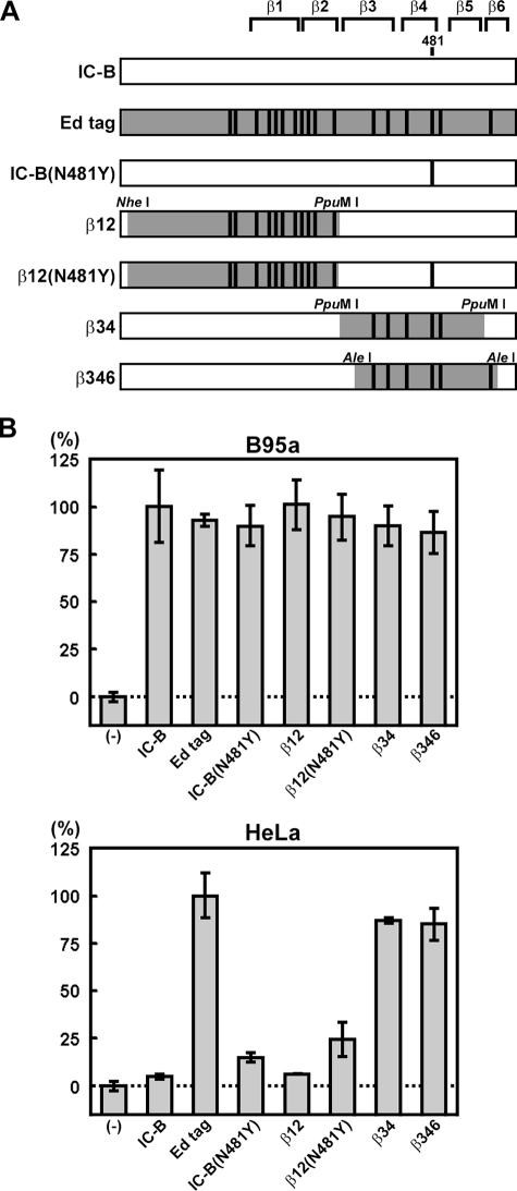
Fusion-inducing activities of chimeric H proteins, with or without N481Y substitution. (A) Diagrams of chimeric H proteins. There were 17 amino acid differences (shown by vertical lines) between the H proteins of the IC-B and Edmonston tag (Ed tag) strains. Regions derived from the Edmonston tag H protein are shaded, and those derived from the IC-B H protein remain white. Restriction enzyme recognition sites used for the construction of chimeric H proteins are indicated. Amino acid position 481 and regions of the six β-sheets (β1 to β6) are indicated. (B) Quantification of membrane fusion activity. B95a and HeLa cells transfected with pG1NT7βgal were incubated with LO-T7-1-infected CHO cells expressing MV H and F proteins, and cell-to-cell fusion was quantified by measuring β-galactosidase activity. β-Galactosidase activity with the IC-B H protein was set to 100% for B95a cells, while that with the Edmonston tag H protein was set to 100% for HeLa cells. β-Galactosidase activity in CHO cells expressing the MV F protein, but not the H protein, was set to 0% (−). The bars indicate the means ± standard deviations for triplicate samples.
FIG. 3.
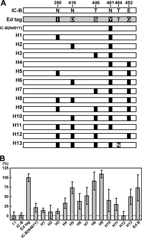
Fusion-inducing activities of H protein mutants. (A) Diagrams of H protein mutants. Regions encompassing β-sheets 3 and 4 of the IC-B H, Edmonston tag H, and mutant H proteins are shown. There were five amino acid differences, at positions 390, 416, 446, 481, and 492, in this region between the H proteins of the IC-B and Edmonston tag strains. The residues at these positions are shown with single letters. The positions of substitutions introduced into the IC-B H protein are shown by black boxes. (B) Quantification of membrane fusion activity. The assay was performed as described in the legend to Fig. 1B. β-Galactosidase activity with the Edmonston tag H protein was set to 100%, and that without the H protein was set to 0% (−). Ed B, Edmonston B.
Quantitative fusion assay.
A quantitative fusion assay was performed using a method described previously (29), with minor modifications. Briefly, monolayers of CHO cells (effector cells) in 24-well cluster plates were infected with LO-T7-1 at a multiplicity of infection (MOI) of 0.5, incubated for 1 h at 37°C, and then transfected with 0.2 μg of an appropriate plasmid (pCA7 plasmid encoding the IC-B, Edmonston tag, Edmonston B, or other mutant H protein) together with 0.2 μg of the IC-B F protein-encoding plasmid, pCA7-ICF, per well, using Lipofectamine 2000 reagent (Invitrogen, Carlsbad, CA). Monolayers of B95a or HeLa cells (target cells) in 24-well cluster plates were transfected with 0.5 μg of pG1NT7βgal (a gift from E. A. Berger), a plasmid containing the lacZ gene under the control of the T7 promoter (29). Twelve hours after transfection, the target cells were harvested, suspended in RPMI medium containing 7.5% fetal calf serum, and transferred to the monolayers of effector cells. After 7 h, β-galactosidase activity in the cells was quantified by a chemiluminescence assay (Roche Diagnostics, Indianapolis, IN).
Virus titration.
Monolayers of Vero/hSLAM cells in 24-well cluster plates were incubated with 50-μl serially diluted virus samples for 1 h at 37°C. After a 1-h incubation, 150 μl of DMEM supplemented with 7.5% FBS and 100 μg/ml fusion block peptide (Z-d-Phe-Phe-Gly) (35) (Peptide Institute Inc., Osaka, Japan) was added to each well to block the second round of infection by progeny viruses. At 36 h postinfection, the number of EGFP-expressing cells was counted under a fluorescence microscope. The number was expressed in cell infectious units (CIU). The number of CIU of each recombinant MV was also determined on HeLa cells and compared with that on Vero/hSLAM cells. The number of CIU of each virus on Vero/hSLAM cells was set to 100%.
Replication kinetics.
HeLa and Jurkat cells in six-well cluster plates were infected with recombinant MVs at an MOI of 0.01 per cell. At various time intervals, cells were harvested in culture medium, and CIU were determined on Vero/hSLAM cells.
RESULTS
Fusion-inducing activities of chimeric H proteins in HeLa cells.
Previous studies have suggested that the MV H protein has a globular ectodomain with six β-sheets (19, 23, 55). There were 17 predicted amino acid differences in the H protein between the wild-type IC-B and Edmonston tag strains (Table 1). Differences were found throughout the ectodomain, except in β-sheet 5. For this report, the predicted locations of respective residues in the tertiary structure of the H protein were based on the model by Vongpunsawad et al. (55). To identify which region of the Edmonston H protein is responsible for its efficient use of CD46, chimeric H proteins were generated using convenient restriction enzyme sites (Fig. 1A). β12, β34, and β346 were chimeric H proteins in which regions containing β-sheets 1 and 2, β-sheets 3 and 4, and β-sheets 3, 4, and 6 of the IC-B H protein were replaced with the corresponding sequences of the Edmonston tag strain (Fig. 1A). β12 also contained two substitutions (positions 174 and 176) outside the β-sheet structures. β34 and β346 possessed one substitution (position 492) in the connecting loop between β-sheets 4 and 5. Furthermore, an N481Y substitution was introduced into the IC-B and β12 H proteins, generating IC-B(N481Y) and β12(N481Y), respectively.
The activities of these H proteins in causing membrane fusion were analyzed using B95a (SLAM+ CD46−) or HeLa (SLAM− CD46+) cells as targets (Fig. 1B). IC-B H, Edmonston tag H, and all mutant H proteins were expressed, together with the IC-B F protein, on the surfaces of CHO cells (effectors) by using expression plasmids. When B95a cells were used as targets, all H proteins induced SLAM-dependent membrane fusion at similar efficiencies, as determined by the quantitative fusion assay (Fig. 1B). When HeLa cells were used as targets, the CD46-dependent fusion-inducing activities of the IC-B and β12 H proteins were marginal. IC-B(N481Y) showed only ∼20% of the activity of the Edmonston tag H protein, consistent with our previous finding that an N481Y substitution alone is not sufficient for the IC-B H protein to use CD46 efficiently (42). β12(N481Y) exhibited activity similar to that of IC-B(N481Y). In contrast, β34 and β346 showed strong fusion-inducing activities almost equivalent to that of the Edmonston tag H protein. These results indicate that the region encompassing β-sheets 3 and 4 of the Edmonston tag H protein is responsible for strong fusion-inducing activity in HeLa cells.
Entry and growth of recombinant MVs with chimeric H proteins.
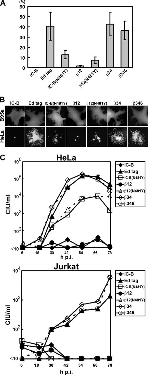
To further characterize the usage of CD46, the EGFP-expressing recombinant MVs with chimeric H proteins described above (Fig. 1A) were generated using a reverse genetics method. The entry of each recombinant MV into HeLa cells was determined by counting the number of EGFP-expressing cells after infection, as described previously (10, 42, 45) (entry into Vero/hSLAM cells was set to 100%) (Fig. 2A). In this assay, secondary infections were blocked with a fusion block peptide. The recombinant MV with the Edmonston tag H protein entered HeLa cells efficiently (∼40%), as it possessed the H protein that can use both SLAM and CD46 as receptors (54, 59, 60). The virus with the β12 H protein entered HeLa cells very poorly (∼2%), like the parental virus with the IC-B H protein. The entry efficiencies of the viruses with the IC-B(N481Y) and β12(N481Y) H proteins were 13% and 8%, respectively, which are much lower than that of the virus with the Edmonston tag H protein. In agreement with the results of the fusion assay, the recombinant MVs with β34 and β346 H proteins entered HeLa cells as efficiently as the one with the Edmonston tag H protein.
FIG. 2.
Characterization of recombinant MVs containing chimeric H proteins, with or without N481Y substitution. (A) Entry efficiencies of recombinant MVs in HeLa cells. The number of CIU of each virus stock was determined in HeLa and Vero/hSLAM cells, and the CIU in HeLa cells was compared with that in Vero/hSLAM cells. The CIU in Vero/hSLAM cells was set to 100%. (B) EGFP autofluorescence in MV-infected cell monolayers. B95a and HeLa cells were infected with recombinant MVs at an MOI of 0.01. Panels show representative images captured with a fluorescence microscope 2 days after infection. (C) Replication kinetics of recombinant MVs. HeLa and Jurkat cells were infected with recombinant MVs at an MOI of 0.01. At various time intervals, cells were harvested in culture medium, and CIU were determined in Vero/hSLAM cells. Average CIU for duplicate experiments are shown.
The cytopathic effect (CPE) produced by recombinant MVs was then examined (Fig. 2B). All recombinant MVs formed extensive syncytia in B95a cells, and no apparent difference was found among the viruses. In contrast, the viruses exhibited different CPEs in HeLa cells. The virus with the Edmonston tag H protein caused extensive syncytium formation in HeLa cells, whereas the one with the IC-B H protein failed to produce syncytia. The results were consistent with previous findings observed in Vero cells (10, 42, 52). The virus with the IC-B(N481Y) H protein produced syncytia, but the number and size of these were much smaller than those produced by the virus with the Edmonston tag H protein, as observed in Vero cells (42). The viruses with the β12 and β12(N481Y) H proteins caused CPEs similar to those caused by the viruses with the IC-B and IC-B(N481Y) H proteins, respectively. In contrast, the viruses with the β34 and β346 H proteins produced large syncytia like those produced by the virus with the Edmonston tag H protein. Syncytium formation in HeLa cells by these recombinant MVs (except for those with the IC-B or β12 H protein, which did not produce syncytia) was completely blocked by a monoclonal antibody against CD46 (clone M75, a gift from T. Seya) (data not shown), indicating that these infections and fusions were CD46 dependent.
Next, the replication kinetics of these recombinant viruses was analyzed in HeLa and Jurkat cells (Fig. 2C). These cell lines express CD46 but not SLAM, and the expression level of CD46 on Jurkat cells is lower than that on HeLa cells (42). The virus with the Edmonston tag H protein replicated well in both HeLa and Jurkat cells, whereas that with the IC-B H protein did not. As reported previously (42), virus with the IC-B(N481Y) H protein replicated less efficiently in HeLa cells than did that with the Edmonston tag H protein, and it failed to replicate in Jurkat cells. The virus with the β12 H protein did not grow in either cell line, similar to the virus with the IC-B H protein. The recombinant virus with the β12(N481Y) H protein showed almost the same replication kinetics in both cell types as the virus with the IC-B(N481Y) H protein. The recombinant viruses with the β34 and β346 H proteins replicated in both HeLa and Jurkat cells as efficiently as the virus with the Edmonston tag H protein. Thus, only three viruses, namely, those having the Edmonston tag, β34, and β346 H proteins, were capable of replicating in Jurkat cells.
Fusion-inducing activities of various mutant H proteins in HeLa cells.
The above results indicate that the region encompassing β-sheets 3 and 4 of the Edmonston tag H protein is important for CD46-dependent membrane fusion, virus entry, and virus replication. There are five amino acid differences in this region between the IC-B and Edmonston tag strains, with two in β-sheet 3 (positions 390 and 416), two in β-sheet 4 (positions 446 and 481), and one in the connecting loop between β-sheets 4 and 5 (position 492) (Table 1). The residues at these positions in the IC-B H protein were replaced in various combinations, generating 12 mutant H proteins (H1 to H12) in addition to IC-B(N481Y) (Fig. 3A). CD46-dependent fusion-inducing activities of these H proteins were analyzed quantitatively in HeLa cells by using expression plasmids (Fig. 3B). The activity of the Edmonston tag H protein was set to 100%. The H12 protein, having all of the substitutions except for the one at position 481, had little fusion-inducing activity, similar to the IC-B H protein, confirming that the N481Y substitution is essential for fusion activity in HeLa cells (20). The addition of another substitution, at position 390, 416, or 446 (H1, H2, and H3, respectively), had no enhancing effect on fusion activity. In contrast, the H4 protein, with an E492G substitution in addition to N481Y, had a slightly better fusion-inducing activity (∼30%) than did the IC-B(N481Y) H protein.
A third substitution, at position 390, 416, or 446, in addition to N481Y and E492G, was then introduced into the IC-B H protein (H5, H6, and H7, respectively). Among these three mutant H proteins, the H5 protein exhibited a higher fusion activity (∼73%) than the H4 protein. When one more substitution, at position 416 or 446, was included besides N390I, N481Y, and E492G (H8 and H9, respectively), the H protein exhibited a fusion-inducing activity almost equivalent to that of the Edmonston tag H protein. Omission of N390I or E492G from the β34 H protein (H10 and H11, respectively) greatly reduced the fusion activity. These results indicate that three substitutions (N390I and E492G plus N416D or T446S) in addition to N481Y are necessary for the IC-B H protein to induce membrane fusion in HeLa cells as efficiently as the Edmonston tag H protein.
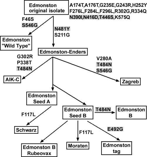
The three substitutions, N390I, N416D, and T446S, are all present in all Edmonston-lineage strains compared with the IC-B strain, whereas the E492G substitution is found only in the Edmonston tag strain (Table 1; Fig. 4) (32, 38). Importantly, many strains of the Edmonston lineage (the Zagreb, AIK-C, and ATCC strains as well as some of the Edmonston B derivatives) are reported to possess an asparagine instead of a threonine at position 484 (32, 38), which may compensate for the absence of the E492G substitution in increasing CD46-dependent fusion activity. Therefore, we also examined the fusion-inducing activity of the IC-B H protein with a T484N substitution in addition to N390I, N416D, T446S, and N481Y (H13) (Fig. 3A) as well as that of the Edmonston B H protein with an asparagine at position 484 (Table 1). The fusion-inducing activities of these H proteins were much higher than that of IC-B(N481Y) but not as efficient as that of the Edmonston tag H protein (Fig. 3B).
FIG. 4.
Lineage of the Edmonston-related MV strains and prediction of amino acid substitutions in their H proteins. Boxes indicate the MV strains, and each arrow connecting the boxes indicates the relationship of a seed strain with its derivative strains. No virus stock of the original isolate of the Edmonston strain exists, and no sequence data are available. By analyzing sequence data for Edmonston-derived MV strains, the H protein of the original isolate of the Edmonston strain was predicted to contain 14 amino acid differences (shown to the right of the box for the Edmonston original isolate) compared with the IC-B strain. Substitutions predicted to be introduced during passages in cell cultures are shown beside the arrows. Substitutions in the shaded boxes were shown to be important for the efficient use of CD46 as a receptor. (Adapted from reference 38 with permission from the publisher.)
Entry and growth of recombinant MVs with various mutant H proteins.
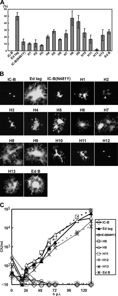
EGFP-expressing recombinant MVs were generated to contain the above mutant H proteins (H1 to H13; viruses were named according to the mutant H proteins they possessed) or the Edmonston B H protein. The entry efficiencies of these recombinant viruses were consistent with the fusion-inducing activities of the H proteins they possessed (Fig. 5A). The H12 virus barely entered HeLa cells, like the parental recombinant virus with the IC-B H protein. In contrast, the H8 and H9 viruses were able to enter HeLa cells almost as efficiently as the virus with the Edmonston tag H protein. Viruses with the H13 or Edmonston B H protein showed higher entry efficiencies than the IC-B(N481Y) virus but lower efficiencies than the virus with the Edmonston tag H protein.
FIG. 5.
Characterization of recombinant MVs with mutant H proteins. (A) Entry efficiencies of recombinant MVs in HeLa cells. The number of CIU in each virus stock was determined in HeLa and Vero/hSLAM cells, and the CIU in HeLa cells was compared with that in Vero/hSLAM cells. The CIU in Vero/hSLAM cells was set to 100%. (B) EGFP autofluorescence in MV-infected monolayers of HeLa cells. Panels show representative images obtained with a fluorescence microscope 2 days after infection. (C) Replication kinetics of recombinant MVs in Jurkat cells. Jurkat cells were infected with recombinant MVs at an MOI of 0.01. At various time intervals, cells were harvested in culture medium, and CIU were determined in Vero/hSLAM cells. Average CIU for duplicate experiments are shown.
Syncytium formation by these recombinant MVs was examined in B95a and HeLa cells. All viruses produced large syncytia in B95a cells (data not shown). In agreement with the data from fusion and entry assays, the H8 and H9 viruses produced large syncytia in HeLa cells, similar to those produced by the virus with the Edmonston tag H protein, whereas the viruses with the IC-B or H12 H protein produced no syncytia (Fig. 5B). Other recombinant viruses produced various sizes of syncytia in HeLa cells. Syncytium formation in HeLa cells by these MVs was completely blocked by anti-CD46 monoclonal antibody M75 (data not shown).
The replication kinetics of these viruses was analyzed in Jurkat cells (Fig. 5C). The H8 and H9 viruses replicated as efficiently as the virus with the Edmonston tag H protein. The H12 virus did not grow at all, like the parental virus with the IC-B H protein. Similar to the virus with the IC-B(N481Y) H protein, the H11 virus did not replicate in Jurkat cells, although it did exhibit some fusion-inducing and entry activities in HeLa cells. The H1, H2, and H3 viruses also did not replicate in Jurkat cells at all (data not shown). The H13 virus, which had an extra T484N substitution compared with the H11 virus, replicated almost as efficiently as the virus with the Edmonston tag H protein. The virus with the Edmonston B H protein also replicated well in Jurkat cells. These results suggest that the T484N substitution has an important role in improving the ability of virus to utilize CD46 as a receptor. The other recombinant viruses grew in Jurkat cells, but not as efficiently as the H8, H9, and H13 viruses (data not shown).
DISCUSSION
Our previous study showed that a single N481Y substitution in the H protein is insufficient to allow the wild-type MV IC-B strain to use CD46 efficiently as a receptor and to grow in CD46+ Jurkat cells (42). In this report, by generating chimeric and mutant H proteins and recombinant viruses harboring them, we show that three substitutions (N390I and E492G plus N416D or T446S) in the H protein, in addition to N481Y, are necessary for the IC-B strain to use CD46 efficiently.
The E492G substitution was found to have the most important role, after the N481Y substitution, in the efficient use of CD46. Notably, this substitution (compared with the sequence of the IC-B H protein) is present only in the Edmonston tag strain, not in other strains of the Edmonston lineage (32, 38). The Edmonston tag strain is an infectious clone generated from cDNAs derived from the Edmonston B strain (34). However, a short segment at the junction of the N and P genes of the full-length genomic cDNA was derived from a different source, the subacute sclerosing panencephalitis-derived IP-3-Ca cell line (2). When the infectious cDNA was constructed, this short segment tag remained (34). There are several discrepancies in the sequence between the Edmonston tag and other Edmonston vaccine strains (our unpublished observation). These mutations were probably introduced into the genome of the Edmonston B strain during passage in cultured cells before preparing genomic RNA for the construction of the full-length genomic cDNA.
The absence of the E492G substitution in the H proteins of other Edmonston vaccine strains might contradict the notion that they use CD46 efficiently. However, the T484N substitution in the H protein, which is not found in the Edmonston tag strain, exists in many Edmonston-lineage strains, including the AIK-C and Zagreb vaccine strains as well as some of the Edmonston B strains. Johnston et al. also reported that their Edmonston tag strain has the T484N substitution in the H protein instead of the E492G substitution (15). Furthermore, non-Edmonston-lineage vaccine strains, such as Leningrad-16, Changchun-47, and Shanghai-191, also have the T484N substitution (38). Therefore, the effect of T484N substitution on the use of CD46 was investigated. Our results show that the T484N substitution, when present together with the N481Y substitution, can enhance the ability of the IC-B H protein to use CD46; however, its effect is not as strong as that of the E492G substitution. Thus, the H protein of the Edmonston tag strain appears to be capable of using CD46 more efficiently than those of other vaccine strains.
All Edmonston-lineage strains have isoleucine, aspartic acid, and serine at positions 390, 416, and 446 of the H protein, respectively, suggesting that these residues were already present in the original isolate (32, 38). Thus, only two additional substitutions, N481Y plus T484N or E492G, were needed for their H proteins to acquire the ability to use CD46 efficiently. The Ma93F strain of genotype C2 was also reported to need two substitutions, at positions 451 and 481 of the H protein, to be capable of using CD46 as a receptor (20). In contrast, the wild-type IC-B strain (genotype D3) has to undergo four substitutions, including N481Y substitution in the H protein, in order to use CD46 efficiently. Examination revealed that most of the reference strains of different genotypes have the same residues as the Edmonston-lineage strains (genotype A) at positions 390, 416, 446, 484, and 492 of the H protein (57). Interestingly, the reference strain of genotype D2 (Johannesburg.SOA/88/1) has an asparagine at position 484 of the H protein in addition to a tyrosine at position 481, whereas that of genotype E (Goettingen.DEU/71 “Braxator”) possesses glycine at positions 492 and 546 of the H protein (57). It is known that S546G substitution in the H protein, like the N481Y substitution, allows MV to use CD46; indeed, some Vero cell-grown MVs have an S546G substitution in the H protein instead of N481Y (13, 14, 21, 24, 27, 32, 36, 37, 43, 46, 56). Thus, these two reference strains seem to have the ability to use CD46 efficiently. At any rate, the requirement of multiple (at least two) amino acid substitutions in the H protein for the efficient use of CD46 might partly explain why many passages in Vero cells are usually required to obtain CD46-using MVs and why CD46-using viruses are barely detected in patients with measles (30).
Recently, two structural models for the MV H protein were reported (23, 55). A series of H protein mutants were examined for SLAM- or CD46-dependent fusion-inducing activity, and the residues important for the interaction with SLAM and CD46 were identified (23, 24, 55). Those studies showed that CD46-relevant residues are mainly located in β-sheet 4 (positions 428, 431, 451, 452, 464, and 481) and the connecting loop between β-sheets 4 and 5 (positions 486 and 487) of the ectodomain, and some residues might reside in β-sheet 5 (positions 527, 546, 548, and 549). Using a different approach, we showed that five residues, at positions 390, 416, 446, 484, and 492 (not identified by the above studies), also have a role in the efficient use of CD46, in addition to the one at position 481. Residues at positions 390 and 416 are situated in β-sheet 3, those at positions 446 and 481 are in β-sheet 4, and those at positions 484 and 492 are in the connecting loop between β-sheets 4 and 5. Thus, CD46-relevant residues identified by previous and present studies are localized over a wide range of domains in the H protein (3, 24, 33, 55). It is possible that some of the residues identified are involved in direct CD46 binding, whereas others affect the structure of the H protein such that its binding site can interact with CD46 efficiently. In fact, N416D substitution might affect the N-glycosylation pattern of the H protein (37, 39). The observation that many of these CD46-relevant residues might not be localized on the surface of the H protein (23, 55) is consistent with this interpretation. Determination of the crystal structure of the H protein, which is in progress in our laboratory, will reveal the roles of these CD46-relevant residues in its interaction with CD46.
During passage in a variety of cultured cells, the Edmonston-lineage strains must also have accumulated mutations in other genes (31, 32). Recently, we showed that amino acid substitutions in the M protein (P64S and E89K) allowed the wild-type IC-B strain to grow well in Vero cells (45). Since the virus with such an Edmonston-like M protein still entered Vero cells very poorly, these substitutions likely enhance virus replication at a postentry step(s). Importantly, these substitutions in the M protein inhibit cell-to-cell fusion and attenuate MV growth in SLAM-positive lymphoid cells (45). Substitutions in the L protein (found in the Edmonston-lineage strains) were also shown to contribute to efficient MV growth in Vero cells (45). These substitutions in the L protein also attenuate virus growth in lymphoid cells. In contrast, the substitutions in the H protein neither compromise the ability to use SLAM nor attenuate MV growth in SLAM-positive cells. Therefore, MV strains that have acquired the ability to use CD46 might gain a growth advantage in human and monkey cells. However, the CD46-using phenotype might be disadvantageous for MV to spread in patients, as it causes downregulation of CD46 from infected cells so that these might be subject to complement-mediated cell lysis (41). Animal experiments might provide clues to the effects of different receptor usages on MV pathogenicity and vaccine attenuation.
Acknowledgments
We thank E. A. Berger, M. A. Billeter, and K. Takeuchi for providing pG1NT7β gal, p(+)MV2A, and pCA-Ed-H, respectively. We also thank M. Kohara, B. Moss, and T. Seya for providing recombinant vaccinia viruses (LO-T7-1 and vTF7-3) and anti-CD46 monoclonal antibody M75, respectively.
This work was supported by grants from the Ministry of Education, Science and Culture of Japan and the Ministry of Health, Labor and Welfare of Japan.
Footnotes
Published ahead of print on 20 December 2006.
REFERENCES
- 1.Aversa, G., J. Carballido, J. Punnonen, C.-C. J. Chang, T. Hauser, B. G. Cocks, and J. E. de Vries. 1997. SLAM and its role in T cell activation and Th cell responses. Immunol. Cell Biol. 75:202-205. [DOI] [PubMed] [Google Scholar]
- 2.Ballart, I., D. Eschle, R. Cattaneo, A. Schmid, M. Metzler, J. Chan, S. Pifko-Hirst, S. A. Udem, and M. A. Billeter. 1990. Infectious measles virus from cloned cDNA. EMBO J. 9:379-384. [DOI] [PMC free article] [PubMed] [Google Scholar] [Retracted]
- 3.Bartz, R., U. Brinckmann, L. M. Dunster, B. Rima, V. ter Meulen, and J. Schneider-Schaulies. 1996. Mapping amino acids of the measles virus hemagglutinin responsible for receptor (CD46) downregulation. Virology 224:334-337. [DOI] [PubMed] [Google Scholar]
- 4.Cocks, B. G., C.-C. J. Chang, J. M. Carballido, H. Yssel, J. E. de Vries, and G. Aversa. 1995. A novel receptor involved in T-cell activation. Nature 376:260-263. [DOI] [PubMed] [Google Scholar]
- 5.Dorig, R. E., A. Marcil, A. Chopra, and C. D. Richardson. 1993. The human CD46 molecule is a receptor for measles virus (Edmonston strain). Cell 75:295-305. [DOI] [PubMed] [Google Scholar]
- 6.Enders, J. F., and T. C. Peebles. 1954. Propagation in tissue cultures of cytopathic agents from patients with measles. Proc. Soc. Exp. Biol. Med. 86:277-286. [DOI] [PubMed] [Google Scholar]
- 7.Erlenhoefer, C., W. J. Wurzer, S. Loffler, S. Schneider-Schaulies, V. ter Meulen, and J. Schneider-Schaulies. 2001. CD150 (SLAM) is a receptor for measles virus but is not involved in viral contact-mediated proliferation inhibition. J. Virol. 75:4499-4505. [DOI] [PMC free article] [PubMed] [Google Scholar]
- 8.Erlenhofer, C., W. Duprex, B. Rima, V. ter Meulen, and J. Schneider-Schaulies. 2002. Analysis of receptor (CD46, CD150) usage by measles virus. J. Gen. Virol. 83:1431-1436. [DOI] [PubMed] [Google Scholar]
- 9.Griffin, D. E. 2001. Measles virus, p. 1401-1441. In D. M. Knipe, P. M. Howley, D. E. Griffin, R. A. Lamb, M. A. Martin, B. Roizman, and S. E. Straus (ed.), Fields virology, 4th ed. Lippincott Williams & Wilkins, Philadelphia, PA.
- 10.Hashimoto, K., N. Ono, H. Tatsuo, H. Minagawa, M. Takeda, K. Takeuchi, and Y. Yanagi. 2002. SLAM (CD150)-independent measles virus entry as revealed by recombinant virus expressing green fluorescent protein. J. Virol. 76:6743-6749. [DOI] [PMC free article] [PubMed] [Google Scholar]
- 11.Hsu, E., C. Iorio, F. Sarangi, A. Khine, and C. Richardson. 2001. CDw150(SLAM) is a receptor for a lymphotropic strain of measles virus and may account for the immunosuppressive properties of this virus. Virology 279:9-21. [DOI] [PubMed] [Google Scholar]
- 12.Hsu, E. C., F. Sarangi, C. Iorio, M. S. Sidhu, S. A. Udem, D. L. Dillehay, W. Xu, P. A. Rota, W. J. Bellini, and C. D. Richardson. 1998. A single amino acid change in the hemagglutinin protein of measles virus determines its ability to bind CD46 and reveals another receptor on marmoset B cells. J. Virol. 72:2905-2916. [DOI] [PMC free article] [PubMed] [Google Scholar]
- 13.Hummel, K. B., and W. J. Bellini. 1995. Localization of monoclonal antibody epitopes and functional domains in the hemagglutinin protein of measles virus. J. Virol. 69:1913-1916. [DOI] [PMC free article] [PubMed] [Google Scholar]
- 14.Hummel, K. B., J. A. Vanchiere, and W. J. Bellini. 1994. Restriction of fusion protein mRNA as a mechanism of measles virus persistence. Virology 202:665-672. [DOI] [PubMed] [Google Scholar]
- 15.Johnston, I. C. D., V. ter Meulen, J. Schneider-Schaulies, and S. Schneider-Schaulies. 1999. A recombinant measles vaccine virus expressing wild-type glycoproteins: consequences for viral spread and cell tropism. J. Virol. 73:6903-6915. [DOI] [PMC free article] [PubMed] [Google Scholar]
- 16.Kobune, F., H. Sakata, and A. Sugiura. 1990. Marmoset lymphoblastoid cells as a sensitive host for isolation of measles virus. J. Virol. 64:700-705. [DOI] [PMC free article] [PubMed] [Google Scholar]
- 17.Kobune, F., H. Takahashi, K. Terao, T. Ohkawa, Y. Ami, Y. Suzaki, N. Nagata, H. Sakata, K. Yamanouchi, and C. Kai. 1996. Nonhuman primate models of measles. Lab. Anim. Sci. 46:315-320. [PubMed] [Google Scholar]
- 18.Lamb, R. A. 1993. Paramyxovirus fusion: a hypothesis for changes. Virology 197:1-11. [DOI] [PubMed] [Google Scholar]
- 19.Langedijk, J. P., F. J. Daus, and J. T. van Oirschot. 1997. Sequence and structure alignment of Paramyxoviridae attachment proteins and discovery of enzymatic activity for a morbillivirus hemagglutinin. J. Virol. 71:6155-6167. [DOI] [PMC free article] [PubMed] [Google Scholar]
- 20.Lecouturier, V., J. Fayolle, M. Caballero, J. Carabana, M. L. Celma, R. Fernandez-Munoz, T. F. Wild, and R. Buckland. 1996. Identification of two amino acids in the hemagglutinin glycoprotein of measles virus (MV) that govern hemadsorption, HeLa cell fusion, and CD46 downregulation: phenotypic markers that differentiate vaccine and wild-type MV strains. J. Virol. 70:4200-4204. [DOI] [PMC free article] [PubMed] [Google Scholar]
- 21.Li, L., and Y. Qi. 2002. A novel amino acid position in hemagglutinin glycoprotein of measles virus is responsible for hemadsorption and CD46 binding. Arch. Virol. 147:775-786. [DOI] [PubMed] [Google Scholar]
- 22.Liszewski, M. K., T. W. Post, and J. P. Atkinson. 1991. Membrane cofactor protein (MCP or CD46): newest member of the regulators of complement activation gene cluster. Annu. Rev. Immunol. 9:431-455. [DOI] [PubMed] [Google Scholar]
- 23.Masse, N., M. Ainouze, B. Neel, T. F. Wild, R. Buckland, and J. P. Langedijk. 2004. Measles virus (MV) hemagglutinin: evidence that attachment sites for MV receptors SLAM and CD46 overlap on the globular head. J. Virol. 78:9051-9063. [DOI] [PMC free article] [PubMed] [Google Scholar]
- 24.Masse, N., T. Barrett, C. P. Muller, T. F. Wild, and R. Buckland. 2002. Identification of a second major site for CD46 binding in the hemagglutinin protein from a laboratory strain of measles virus (MV): potential consequences for wild-type MV infection. J. Virol. 76:13034-13038. [DOI] [PMC free article] [PubMed] [Google Scholar]
- 25.Nakatsu, Y., M. Takeda, M. Kidokoro, M. Kohara, and Y. Yanagi. 2006. Rescue system for measles virus from cloned cDNA driven by vaccinia virus Lister vaccine strain. J. Virol. Methods 137:152-155. [DOI] [PubMed] [Google Scholar]
- 26.Naniche, D., G. Varior-Krishnan, F. Cervoni, T. F. Wild, B. Rossi, C. Rabourdin-Combe, and D. Gerlier. 1993. Human membrane cofactor protein (CD46) acts as a cellular receptor for measles virus. J. Virol. 67:6025-6032. [DOI] [PMC free article] [PubMed] [Google Scholar]
- 27.Nielsen, L., M. Blixenkrone-Moller, M. Thylstrup, N. J. Hansen, and G. Bolt. 2001. Adaptation of wild-type measles virus to CD46 receptor usage. Arch. Virol. 146:197-208. [DOI] [PubMed] [Google Scholar]
- 28.Niwa, H., K. Yamamura, and J. Miyazaki. 1991. Efficient selection for high-expression transfectants with a novel eukaryotic vector. Gene 108:193-199. [DOI] [PubMed] [Google Scholar]
- 29.Nussbaum, O., C. C. Broder, and E. A. Berger. 1994. Fusogenic mechanisms of enveloped-virus glycoproteins analyzed by a novel recombinant vaccinia virus-based assay quantitating cell fusion-dependent reporter gene activation. J. Virol. 68:5411-5422. [DOI] [PMC free article] [PubMed] [Google Scholar]
- 30.Ono, N., H. Tatsuo, Y. Hidaka, T. Aoki, H. Minagawa, and Y. Yanagi. 2001. Measles viruses on throat swabs from measles patients use signaling lymphocytic activation molecule (CDw150) but not CD46 as a cellular receptor. J. Virol. 75:4399-4401. [DOI] [PMC free article] [PubMed] [Google Scholar]
- 31.Parks, C. L., R. A. Lerch, P. Walpita, H. P. Wang, M. S. Sidhu, and S. A. Udem. 2001. Analysis of the noncoding regions of measles virus strains in the Edmonston vaccine lineage. J. Virol. 75:921-933. [DOI] [PMC free article] [PubMed] [Google Scholar]
- 32.Parks, C. L., R. A. Lerch, P. Walpita, H. P. Wang, M. S. Sidhu, and S. A. Udem. 2001. Comparison of predicted amino acid sequences of measles virus strains in the Edmonston vaccine lineage. J. Virol. 75:910-920. [DOI] [PMC free article] [PubMed] [Google Scholar]
- 33.Patterson, J. B., F. Scheiflinger, M. Manchester, T. Yilma, and M. B. Oldstone. 1999. Structural and functional studies of the measles virus hemagglutinin: identification of a novel site required for CD46 interaction. Virology 256:142-151. [DOI] [PubMed] [Google Scholar]
- 34.Radecke, F., P. Spielhofer, H. Schneider, K. Kaelin, M. Huber, C. Dotsch, G. Christiansen, and M. A. Billeter. 1995. Rescue of measles viruses from cloned DNA. EMBO J. 14:5773-5784. [DOI] [PMC free article] [PubMed] [Google Scholar]
- 35.Richardson, C. D., A. Scheid, and P. W. Choppin. 1980. Specific inhibition of paramyxovirus and myxovirus replication by oligopeptides with amino acid sequences similar to those at the N-termini of the F1 or HA2 viral polypeptides. Virology 105:205-222. [DOI] [PubMed] [Google Scholar]
- 36.Rima, B. K., J. A. P. Earle, K. Baczko, V. ter Meulen, U. G. Liebert, C. Carstens, J. Carabana, M. Caballero, M. L. Celma, and R. Fernandez-Munoz. 1997. Sequence divergence of measles virus haemagglutinin during natural evolution and adaptation to cell culture. J. Gen. Virol. 78:97-106. [DOI] [PubMed] [Google Scholar]
- 37.Rota, J. S., K. B. Hummel, P. A. Rota, and W. J. Bellini. 1992. Genetic variability of the glycoprotein genes of current wild-type measles isolates. Virology 188:135-142. [DOI] [PubMed] [Google Scholar]
- 38.Rota, J. S., Z. D. Wang, P. A. Rota, and W. J. Bellini. 1994. Comparison of sequences of the H, F, and N coding genes of measles virus vaccine strains. Virus Res. 31:317-330. [DOI] [PubMed] [Google Scholar]
- 39.Sakata, H., F. Kobune, T. A. Sato, K. Tanabayashi, A. Yamada, and A. Sugiura. 1993. Variation in field isolates of measles virus during an 8-year period in Japan. Microbiol. Immunol. 37:233-237. [DOI] [PubMed] [Google Scholar]
- 40.Schneider-Schaulies, J., J.-J. Schnorr, U. Brinckmann, L. M. Dunster, K. Baczko, U. G. Liebert, S. Schneider-Schaulies, and V. ter Meulen. 1995. Receptor usage and differential downregulation of CD46 by measles virus wild-type and vaccine strains. Proc. Natl. Acad. Sci. USA 92:3943-3947. [DOI] [PMC free article] [PubMed] [Google Scholar]
- 41.Schnorr, J., L. Dunster, R. Nanan, J. Schneider-Schaulies, S. Schneider-Schaulies, and V. ter Meulen. 1995. Measles virus-induced down-regulation of CD46 is associated with enhanced sensitivity to complement-mediated lysis of infected cells. Eur. J. Immunol. 25:976-984. [DOI] [PubMed] [Google Scholar]
- 42.Seki, F., M. Takeda, H. Minagawa, and Y. Yanagi. 2006. Recombinant wild-type measles virus containing a single N481Y substitution in its haemagglutinin cannot use receptor CD46 as efficiently as that having the haemagglutinin of the Edmonston laboratory strain. J. Gen. Virol. 87:1643-1648. [DOI] [PubMed] [Google Scholar]
- 43.Shibahara, K., H. Hotta, Y. Katayama, and M. Homma. 1994. Increased binding activity of measles virus to monkey red blood cells after long-term passage in Vero cell cultures. J. Gen. Virol. 75:3511-3516. [DOI] [PubMed] [Google Scholar]
- 44.Sidorenko, S. P., and E. A. Clark. 1993. Characterization of a cell surface glycoprotein IPO-3, expressed on activated human B and T lymphocytes. J. Immunol. 151:4614-4624. [PubMed] [Google Scholar]
- 45.Tahara, M., M. Takeda, and Y. Yanagi. 2005. Contributions of matrix and large protein genes of the measles virus Edmonston strain to growth in cultured cells as revealed by recombinant viruses. J. Virol. 79:15218-15225. [DOI] [PMC free article] [PubMed] [Google Scholar]
- 46.Takeda, M., A. Kato, F. Kobune, H. Sakata, Y. Li, T. Shioda, Y. Sakai, M. Asakawa, and Y. Nagai. 1998. Measles virus attenuation associated with transcriptional impediment and a few amino acid changes in the polymerase and accessory proteins. J. Virol. 72:8690-8696. [DOI] [PMC free article] [PubMed] [Google Scholar]
- 47.Takeda, M., S. Ohno, F. Seki, K. Hashimoto, N. Miyajima, K. Takeuchi, and Y. Yanagi. 2005. Efficient rescue of measles virus from cloned cDNA using SLAM-expressing Chinese hamster ovary cells. Virus Res. 108:161-165. [DOI] [PubMed] [Google Scholar]
- 48.Takeda, M., S. Ohno, F. Seki, Y. Nakatsu, M. Tahara, and Y. Yanagi. 2005. Long untranslated regions of the measles virus M and F genes control virus replication and cytopathogenicity. J. Virol. 79:14346-14354. [DOI] [PMC free article] [PubMed] [Google Scholar]
- 49.Takeda, M., T. Sakaguchi, Y. Li, F. Kobune, A. Kato, and Y. Nagai. 1999. The genome nucleotide sequence of a contemporary wild strain of measles virus and its comparison with the classical Edmonston strain genome. Virology 256:340-350. [DOI] [PubMed] [Google Scholar]
- 50.Takeda, M., K. Takeuchi, N. Miyajima, F. Kobune, Y. Ami, N. Nagata, Y. Suzaki, Y. Nagai, and M. Tashiro. 2000. Recovery of pathogenic measles virus from cloned cDNA. J. Virol. 74:6643-6647. [DOI] [PMC free article] [PubMed] [Google Scholar]
- 51.Takeuchi, K., N. Miyajima, F. Kobune, and M. Tashiro. 2000. Comparative nucleotide sequence analysis of the entire genomes of B95a cell-isolated and Vero cell-isolated measles viruses from the same patient. Virus Genes 20:253-257. [DOI] [PubMed] [Google Scholar]
- 52.Takeuchi, K., M. Takeda, N. Miyajima, F. Kobune, K. Tanabayashi, and M. Tashiro. 2002. Recombinant wild-type and Edmonston strain measles viruses bearing heterologous H proteins: role of H protein in cell fusion and host cell specificity. J. Virol. 76:4891-4900. [DOI] [PMC free article] [PubMed] [Google Scholar]
- 53.Tanaka, K., M. Xie, and Y. Yanagi. 1998. The hemagglutinin of recent measles virus isolates induces cell fusion in a marmoset cell line, but not in other CD46-positive human and monkey cell lines, when expressed together with the F protein. Arch. Virol. 143:213-225. [DOI] [PubMed] [Google Scholar]
- 54.Tatsuo, H., N. Ono, K. Tanaka, and Y. Yanagi. 2000. SLAM (CDw150) is a cellular receptor for measles virus. Nature 406:893-897. [DOI] [PubMed] [Google Scholar]
- 55.Vongpunsawad, S., N. Oezgun, W. Braun, and R. Cattaneo. 2004. Selectively receptor-blind measles viruses: identification of residues necessary for SLAM- or CD46-induced fusion and their localization on a new hemagglutinin structural model. J. Virol. 78:302-313. [DOI] [PMC free article] [PubMed] [Google Scholar]
- 56.Woelk, C. H., L. Jin, E. C. Holmes, and D. W. Brown. 2001. Immune and artificial selection in the haemagglutinin (H) glycoprotein of measles virus. J. Gen. Virol. 82:2463-2474. [DOI] [PubMed] [Google Scholar]
- 57.World Health Organization. 2003. Update of the nomenclature for describing the genetic characteristics of wild-type measles viruses: new genotypes and reference strains. Wkly. Epidemiol. Rec. 78:229-232. [PubMed] [Google Scholar]
- 58.Xie, M.-F., K. Tanaka, N. Ono, H. Minagawa, and Y. Yanagi. 1999. Amino acid substitutions at position 481 differently affect the ability of the measles virus hemagglutinin to induce cell fusion in monkey and marmoset cells co-expressing the fusion protein. Arch. Virol. 144:1689-1699. [DOI] [PubMed] [Google Scholar]
- 59.Yanagi, Y., N. Ono, H. Tatsuo, K. Hashimoto, and H. Minagawa. 2002. Measles virus receptor SLAM (CD150). Virology 299:155-161. [DOI] [PubMed] [Google Scholar]
- 60.Yanagi, Y., M. Takeda, and S. Ohno. 2006. Measles virus: cellular receptors, tropism and pathogenesis. J. Gen. Virol. 87:2767-2779. [DOI] [PubMed] [Google Scholar]


