Abstract
Nested PCR was used to amplify envelope V3-V6 gene fragments of feline immunodeficiency virus (FIV) from New Zealand cats. Phylogenetic analyses established that subtypes A and C predominate among New Zealand cats, with clear evidence of intersubtype recombination. In addition, 17 sequences were identified that were distinct from all known FIV clades, and we tentatively suggest these belong to a novel subtype.
Feline immunodeficiency virus (FIV) is a lentivirus that infects the domestic cat (Felis catus), causing progressive immunodeficiency analogous to AIDS in humans (21). Five distinct FIV subtypes have been identified, based on sequence diversity within the V3 to V5 region of the envelope gene (13, 20, 29). A recent study identified a distinct group of FIV isolates from Texas that possibly represent a new subtype (32). In addition, intersubtype recombination has been detected in natural populations (1, 4, 23).
The only previously published study on FIV in New Zealand cats examined the infection status of 250 domestic cats of different sex, age, breed, and health condition (30). In the present study, New Zealand cats were sampled from April 2003 to January 2006. Lymph nodes were dissected from 334 New Zealand feral cats and 28 New Zealand stray cats obtained from seven locations throughout both the North and South Islands of New Zealand. Feral cats are defined as unowned cats that inhabit rural areas, including wooded countryside and parkland, whereas stray cats inhabit urban areas. Blood samples from 48 FIV-symptomatic domestic New Zealand cats were also obtained from a local veterinary diagnostic facility, and a further 29 seropositive New Zealand domestic cat blood samples were sourced from veterinarians. Genomic DNA was extracted from lymph nodes by using QIAamp DNA Minikit (QIAGEN) and from blood samples by using QIAamp DNA blood minikit (QIAGEN).
Nested PCR was used to amplify 858 bp of the FIV envelope gene within the V3 to V6 region. Briefly, a 25-μl reaction containing 0.4 μM concentrations of each primer, 0.2 mM concentrations of each deoxynucleoside triphosphate, and 0.625 U of Platinum Taq DNA polymerase was used. In the first round, 2 μl of genomic DNA was added. In the second round, 1 μl from the first round tube was added. The reactions were run on a Biometra T1 thermal cycler. The first-round primers, VE1R and VE1S, amplified a 1,230-bp fragment containing the annealing sites for the second-round primers, VE2R and VE2S (17). Thermal cycling parameters used were as follows: 3 min at 94°C, followed by 5 cycles of 1 min at 94°C, 1 min at 57°C, and 2 min at 72°C, followed in turn by 31 cycles of 15 s at 94°C, 45 s at 57°C, and 1 min at 72°C, followed finally by extension for 2 min at 72°C. PCR products were visualized by electrophoresis on a 1.6% agarose gel at 100 V.
Direct sequencing of PCR products was performed by using a BigDye terminator version 3.1 ready reaction cycle sequencing kit and an Applied Biosystems genetic analyzer AB13730. Sequences were edited in Sequencher v4.1 (Genecodes Corp.). A multiple alignment was constructed by using CLUSTALX v1.81 (11, 12). In Se-Al v2.0a11 Carbon (A. Rambaut, University of Oxford) a 33-bp ambiguously aligned region of V5 was deleted to exclude it from subsequent analyses. The best model of evolution for our data was determined by using Modeltest v3.7 (22). This model, TVM+I+G, allowing for variable rates across sites and a proportion of invariant sites, was used to construct a neighbor-joining (NJ) tree (27) with PAUP* v4.0b10 (31). A nonparametric bootstrap (6) with 1,000 replicates was conducted.
Novel sequences from New Zealand cats were submitted to GenBank (accession numbers EF153955 to EF154083).
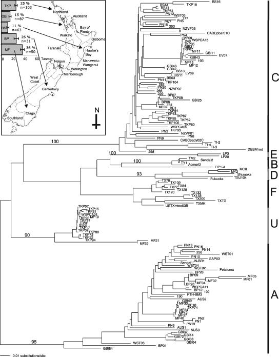
FIV subtypes A and C predominate within infected New Zealand cats (Fig. 1). The principal subtype is C, comprising 48% (±4.4%) of all sequences (Table 1) . Although there is some clustering of sequences from the same location, such as that seen from Great Barrier Island in subtype A, there is no obvious geographical pattern to the subtypes found throughout NZ. Australia, the country closest to New Zealand, has only subtypes A and, more rarely, B (14). Thus, the high prevalence of FIV-C in New Zealand cats was unexpected. FIV subtype A sequences from three infected Australian cats were included in Fig. 1 to determine whether Australia is a potential country of origin of New Zealand FIV-A. These Australian sequences group within the New Zealand subtype A clade, as did reference FIV-A sequences from other countries; thus, there is no firm evidence that Australia is the origin of New Zealand FIV-A.
FIG. 1.
Midpoint-rooted neighbor-joining tree with map of New Zealand showing regions and feral cat prevalence data. Subtypes are shown along the right side of the tree. F represents sequences from a recent study in Texas (32) with accession numbers U02422, AY139094, AY139095, AY139096, AY139097, AY139098, AY139099, AY139100, AY139101, AY139102, AY139103, and AY139104. U represents sequences from New Zealand that do not group with a known subtype. Three subtype A Australian sequences are included (AUS1, AUS2, and AUS3). The reference sequences downloaded from GenBank include subtype A: JN-BR1 (D67052, Japan), Petaluma (M25381, United States), PTH-BM3 (AB010401, Japan), SAP03 (AB010404, Japan), Sendai1 (D37813, Japan); subtype B: Aomori2 (D37817, Japan), RP1-A (AJ304988, Portugal), Sendai2 (D37814, Japan), TM2 (M59418, Japan), and TY1 (D67064, Japan); subtype C: CABCpady02C (U02392, Canada), CABCpbar01C (U02393, Canada), DEBAfred (U57020, Germany), TI-2 (AB016026, Taiwan), TI-3 (AB016027, Taiwan), and TI-4 (AB016028, Taiwan); subtype D: Fukuoka (D37815, Japan), MC8 (D67062, Japan), MY2 (D67063, Japan), Shizuoka (D37811, Japan), and TSU104 (AB021111, Japan); subtype E: LP3 (D84496, Argentina) and LP20 (D84498, Argentina). Numbers are bootstrap values based on 1,000 replicates.
TABLE 1.
FIV-infected NZ cat samples, showing cat lifestyle, location, and FIV subtype
| Sample | Lifestyle | Locationa | Subtypeb | Sample | Lifestyle | Locationa | Subtypeb | |
|---|---|---|---|---|---|---|---|---|
| 190 | Domestic | Auckland | A | EV09 | Stray | Auckland | C | |
| 192 | Domestic | Canterbury | A | WSPCA05 | Stray | Northland | C | |
| 256 | Domestic | Hawke's Bay | A | WSPCA15 | Stray | Northland | C | |
| PN1 | Domestic | Wellington | A | BP07 | Feral | Canterbury | C | |
| PN2 | Domestic | Wellington | A | BP08 | Feral | Canterbury | C | |
| PN6 | Domestic | Manawatu-Wanganui | A | BS03 | Feral | Hawke's Bay | C | |
| PN10 | Domestic | Wellington | A | BS11 | Feral | Hawke's Bay | C | |
| PN13 | Domestic | Taranaki | A | BS13 | Feral | Hawke's Bay | C | |
| PN14 | Domestic | Manawatu-Wanganui | A | BS14 | Feral | Hawke's Bay | C | |
| PN16 | Domestic | Taranaki | A | BS16 | Feral | Hawke's Bay | C | |
| PN19 | Domestic | Wellington | A | BS44 | Feral | Hawke's Bay | C | |
| WST01 | Domestic | West Coast | A | GBI11 | Feral | Auckland | C | |
| WST02 | Domestic | West Coast | A | GBI25 | Feral | Auckland | C | |
| WST04 | Domestic | NA | A | GBI31 | Feral | Auckland | C | |
| WST05 | Domestic | NA | A | GBI43 | Feral | Auckland | C | |
| WSPCA11 | Stray | Northland | A | GBI46 | Feral | Auckland | C | |
| BP01 | Feral | Canterbury | A | GBI47 | Feral | Auckland | C | |
| BP09 | Feral | Canterbury | A | MF07 | Feral | Otago | C | |
| BP12 | Feral | Canterbury | A | MF09 | Feral | Otago | C | |
| BP18 | Feral | Canterbury | A | MF11 | Feral | Otago | C | |
| BP26 | Feral | Canterbury | A | MF12 | Feral | Otago | C | |
| BP28 | Feral | Canterbury | A | MF16 | Feral | Otago | C | |
| GBI04 | Feral | Auckland | A | TKP05 | Feral | Northland | C | |
| GBI08 | Feral | Auckland | A | TKP07 | Feral | Northland | C | |
| GBI14 | Feral | Auckland | A | TKP08 | Feral | Northland | C | |
| GBI37 | Feral | Auckland | A | TKP18 | Feral | Northland | C | |
| GBI49 | Feral | Auckland | A | TKP43 | Feral | Northland | C | |
| GBI72 | Feral | Auckland | A | TKP52 | Feral | Northland | C | |
| GBI84 | Feral | Auckland | A | TKP54 | Feral | Northland | C | |
| MF01 | Feral | Otago | A | TKP60 | Feral | Northland | C | |
| MF02 | Feral | Otago | A | TKP64 | Feral | Northland | C | |
| MF04 | Feral | Otago | A | TKP87 | Feral | Northland | C | |
| MF05 | Feral | Otago | A | TKP93 | Feral | Northland | C | |
| MF33 | Feral | Otago | A | TKP95 | Feral | Northland | C | |
| MF34 | Feral | Otago | A | TKP104 | Feral | Northland | C | |
| MF37 | Feral | Otago | A | TKP105 | Feral | Northland | C | |
| MF40 | Feral | Otago | A | 168 | Domestic | West Coast | PR | |
| MF42 | Feral | Otago | A | 197 | Domestic | Nelson | PR | |
| TKP74 | Feral | Northland | A | 214 | Domestic | Waikato | PR | |
| 164 | Domestic | Wellington | C | 258 | Domestic | Bay of Plenty | PR | |
| 177 | Domestic | Southland | C | 259 | Domestic | Taranaki | PR | |
| 193 | Domestic | Canterbury | C | 260 | Domestic | Bay of Plenty | O | |
| 229 | Domestic | Auckland | C | PN17 | Domestic | Wellington | PR | |
| 240 | Domestic | NA | C | PN21 | Domestic | Manawatu-Wanganui | PR | |
| 253 | Domestic | Waikato | C | PN22 | Domestic | Manawatu-Wanganu | O | |
| 266 | Domestic | Taranaki | C | PN23 | Domestic | Wellington | PR | |
| 282 | Domestic | NA | C | MF14 | Feral | Otago | PR | |
| 298 | Domestic | NA | C | EV01 | Stray | Auckland | U | |
| B | Domestic | Auckland | C | WSPCA01 | Stray | Northland | U | |
| BD1 | Domestic | Auckland | C | BS08 | Feral | Hawke's Bay | U | |
| NZVP01 | Domestic | Wellington | C | MF19 | Feral | Otago | U | |
| NZVP02 | Domestic | Wellington | C | MF21 | Feral | Otago | U | |
| NZVP03 | Domestic | NA | C | MF29 | Feral | Otago | U | |
| PN3 | Domestic | Hawke's Bay | C | TKP02 | Feral | Northland | U | |
| PN4 | Domestic | NA | C | TKP14 | Feral | Northland | U | |
| PN5 | Domestic | Wellington | C | TKP15 | Feral | Northland | U | |
| PN7 | Domestic | Wellington | C | TKP17 | Feral | Northland | U | |
| PN8 | Domestic | Manawatu-Wanganui | C | TKP20 | Feral | Northland | U | |
| PN9 | Domestic | Manawatu-Wanganui | C | TKP21 | Feral | Northland | U | |
| PN11 | Domestic | Wellington | C | TKP22 | Feral | Northland | U | |
| PN12 | Domestic | Manawatu-Wanganui | C | TKP57 | Feral | Northland | U | |
| PN15 | Domestic | Wellington | C | TKP73 | Feral | Northland | U | |
| PN20 | Domestic | Wellington | C | TKP88 | Feral | Northland | U | |
| WST03 | Domestic | West Coast | C | TKP94 | Feral | Northland | U | |
| EV07 | Stray | Auckland | C |
NA, not available. Locations by region are shown on the map in Fig. 1.
U, unknown subtype; O, outlier but not labeled putative recombinant since not significant as determined by KH test; PR, putative recombinant.
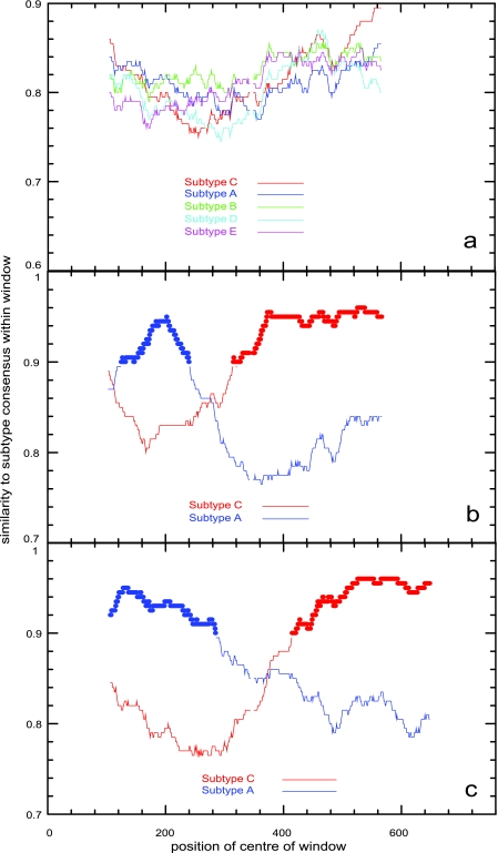
A group of 17 New Zealand sequences, 11 of which are from feral cats from one location, did not group with any known subtype on the phylogenetic tree. Fifteen sequences from this group are very closely related (91.0% similarity), suggesting their recent spread in New Zealand cats. All 17 sequences were analyzed by using a recombination identification program (RIP) (28; http://hivweb.lanl.gov/RIP/RIPsubmit.html) to test for recombination. None of these sequences showed any significant similarity to any previously described subtype (Fig. 2a), nor did they appear to be recombinants of known subtypes. Two lymph node samples, one that yielded FIV sequence of subtype C and another of the “unknown” subtype, were amplified and sequenced independently at the School of Veterinary Science, University of Queensland, in a blind, independent check for contamination. These sequences were almost identical (99%) to those we obtained, thus confirming the novelty of the “unknown” group. We note that the suggested requirements for designating a new human immunodeficiency virus subtype requires three genome sequences from epidemiologically independent infected individuals (24). While FIV subtype designation has been based on V3-V5 env sequence (29), we are currently sequencing the pol and gag genes to provide further sequence data to validate the “unknown” group as a novel NZ-specific subtype.
FIG. 2.
RIP graphical outputs of TKP21, a sequence from the “unknown” group (a); 214 (b) and 197 (c), two outlier sequences. The plots represent the distance between the query sequence and the subtype A (blue) or subtype C (red) reference sequence, over a sliding window of size 200 bp. In panels b and c, there is a clear point (the putative recombinant crossover point) at which the query sequences cease to be closer to A and become closer to C. In panel a, all five known subtypes are compared to the query sequence, with no significant similarity, although at the very end of the sequenced region there is some similarity to subtype C.
Eleven other sequences that did not group with any subtype were tested for recombination by using RIP (Fig. 2b and c). A Kishino-Hasegawa (KH) test (9, 10, 15) implemented in PAUP* was used to further investigate recombination. Thirteen reference sequences were used in each KH test: five subtype A sequences, five subtype C sequences, and one sequence of each subtype B, D, and E. The results from this test (Table 2) show that, conservatively, 9 of the 11 samples can be assigned as putative recombinants of subtypes A and C.
TABLE 2.
Kishino-Hasegawa test results of the eleven sequences ambiguously positioned on the phylogenetic tree
| Sequencea | Diff −lnLb | Pc |
|---|---|---|
| Firstd | ||
| 168† | 67.635 | 0.000* |
| 197 | 85.935 | 0.000* |
| 214 | 28.094 | 0.006* |
| 258† | 108.153 | 0.000* |
| 259† | 68.407 | 0.000* |
| 260 | 2.656 | 0.344 |
| MF14 | 29.355 | 0.005* |
| PN17 | 72.269 | 0.000* |
| PN21† | 35.537 | 0.002* |
| PN22 | 62.599 | 0.000* |
| PN23† | 66.752 | 0.000* |
| Seconde | ||
| 168 | 31.741 | 0.006* |
| 197 | 51.234 | 0.000* |
| 214 | 117.730 | 0.000* |
| 258 | 31.743 | 0.002* |
| 259 | 25.720 | 0.035* |
| 260 | 40.658 | 0.000* |
| MF14 | 116.522 | 0.000* |
| PN17 | 23.612 | 0.047* |
| PN21 | 61.732 | 0.000* |
| PN22 | 20.601 | 0.092 |
| PN23 | 27.922 | 0.019* |
†, Samples for which endpoint dilutions were performed.
The difference in log-likelihood scores between the best tree for that subalignment against the best tree for the alternative subalignment.
The probability of obtaining that difference in log-likelihoods by chance alone, given that the true trees are identical. *, P values significant at the 0.05 level. Note that, to be conservative, we only identify a sequence as a putative recombinant if both subalignments produce trees that are mutually (statistically) different.
The subalignment of sequences using the query sequence before the putative crossover point identified by RIP.
The subalignment of sequences using the query sequence after the putative crossover point identified by RIP.
To exclude PCR-mediated recombination as a potential source for these putative recombinants (16, 18), we used endpoint dilution (25, 26) on representative samples from which recombinants were detected. Because of limited sample availability, we were only able to perform these tests on five of the nine putative recombinants (Table 2). A fivefold serial dilution was used, and the PCR results were entered into QUALITY (http://ubik.microbiol.washington.edu/computing/quality/jquality.htm) to determine the proviral copy number. Samples were then diluted to an endpoint and amplified to give at least five sequences. In all cases, the endpoint sequences were at least 98% similar to the respective consensus sequences and to each other. Consequently, these recombinants are confirmed as authentic viral sequences, and there is no evidence of dual infection.
Of the 334 feral cats tested, 72 (21.5% ± 2.25%) were positive for FIV proviral DNA. This is generally higher than other feral cat populations globally (2, 8, 19, 33; however, see also reference 3). The FIV prevalence in feral cats from the five New Zealand locations ranges from 11 to 36%, with no obvious geographic pattern (Fig. 1).
More than two-thirds of the FIV-infected feral cats were male, a statistically significant difference (χ2 = 14.16, df = 1, P ≤ 0.001), and adult male cats had the highest prevalence of 51% (n = 70). This trend is expected because mature male cats demonstrate higher levels of social aggression, caused by competition over mates and territories (5, 7). Thus, mature male cats tend to have a greater number of biting encounters, the typical mode of transmission of FIV (34).
This study is the first to use phylogenetics to analyze FIV in New Zealand cats. Two subtypes and nine putative A/C recombinant strains are identified in New Zealand cats. A novel clade was detected, possibly representing a New Zealand-specific subtype.
Acknowledgments
We thank the New Zealand Department of Conservation; the Whangarei branch of the Society for the Prevention of Cruelty to Animals; the Lonely Miaow Association, Inc.; the Epsom Central Veterinary Centre; Gribbles Laboratories; Gribbles-Alpha (formerly Alpha Scientific, Hamilton); the Blockhouse Bay Veterinary Centre; Animals! The Vets!; Buller Veterinary Services; and New Zealand Veterinary Pathology, Palmerston North. We are grateful to Zainab Issa for initial help with labwork. Sequencing was carried out at the Allan Wilson Centre sequencing facility, Massey University, Albany, New Zealand.
This research was funded by a grant from the National Institutes of Health and with support from both the Allan Wilson Centre for Molecular Ecology and Evolution (New Zealand) and the University of Auckland, Auckland, New Zealand.
Footnotes
Published ahead of print on 27 December 2006.
REFERENCES
- 1.Bachmann, M. H., C. Mathiason-Dubard, G. H. Learn, A. G. Rodrigo, D. L. Sodora, P. Mazzetti, E. A. Hoover, and J. I. Mullins. 1997. Genetic diversity of feline immunodeficiency virus: dual infection, recombination, and distinct evolutionary rates among envelope sequence clades. J. Virol. 71:4241-4253. [DOI] [PMC free article] [PubMed] [Google Scholar]
- 2.Baneth, G., P. H. Kazz, D. Steinfeld, and M. Besser. 1999. A seroepidemiological study of feline coronavirus, feline immunodeficiency virus and feline leukemia virus among cats in Israel. Israel J. Vet. Med. 70:55-56. [Google Scholar]
- 3.Bennett, M., C. McCracken, H. Lutz, C. J. Gaskell, R. M. Gaskell, A. Brown, and J. O. Knowles. 1989. Prevalence of antibody to feline immunodeficiency virus in some cat populations. Vet. Rec. 124:397-398. [DOI] [PubMed] [Google Scholar]
- 4.Carpenter, M. A., E. W. Brown, D. W. MacDonald, and S. J. O'Brien. 1998. Phylogeographic patterns of feline immunodeficiency virus genetic diversity in the domestic cat. Virology 251:234-243. [DOI] [PubMed] [Google Scholar]
- 5.Courchamp, F., N. G. Yoccoz, M. Artois, and D. Pontier. 1998. At-risk individuals in feline immunodeficiency virus epidemiology: evidence from a multivariate approach in a natural population of domestic cats (Felis catus). Epidemiol. Infect. 121:227-236. [DOI] [PMC free article] [PubMed] [Google Scholar]
- 6.Felsenstein, J. 1985. Confidence limits on phylogenies: an approach using the bootstrap. Evolution 39:783-791. [DOI] [PubMed] [Google Scholar]
- 7.Fromont, E., F. Courchamp, M. Artois, and D. Pontier. 1997. Infection strategies of retroviruses and social grouping of domestic cats. Can. J. Zool. 75:1994-2002. [Google Scholar]
- 8.Gibson, K. L., K. Keizer, and C. Golding. 2002. A trap, neuter, and release program for feral cats on Prince Edward Island. Can. Vet. Med. Assoc. 43:695-698. [PMC free article] [PubMed] [Google Scholar]
- 9.Goldman, N., J. P. Anderson, and A. G. Rodrigo. 2000. Likelihood-based tests of topologies in phylogenetics. Syst. Biol. 49:652-670. [DOI] [PubMed] [Google Scholar]
- 10.Hasegawa, M., and H. Kishino. 1989. Confidence limits on the maximum-likelihood estimate of the hominoid tree from mitochondrial-DNA sequences. Evolution 43:672-677. [DOI] [PubMed] [Google Scholar]
- 11.Higgins, D. G., and P. M. Sharp. 1988. CLUSTAL: a package for performing multiple sequence alignment on a microcomputer. Gene 73:237-244. [DOI] [PubMed] [Google Scholar]
- 12.Jeanmougin, F., J. D. Thompson, M. Gouy, D. G. Higgins, and T. J. Gibson. 1998. Multiple sequence alignment with CLUSTAL X. Trends Biochem. Sci. 23:403-405. [DOI] [PubMed] [Google Scholar]
- 13.Kakinuma, S., K. Motokawa, T. Hohdatsu, J. K. Yamamoto, H. Koyama, and H. Hashimoto. 1995. Nucleotide sequence of feline immunodeficiency virus: classification of Japanese isolates into two subtypes which are distinct from non-Japanese subtypes. J. Virol. 69:3639-3646. [DOI] [PMC free article] [PubMed] [Google Scholar]
- 14.Kann, R. K. C., M. T. Kyaw-Tanner, J. M. Seddon, P. R. Lehrbach, R. J. G. Zwijnenberg, and J. Meers. 2006. Molecular subtyping of feline immunodeficiency virus from domestic cats in Australia. Aust. Vet. J. 84:112-116. [DOI] [PubMed] [Google Scholar]
- 15.Kishino, H., and M. Hasegawa. 1989. Evaluation of the maximum-likelihood estimate of the evolutionary tree topologies from DNA sequence data, and the branching order in Hominoidea. J. Mol. Evol. 29:170-179. [DOI] [PubMed] [Google Scholar]
- 16.Meyerhans, A., J. P. Vartanian, and S. Wain-Hobson. 1990. DNA recombination during PCR. Nucleic Acids Res. 18:1687-1691. [DOI] [PMC free article] [PubMed] [Google Scholar]
- 17.Nishimura, Y., S. Nakamura, N. Goto, T. Hasegawa, H. Pang, Y. Goto, H. Kato, H. Y. Youn, Y. Endo, T. Mizuno, Y. Momoi, K. Ohno, T. Watari, H. Tsujimoto, and A. Hasegawa. 1996. Molecular characterization of feline immunodeficiency virus genome obtained directly from organs of a naturally infected cat with marked neurological symptoms and encephalitis. Arch. Virol. 141:1933-1948. [DOI] [PubMed] [Google Scholar]
- 18.Odelberg, S. J., R. B. Weiss, A. Hata, and R. White. 1995. Template-switching during DNA synthesis by Thermus aquaticus DNA polymerase I. Nucleic Acids Res. 23:2049-2057. [DOI] [PMC free article] [PubMed] [Google Scholar]
- 19.Ostrowski, S., M. Van Vuuren, D. M. Lenain, and A. Durand. 2003. A serologic survey of wild felids from central west Saudi Arabia. J. Wildl. Dis. 39:696-701. [DOI] [PubMed] [Google Scholar]
- 20.Pecoraro, M. R., K. Tomonaga, T. Miyazawa, Y. Kawaguchi, S. Sugita, Y. Tohya, C. Kai, M. E. Etcheverrigaray, and T. Mikami. 1996. Genetic diversity of Argentine isolates of feline immunodeficiency virus. J. Genet. Virol. 77:2031-2035. [DOI] [PubMed] [Google Scholar]
- 21.Pedersen, N. C., and J. E. Barlough. 1991. Clinical overview of feline immunodeficiency virus. J. Am. Vet. Med. Assoc. 199:1298-1305. [PubMed] [Google Scholar]
- 22.Posada, D., and K. A. Crandall. 1998. MODELTEST: testing the model of DNA substitution. Bioinformatics 14:817-818. [DOI] [PubMed] [Google Scholar]
- 23.Reggeti, F., and D. Bienzle. 2004. Feline immunodeficiency virus subtypes A, B and C and intersubtype recombinants in Ontario, Canada. J. Gen. Virol. 85:1843-1852. [DOI] [PubMed] [Google Scholar]
- 24.Robertson, D. L., J. P. Anderson, J. A. Bradac, J. K. Carr, B. Foley, R. K. Funkhouser, F. Gao, B. H. Hahn, M. L. Kalish, C. Kuiken, G. H. Learn, T. Leitner, F. McCutchan, S. Osmanov, M. Peeters, D. Pieniazek, M. Salminen, P. M. Sharp, S. Wolinsky, and B. Korber. 2000. HIV-1 nomenclature proposal. Science 288:55-57. [DOI] [PubMed] [Google Scholar]
- 25.Rodrigo, A. G., P. C. Goracke, K. Rowhanain, and J. I. Mullins. 1997. Quantitation of target molecules from polymerase chain reaction-based limiting dilution assays. AIDS Res. Hum. Retrovir. 13:737-742. [DOI] [PubMed] [Google Scholar]
- 26.Rodrigo, A. G., E. W. Hanley, P. C. Goracke, and G. H. Learn. 2001. Sampling and processing HIV molecular sequences: a computational evolutionary biologist's perspective, p. 300. In A. G. Rodrigo and G. H. Learn (ed.), Computational and evolutionary analysis of HIV molecular sequences. Kluwer Academic Publishers, Boston, MA.
- 27.Saitou, N., and M. Nei. 1987. The neighbor-joining method: a new method for reconstructing phylogenetic trees. Mol. Biol. Evol. 4:406-425. [DOI] [PubMed] [Google Scholar]
- 28.Siepel, A. C., A. L. Halpern, C. Macken, and B. T. M. Korber. 1995. A computer program designed to rapidly screen for HIV-1 intersubtype recombinant sequences. AIDS Res. Hum. Retrovir. 11:1413-1416. [DOI] [PubMed] [Google Scholar]
- 29.Sodora, D. L., E. G. Shpaer, B. E. Kitchell, S. W. Dow, E. A. Hoover, and J. I. Mullins. 1994. Identification of three feline immunodeficiency virus (FIV) env gene subtypes and comparison of the FIV and human immunodeficiency virus type 1 evolutionary patterns. J. Virol. 68:2230-2238. [DOI] [PMC free article] [PubMed] [Google Scholar]
- 30.Swinney, G. R., J. V. Pauli, B. R. Jones, and C. R. Wilks. 1989. Feline t-lymphotropic virus (FTLV) (feline immunodeficiency virus infection) in cats in New Zealand. New Zealand Vet. J. 37:41-43. [DOI] [PubMed] [Google Scholar]
- 31.Swofford, D. L. 2002. PAUP*. Phylogenetic analysis using parsimony (*and other methods), version. 4. Sinauer Associates, Sunderland, MA.
- 32.Weaver, E. A., E. W. Collisson, M. Slater, and G. Zhu. 2004. Phylogenetic analyses of Texas isolates indicate an evolving subtype of the clade B feline immunodeficiency viruses. J. Virol. 78:2158-2163. [DOI] [PMC free article] [PubMed] [Google Scholar]
- 33.Winkler, I. G., M. Löchelt, and R. L. P. Flower. 1999. Epidemiology of feline foamy virus and feline immunodeficiency virus infections in domestic and feral cats: a seroepidemiological study. J. Clin. Microbiol. 37:2848-2851. [DOI] [PMC free article] [PubMed] [Google Scholar]
- 34.Yamamoto, J. K., H. Hansen, E. W. Ho, T. Y. Morishita, T. Okuda, T. R. Sawa, R. M. Nakamura, and N. C. Pedersen. 1989. Epidemiologic and clinical aspects of feline immunodeficiency virus infection in cats from the continental United States and Canada and possible mode of transmission. J. Am. Vet. Med. Assoc. 194:213-220. [PubMed] [Google Scholar]


