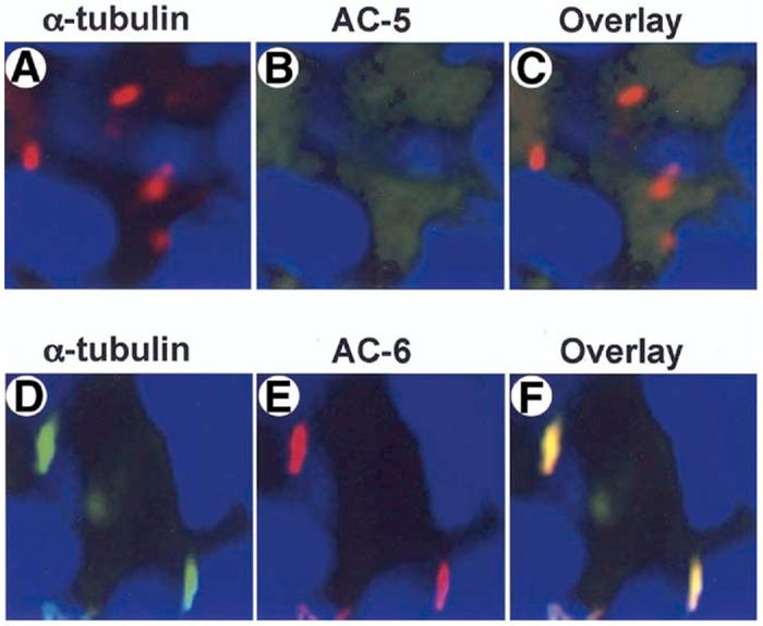
Figure 8.

AC6 is expressed in cholangiocyte cilia. Cholangiocytes of isolated IBDUs were stained with an antibody to ciliary marker, acetylated a-tubulin (red) (A, D) and antibodies to AC5 and AC6 (green) (B, E). When the two images were overlay (C, F) co-localization of acetylated a-tubulin and AC6 (yellow, F) but not AC5 (red, C) was seen, indicating that Ca2+-inhibitable AC6 is expressed in cholangiocyte cilia. Nuclei were visualized by staining with DAPI (blue).
