Abstract
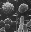
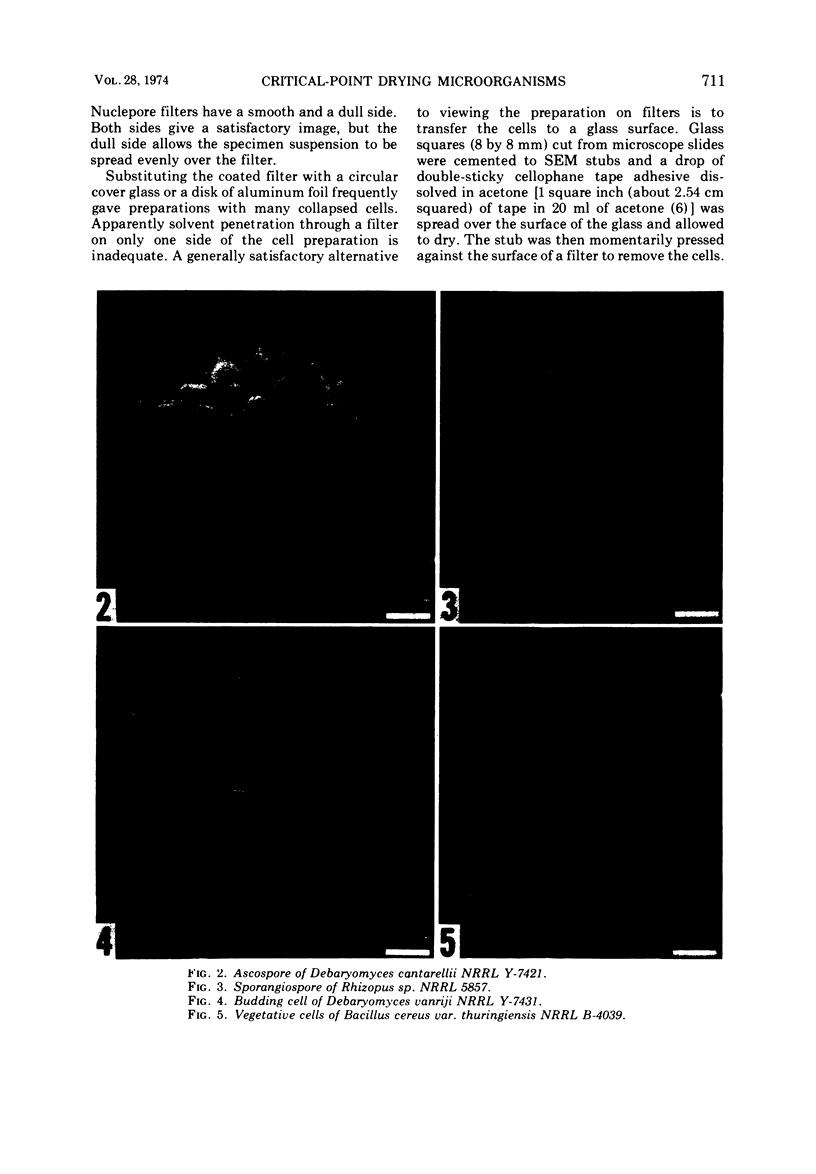
Critical-point drying of microorganisms for scanning electron microscopy can be rapidly and effectively accomplished by use of a newly described specimen holder. Up to eight different samples of spores or vegetative cells are placed between polycarbonate membrane filters in the holder and processed through solvent dehydration and critical-point drying using carbon dioxide without loss or cross contamination of microorganisms. Yeasts, molds, bacteria, and actinomycetes have been successfully processed.
Full text
PDF
Images in this article
Selected References
These references are in PubMed. This may not be the complete list of references from this article.
- Small E. B., Marszalek D. S. Scanning electron microscopy of fixed, frozen, and dried protozoa. Science. 1969 Mar 7;163(3871):1064–1065. doi: 10.1126/science.163.3871.1064. [DOI] [PubMed] [Google Scholar]
- Talens L. T., Miranda M., Miller M. W. Electron Micrography of Bud Formation in Metschnikowia krissii. J Bacteriol. 1973 Apr;114(1):413–423. doi: 10.1128/jb.114.1.413-423.1973. [DOI] [PMC free article] [PubMed] [Google Scholar]


