Abstract
Postmortem and in vivo vibration responses to sound of the stapes and the umbo of human ears are surveyed. The magnitudes of umbo velocity responses recorded postmortem decay between 1 and 5 or 10 kHz at rates between 0 and −3 dB/octave. In contrast, the magnitudes of in vivo umbo vibration are relatively invariant over a wide frequency range, amply exceeding the bandwidth of the audiogram according to one report. Similarly, most studies of postmortem stapes vibration report velocities tuned to about 1 kHz, with magnitudes that decay at a rate of about −6 dB/octave at higher frequencies. In contrast, in vivo stapes responses are apparently only mildly tuned. We conjecture that the bandwidth of stapes vibration velocity in humans will eventually be shown to exceed the bandwidth of the audiogram, in line with findings in other amniotic vertebrates.
Introduction
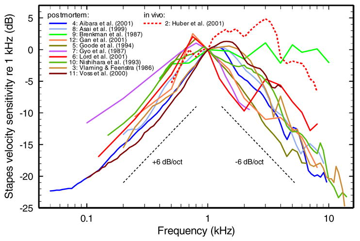
In a recent review, we showed that the magnitudes of stapes (or columella) vibration velocity in the middle ears of several amniotic vertebrates (i.e., mammals, birds, and reptiles) are relatively invariant over a wide frequency range and that their bandwidths generally exceed the bandwidths of behavioral thresholds 1. In apparent contrast, most studies of stapes vibration velocity in humans report that the middle ear acts as a resonant system tuned to a frequency between 700 and 1200 Hz, with responses’ magnitudes decaying at rates of +6 and −6 dB/octave, respectively, at lower and higher frequencies (Fig. 1). At face value, these results imply that the human middle ear is exceptional among those of amniotic vertebrates in that it severely limits the bandwidth of hearing.
Fig. 1.

The frequency dependence of stapes vibration velocity in human ears, in vivo and postmortem. The magnitudes of stapes velocity were normalized to 1 kHz and plotted as a function of frequency using a decibel scale. The in vivo data (red dashed line) were taken from 2, Fig. 5. The curve from Vlaming and Feenstra is a median computed from 4 individual curves presented in their Fig. 5 3. Other postmortem data (solid lines) were taken from 4, Fig. 3; 5, Fig. 1; 6, Fig. 2; 7, Fig. 4; 8, Fig. 5; 9, Fig. 7; 10, Fig. 2; 11, Fig. 4; 12, Fig. 5. (Those reading a black-and-white version of the paper should refer to the color version of this figure in the online archival version.)
Here we compare the magnitudes of postmortem and in vivo vibrations of the stapes and the umbo of the tympanic membrane in the ears of humans. This comparison leads us to conjecture that most postmortem recordings of ossicular vibrations are flawed and that middle-ear transmission in humans, in fact, does not severely limit the bandwidth of hearing.
Methods
Our survey of ossicular vibration data (Figs. 1 and 2) is extensive but not exhaustive. It includes all available data from the last decade, as well as some older data selected so as to include the work of additional laboratories. The data were extracted from the original figures by digitization with a resolution of 12 points/octave. Displacement units were converted to velocity units, as needed. Expressing the magnitudes of ossicular vibration in terms of velocity (rather than, say, displacement or acceleration) allows for direct comparison with hearing thresholds, since pressure in scala vestibuli near the stapes (essentially the input signal for sound analysis and transduction in the cochlea) is proportional to stapes velocity over the wide range of frequencies in which cochlear input impedance is resistive 13–17.
Fig. 2.

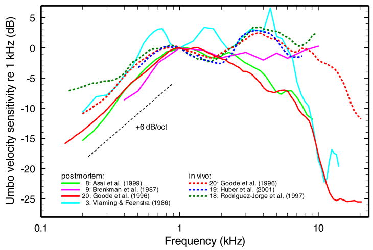
The frequency dependence of umbo vibration velocity in humans, in vivo and postmortem. The magnitudes of umbo velocity were normalized to 1 kHz and plotted as a function of frequency using a decibel scale. The in vivo data (dashed lines) from 18 were computed from their Fig. 6, according to the equation, [(χ + SD) – (χ − SD)]/2, where χ = mean, SD = standard deviation of the mean. Other in vivo data were taken from 19, Fig. 5, and 20, Fig. 4c. The postmortem curve from Fig. 4c of 20 represents data from 3 other studies by the same group: 5,10,21. The postmortem curve from 3 represents medians computed from 4 individual measurements given in their Fig. 2. Other postmortem data (solid lines) were taken from: 8, Fig. 5 and 9, Fig. 7. (Those reading a black-and-white version of the paper should refer to the color version of this figure in the online archival version.)
Results
In vivo stapes vibration
Huber et al. reported in vivo of stapes vibration in humans, recorded from 7 patients undergoing ear surgery 2. Fig. 1 compares the average magnitudes of stapes vibration velocity from that report (red dashed line), with average curves from 10 postmortem studies in temporal bones (continuous lines). At frequencies lower than 1 kHz, the normalized curves follow similar trajectories, regardless of whether they were obtained in vivo or postmortem. Above 1 kHz, however, most postmortem curves follow trajectories with a slope of about −6 dB/octave, whereas the single in vivo curve and one postmortem curve are relatively flat. Inspection of Fig. 1 indicates that, whereas the magnitudes of postmortem velocities, with the indicated single exception 9, are clearly tuned to about 1 kHz, with slopes of +6 dB/octave and −6 dB/octave below and above that frequency respectively, the in vivo velocity data are more broadly tuned, with similar velocity magnitudes between 1 and 3.5 kHz. Measured at −10 dB relative to the peak responses, the in vivo stapes responses have a bandwidth of 8 kHz or about double the 10−dB bandwidth of the postmortem measurements. The exceptional postmortem curve 9 has an even wider bandwidth.
Umbo vibrations in vivo and postmortem
After pioneering investigations measured umbo vibrations in the ears of living humans at frequencies lower than 5 kHz 22,23, three recent studies 18–20 reported in vivo measurements over wider frequency ranges. * Their results (dashed lines) are plotted in Fig. 2 together with umbo response magnitudes measured postmortem in temporal bones (continuous lines). In vivo velocity responses of the umbo are nearly flat (± 3 dB) over a wide frequency range extending from 400 Hz to nearly 10 kHz and in one case 20 almost 15 kHz. Measured at −10 dB relative to the peak, the bandwidth of umbo velocity responses is nearly 19 kHz. In contrast, postmortem responses vary widely above 2 kHz: some studies 8,20 reported velocity responses that decay with frequency at a rate of about −3 dB/octave and others 3,9 reported responses that remain relatively invariant up to 6 or 10 kHz.
The differences between postmortem and in vivo ossicular vibrations
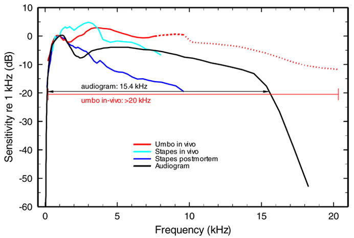
Fig. 3 summarizes the data of Figs. 1 and 2 and compares the human audiogram 25 with the velocity-vs.-frequency curves of ossicular responses. Fig. 3 shows that ossicular velocity responses, both in vivo and postmortem, and audiometric thresholds vary in direct proportion to one another at frequencies lower than 1 kHz (the frequency at which the curves were normalized). Between 1 and 8 kHz, both the audiogram and the ossicular-velocity response curves measured in vivo are relatively flat as a function of frequency. In one case (red dashed line), the 10−dB bandwidth of in vivo umbo vibration velocity extends to 20 kHz, amply exceeding the bandwidth of the audiogram 20.
Fig. 3.

A comparison between the human behavioral audiogram and the magnitudes of vibration velocity of the stapes and umbo, in vivo and postmortem. Black line: human behavioral audiogram from 25. Red line: in vivo umbo data of Fig. 2: the solid segment indicates medians computed from three studies (Fig. 2); the thick-dash line segment represents two studies; the dotted line represents one study. Light blue line: the in vivo stapes data of Fig. 1 2. Dark blue line: group medians for the postmortem stapes data of Fig. 1. (Those reading a black-and-white version of the paper should refer to the color version of this figure in the online archival version.)
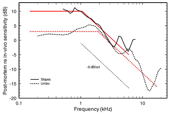
Two groups of experimenters have published both in vivo and postmortem vibration data for human ears obtained and analyzed with similar techniques. Their data can therefore be used to advantage in a quantitative evaluation of the differences between postmortem and in vivo responses. Fig. 4 plots the ratios (expressed in decibels) between the magnitudes of postmortem and in vivo velocity responses of the umbo and the stapes reported by the Goode 20 and Huber 2,8 groups, respectively. This figure suggests that the postmortem effects on both stapes and umbo vibrations resemble low-pass filtering with corner frequencies between 1 and 2 kHz and high- frequency slopes of roughly −6 dB/octave.
Fig. 4.

Relative magnitudes of postmortem and in vivo vibrations of the stapes and umbo. The magnitudes of postmortem vibration velocity are expressed in decibels relative to in vivo velocity. The relative magnitudes of umbo and stapes velocity are indicated by the solid and dashed lines, respectively. The umbo data are from 20; the stapes data are from 2,8. The red lines represent low-pass filters with corner frequency between 1 and 2 kHz and high-frequency slopes of −6dB/octave. (Those reading a black-and-white version of the paper should refer to the color version of this figure in the online archival version.)
Conclusions and speculations
The literature contains several statements to the effect that ossicular vibrations in cadaveric temporal bones are representative of ossicular vibrations in living humans. For example: in a report on umbo vibrations, Nishihara and Goode commented that “the human temporal bones showed very similar results to the normal … ears” (26, p. 91). Similarly, in their investigation of stapes vibration, Huber et al., found “no major differences in the results from the live human subjects and temporal bone preparations” (2, p. 31) and concluded that “the temporal bone is a very good model for studying middle ear mechanics” (27, p. 13). The present review disputes that belief, presenting evidence that, in fact, there are substantial differences between in vivo and most postmortem measurements.
We conjecture that the finding of wide-band umbo vibration velocities (red dashed lines in Figs. 2 and 3) will be replicated. We also conjecture that when stapes velocities are measured in vivo at sufficiently high frequencies, their bandwidth will also be found to be comparable to that of the audiogram. In other words, the human middle ear is not exceptional among those of amniotic vertebrates and resembles them in acting as a wide-band pressure transformer which only minimally limits the performance of cochlear frequency analysis. Finally, we note that the present results support the hypothesis that high-frequency hearing in humans is determined by the high-frequency slopes of the auditory-nerve fibers with the highest characteristic frequencies 28,29, as is it appears to be the case in other amniotic vertebrates 1.
Acknowledgments
Work supported by Grant DC-00419 from the National Institute on Deafness and other Communication Disorders.
Footnotes
The 1993 measurements of Goode et al. 21 are not shown here because they were superseded by a much more extensive dataset from the same laboratory 20. The 1993 data 21 were incorrectly reproduced in Fig. 9–16 of 24, giving the impression that the magnitude of umbo velocity in vivo decays above 500 Hz at a rate of −6 dB/octave.
References
- 1.Ruggero MA, Temchin AN. The roles of the external, middle, and inner ears in determining the bandwidth of hearing. Proc Natl Acad Sci USA. 2002;99:13206–13210. doi: 10.1073/pnas.202492699. [DOI] [PMC free article] [PubMed] [Google Scholar]
- 2.Huber A, Linder T, Ferrazzini M, Schmid S, Dillier N, Stoeckli S, Fisch U. Intraoperative assessment of stapes movement. Ann Otol Rhinol Laryngol. 2001;110:31–35. doi: 10.1177/000348940111000106. [DOI] [PubMed] [Google Scholar]
- 3.Vlaming MS, Feenstra L. Studies on the mechanics of the normal human middle ear. Clin Otolaryngol. 1986;11:353–363. doi: 10.1111/j.1365-2273.1986.tb00137.x. [DOI] [PubMed] [Google Scholar]
- 4.Aibara R, Welsh JT, Puria S, Goode RL. Human middle-ear sound transfer function and cochlear input impedance. Hear Res. 2001;152:100–109. doi: 10.1016/s0378-5955(00)00240-9. [DOI] [PubMed] [Google Scholar]
- 5.Goode RL, Killion M, Nakamura K, Nishihara S. New knowledge about the function of the human middle ear: development of an improved analog model. Am J Otolaryngol. 1994;15:145–154. [PubMed] [Google Scholar]
- 6.Lord RM, Abel EW, Wang Z, Mills RP. Effects of draining cochlear fluids on stapes displacement in human middle-ear models. J Acoust Soc Am. 2001;110:3132–3139. doi: 10.1121/1.1419095. [DOI] [PubMed] [Google Scholar]
- 7.Gyo K, Aritomo H, Goode RL. Measurement of the ossicular vibration ratio in human temporal bones by use of a video measuring system. Acta Otolaryngol. 1987;103:87–95. doi: 10.3109/00016488709134702. [DOI] [PubMed] [Google Scholar]
- 8.Asai M, Huber AM, Goode RL. Analysis of the best site on the stapes footplate for ossicular chain reconstruction. Acta Otolaryngol. 1999;119:356–361. doi: 10.1080/00016489950181396. [DOI] [PubMed] [Google Scholar]
- 9.Brenkman CJ, Grote JJ, Rutten WL. Acoustic transfer characteristics in human middle ears studied by a SQUID magnetometer method. J Acoust Soc Am. 1987;82:1646–1654. doi: 10.1121/1.395156. [DOI] [PubMed] [Google Scholar]
- 10.Nishihara S, Aritomo H, Goode RL. Effect of changes in mass on middle ear function. Otolaryngol Head Neck Surg. 1993;109:899–910. doi: 10.1177/019459989310900520. [DOI] [PubMed] [Google Scholar]
- 11.Voss SE, Rosowski JJ, Merchant SN, Peake WT. Acoustic responses of the human middle ear. Hear Res. 2000;150:43–69. doi: 10.1016/s0378-5955(00)00177-5. [DOI] [PubMed] [Google Scholar]
- 12.Gan RZ, Dyer RK, Wood MW, Dormer KJ. Mass loading on the ossicles and middle ear function. Ann Otol Rhinol Laryngol. 2001;110:478–485. doi: 10.1177/000348940111000515. [DOI] [PubMed] [Google Scholar]
- 13.Zwislocki JJ. Theorie der Scheneckenmechanik: qualitative und quantitative analyse. Acta Otolaryngol Suppl. 1948;72:1–76. [Google Scholar]
- 14.Zwislocki JJ. In: “Analysis of some auditory characteristics,” in Handbook of Mathematical Psychology. Luce RC, Bush RR, Galanter E, editors. Wiley: New York; 1965. pp. 1–97. [Google Scholar]
- 15.Dallos P. Low-frequency auditory characteristics: Species dependence. J Acoust Soc Am. 1970;48:489–499. doi: 10.1121/1.1912163. [DOI] [PubMed] [Google Scholar]
- 16.Weiss TF, Peake WT, Sohmer HS. Intracochlear potential recorded with micropipets. III. Relation of cochlear microphonic potential to stapes velocity. J Acoust Soc Am. 1971;50:602–615. doi: 10.1121/1.1912676. [DOI] [PubMed] [Google Scholar]
- 17.Zwislocki JJ. Auditory Sound Transmission - An Autobiographical Perspective. Lawrence Erlbaum Associates; Mahwah, NJ: 2002. [Google Scholar]
- 18.Rodriguez-Jorge J, Zenner HP, Hemmert W, Burkhardt C, Gummer AW. Laservibrometrie: Ein Mittelohr und Kochleaanalysator zur nicht invasiven Untersuchung von Mittel und Innenohrfunktionsstörungen. HNO. 1997;45:997–1007. doi: 10.1007/s001060050185. [DOI] [PubMed] [Google Scholar]
- 19.Huber AM, Schwab C, Linder T, Stoeckli SJ, Ferrazzini M, Dillier N, Fisch U. Evaluation of eardrum laser doppler interferometry as a diagnostic tool. Laryngoscope. 2001;111:501–507. doi: 10.1097/00005537-200103000-00022. [DOI] [PubMed] [Google Scholar]
- 20.Goode RL, Ball G, Nishihara S, Nakamura K. Laser Doppler vibrometer (LDV)--a new clinical tool for the otologist. Am J Otolaryngol. 1996;17:813–822. [PubMed] [Google Scholar]
- 21.Goode RL, Ball G, Nishihara S. Measurement of umbo vibration in human subjects--method and possible clinical applications. Am J Otolaryngol. 1993;14:247–251. [PubMed] [Google Scholar]
- 22.Løkberg OJ, Høgmoen K, Gundersen T. Vibration measurement of the human tympanic membrane - in vivo. Acta Otolaryngol. 1980;89:37–42. doi: 10.3109/00016488009127106. [DOI] [PubMed] [Google Scholar]
- 23.Svane-Knudsen V, Michelsen A. Effect of crossed stapedius reflec on vibration of mallear handle in man. Acta Otolaryngol. 1989;107:219–224. doi: 10.3109/00016488909127501. [DOI] [PubMed] [Google Scholar]
- 24.Rosowski JJ, Relkin EM. Introduction to the analysis of middle ear function. In: Jahn AF, Santos-Sacchi J, editors. Physiology of the Ear. Singular; San Diego: 2001. pp. 161–190. [Google Scholar]
- 25.Killion MC. Revised estimate of minimum audible pressure: where is the ‘missing 6 dB’? J Acoust Soc Am. 1978;63:1501–1508. doi: 10.1121/1.381844. [DOI] [PubMed] [Google Scholar]
- 26.Nishihara S, Goode RL. Measurement of tympanic membrane vibration in 99 human ears. In: Hüttenbrink K-B, editor. Middle Ear Mechanics in Research and Otosurgery. Dept. of Oto-Rhino-Laryngology, Dresden University of Technology; Dresden, Germany: 1997. pp. 91–93. [Google Scholar]
- 27.Huber A, Linder T, Ferrazzini M, Schmid ST, Dillier N, Fisch U. Intraoperative scanning laser Doppler interferometry. In: Wada H, Takasaka T, Ikeda K, Ohyama K, Koike T, editors. Recent Developments in Auditory Mechanics. World Scientific; Singapore: 2000. pp. 10–14. [Google Scholar]
- 28.Buus S, Florentine M, Mason CR. Tuning curves at high frequencies and their relation to the absolute threshold curve. In: Moore BCJ, Patterson RD, editors. Auditory Frequency Selectivity. Plenum; New York: 1986. pp. 341–350. [Google Scholar]
- 29.Moore BCJ, Glasberg BR, Baer T. A model for the prediction of thresholds, loudness and partial loudness. J Audio Eng Soc. 1997;45:224–240. [Google Scholar]


