

Abstract
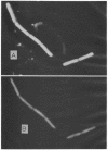
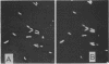
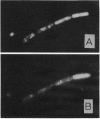
Two fluorescent brighteners were used to stain an isolate of Bacillus cereus var. mycoides and a soil pseudomonad. The stained organisms were fractionated by two procedures to determine which cellular constituents were reacting with the brighteners. Both fractionation procedures provided evidence that the brighteners were adsorbed to proteins within the cells. Microscopy examination of ghost cells of the bacillus showed that cell walls were not being stained. Spheroplasts of the bacillus and the pseudomonad were stained by the brighteners.
Full text
PDF


Images in this article
Selected References
These references are in PubMed. This may not be the complete list of references from this article.
- DARKEN M. A. Absorption and transport of fluorescent brighteners by microorganisms. Appl Microbiol. 1962 Sep;10:387–393. doi: 10.1128/am.10.5.387-393.1962. [DOI] [PMC free article] [PubMed] [Google Scholar]
- DARKEN M. A. Applications of fluorescent brighteners in biological techniques. Science. 1961 May 26;133(3465):1704–1705. doi: 10.1126/science.133.3465.1704. [DOI] [PubMed] [Google Scholar]
- Gull K., Trinci A. P. Detection of areas of wall differentiation in fungi using fluorescent staining. Arch Mikrobiol. 1974 Mar 1;96(1):53–57. doi: 10.1007/BF00590162. [DOI] [PubMed] [Google Scholar]
- Harrington B. J., Raper K. B. Use of a fluorescent brightener to demonstrate cellulose in the cellular slime molds. Appl Microbiol. 1968 Jan;16(1):106–113. doi: 10.1128/am.16.1.106-113.1968. [DOI] [PMC free article] [PubMed] [Google Scholar]
- Weiss R. L., Fraser D. Surface structure of intact cells and spheroplasts of pseudomonas aeruginosa. J Bacteriol. 1973 Feb;113(2):963–968. doi: 10.1128/jb.113.2.963-968.1973. [DOI] [PMC free article] [PubMed] [Google Scholar]