

Abstract
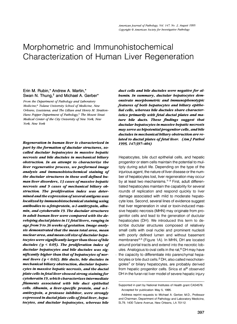
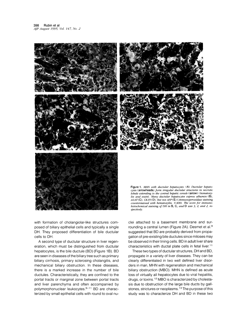
Regeneration in human liver is characterized in part by the formation of ductular structures, so-called ductular hepatocytes in massive hepatic necrosis and bile ductules in mechanical biliary obstruction. In an attempt to characterize the liver regenerative process, we performed image analysis and immunohistochemical staining of the ductular structures in these well defined human liver disorders, 13 cases of massive hepatic necrosis and 9 cases of mechanical biliary obstruction. The proliferation index was determined and the expression of several antigens was localized by immunohistochemical staining using antibodies to alpha-fetoprotein, alpha-1-antitrypsin, albumin, and cytokeratin 19. The ductular structures in adult human liver were compared with the developing ductal plates in 11 fetal livers, ranging in age from 9 to 36 weeks of gestation. Image analysis demonstrated that the mean total area, mean nuclear area, and mean cell size of ductular hepatocytes were significantly larger than those of bile ductules (p < 0.05). The proliferation index of ductular hepatocytes and bile ductules was significantly higher than that of hepatocytes of normal livers (p < 0.02). Bile ducts, bile ductules in mechanical biliary obstruction, ductular hepatocytes in massive hepatic necrosis, and the ductal plate cells in fetal liver showed strong staining for cytokeratin 19, which characterizes intermediate filaments associated with bile duct epithelial cells. Albumin, a liver-specific protein, and alpha-1-antitrypsin, a protease inhibitor, were strongly expressed in ductal plate cells of fetal liver, hepatocytes, and ductular hepatocytes, whereas bile duct cells and bile ductules were negative for albumin. In summary, ductular hepatocytes demonstrate morphometric and immunophenotypic features of both hepatocytes and biliary epithelial cells, whereas bile ductules share characteristics primarily with fetal ductal plates and mature bile ducts. These findings suggest that ductular hepatocytes in massive hepatic necrosis may serve as bipotential progenitor cells, and bile ductules in mechanical biliary obstruction are related to ductal plates of fetal liver.
Full text
PDF
Images in this article
Selected References
These references are in PubMed. This may not be the complete list of references from this article.
- Cattoretti G., Becker M. H., Key G., Duchrow M., Schlüter C., Galle J., Gerdes J. Monoclonal antibodies against recombinant parts of the Ki-67 antigen (MIB 1 and MIB 3) detect proliferating cells in microwave-processed formalin-fixed paraffin sections. J Pathol. 1992 Dec;168(4):357–363. doi: 10.1002/path.1711680404. [DOI] [PubMed] [Google Scholar]
- Desmet V. J. Intrahepatic bile ducts under the lens. J Hepatol. 1985;1(5):545–559. doi: 10.1016/s0168-8278(85)80752-2. [DOI] [PubMed] [Google Scholar]
- Gerber M. A., Thung S. N. Liver stem cells and development. Lab Invest. 1993 Mar;68(3):253–254. [PubMed] [Google Scholar]
- Gerber M. A., Thung S. N., Shen S., Stromeyer F. W., Ishak K. G. Phenotypic characterization of hepatic proliferation. Antigenic expression by proliferating epithelial cells in fetal liver, massive hepatic necrosis, and nodular transformation of the liver. Am J Pathol. 1983 Jan;110(1):70–74. [PMC free article] [PubMed] [Google Scholar]
- Grisham J. W. Migration of hepatocytes along hepatic plates and stem cell-fed hepatocyte lineages. Am J Pathol. 1994 May;144(5):849–854. [PMC free article] [PubMed] [Google Scholar]
- Harrison R. F., Reynolds G. M., Rowlands D. C. Immunohistochemical evidence for the expression of proliferating cell nuclear antigen (PCNA) by non-proliferating hepatocytes adjacent to metastatic tumours and in inflammatory conditions. J Pathol. 1993 Oct;171(2):115–122. doi: 10.1002/path.1711710208. [DOI] [PubMed] [Google Scholar]
- Kawakita N., Seki S., Sakaguchi H., Yanai A., Kuroki T., Mizoguchi Y., Kobayashi K., Monna T. Analysis of proliferating hepatocytes using a monoclonal antibody against proliferating cell nuclear antigen/cyclin in embedded tissues from various liver diseases fixed in formaldehyde. Am J Pathol. 1992 Feb;140(2):513–520. [PMC free article] [PubMed] [Google Scholar]
- Kayano K., Yasunaga M., Kubota M., Takenaka K., Mori K., Yamashita A., Kubo Y., Sakaida I., Okita K., Sanuki K. Detection of proliferating hepatocytes by immunohistochemical staining for proliferating cell nuclear antigen (PCNA) in patients with acute hepatic failure. Liver. 1992 Jun;12(3):132–136. doi: 10.1111/j.1600-0676.1992.tb00571.x. [DOI] [PubMed] [Google Scholar]
- Lai Y. S., Thung S. N., Gerber M. A., Chen M. L., Schaffner F. Expression of cytokeratins in normal and diseased livers and in primary liver carcinomas. Arch Pathol Lab Med. 1989 Feb;113(2):134–138. [PubMed] [Google Scholar]
- Lee W. M. Acute liver failure. N Engl J Med. 1993 Dec 16;329(25):1862–1872. doi: 10.1056/NEJM199312163292508. [DOI] [PubMed] [Google Scholar]
- Lemire J. M., Shiojiri N., Fausto N. Oval cell proliferation and the origin of small hepatocytes in liver injury induced by D-galactosamine. Am J Pathol. 1991 Sep;139(3):535–552. [PMC free article] [PubMed] [Google Scholar]
- Nakanuma Y., Ohta G. Immunohistochemical study on bile ductular proliferation in various hepatobiliary diseases. Liver. 1986 Aug;6(4):205–211. doi: 10.1111/j.1600-0676.1986.tb01067.x. [DOI] [PubMed] [Google Scholar]
- Naval J., Calvo M., Laborda J., Dubouch P., Frain M., Sala-Trepat J. M., Uriel J. Expression of mRNAs for alpha-fetoprotein (AFP) and albumin and incorporation of AFP and docosahexaenoic acid in baboon fetuses. J Biochem. 1992 May;111(5):649–654. doi: 10.1093/oxfordjournals.jbchem.a123813. [DOI] [PubMed] [Google Scholar]
- Nomoto M., Uchikosi Y., Kajikazawa N., Tanaka Y., Asakura H. Appearance of hepatocytelike cells in the interlobular bile ducts of human liver in various liver disease states. Hepatology. 1992 Nov;16(5):1199–1205. [PubMed] [Google Scholar]
- Phillips M. J., Poucell S. Modern aspects of the morphology of viral hepatitis. Hum Pathol. 1981 Dec;12(12):1060–1084. doi: 10.1016/s0046-8177(81)80328-0. [DOI] [PubMed] [Google Scholar]
- Sell S. Is there a liver stem cell? Cancer Res. 1990 Jul 1;50(13):3811–3815. [PubMed] [Google Scholar]
- Shah K. D., Gerber M. A. Development of intrahepatic bile ducts in humans. Immunohistochemical study using monoclonal cytokeratin antibodies. Arch Pathol Lab Med. 1989 Oct;113(10):1135–1138. [PubMed] [Google Scholar]
- Sirica A. E., Gainey T. W., Mumaw V. R. Ductular hepatocytes. Evidence for a bile ductular cell origin in furan-treated rats. Am J Pathol. 1994 Aug;145(2):375–383. [PMC free article] [PubMed] [Google Scholar]
- Sirica A. E., Williams T. W. Appearance of ductular hepatocytes in rat liver after bile duct ligation and subsequent zone 3 necrosis by carbon tetrachloride. Am J Pathol. 1992 Jan;140(1):129–136. [PMC free article] [PubMed] [Google Scholar]
- Stosiek P., Kasper M., Karsten U. Expression of cytokeratin 19 during human liver organogenesis. Liver. 1990 Feb;10(1):59–63. doi: 10.1111/j.1600-0676.1990.tb00436.x. [DOI] [PubMed] [Google Scholar]
- Thung S. N. The development of proliferating ductular structures in liver disease. An immunohistochemical study. Arch Pathol Lab Med. 1990 Apr;114(4):407–411. [PubMed] [Google Scholar]
- Van Eyken P., Sciot R., Callea F., Van der Steen K., Moerman P., Desmet V. J. The development of the intrahepatic bile ducts in man: a keratin-immunohistochemical study. Hepatology. 1988 Nov-Dec;8(6):1586–1595. doi: 10.1002/hep.1840080619. [DOI] [PubMed] [Google Scholar]
- Vandersteenhoven A. M., Burchette J., Michalopoulos G. Characterization of ductular hepatocytes in end-stage cirrhosis. Arch Pathol Lab Med. 1990 Apr;114(4):403–406. [PubMed] [Google Scholar]