Abstract
Penicillamine is an oral agent used to treat intracerebral copper overload in Wilson’s disease. Copper is a known regulator of angiogenesis; copper reduction inhibits experimental glioma growth and invasiveness. This study examined the feasibility, safety, and efficacy of creating a copper deficiency in human glioblastoma multiforme. Forty eligible patients with newly diagnosed glioblastoma multiforme began radiation therapy (6000 cGy in 30 fractions) in conjunction with a low-copper diet and escalating doses of penicillamine. Serum copper was measured at baseline and monthly. The primary end point of this study was overall survival compared to historical controls within the NABTT CNS Consortium database. The 25 males and 15 females who were enrolled had a median age of 54 years and a median Karnofsky performance status of 90. Surgical resection was performed in 83% of these patients. Normal serum copper levels at baseline (median, 130 μg/dl; range, 50–227 μg/dl) fell to the target range of <50 μg/dl (median, 42 μg/dl; range, 12–118 μg/dl) after two months. Penicillamine-induced hypocupremia was well tolerated for months. Drug-related myelosuppression, elevated liver function tests, and skin rash rapidly reversed with copper repletion. Median survival was 11.3 months, and progression-free survival was 7.1 months. Achievement of hypocupremia did not significantly increase survival. Although serum copper was effectively reduced by diet and penicillamine, this antiangiogenesis strategy did not improve survival in patients with glioblastoma multiforme.
Keywords: angiogenesis, brain tumor, copper, glioblastoma, penicillamine
Despite medical and surgical advances during the past 25 years that have improved survival for many types of brain tumors, glioblastoma multiforme remains a therapeutic challenge (Davis et al., 1998). The median survival remains less than 11 months, and less than four months for patients over 65 years of age (Davis et al., 1998). Standard treatment consists of aggressive cytoreductive surgery followed by radiotherapy (Lacroix et al., 2001). A modest survival benefit of chemotherapy has recently been reported (Stupp et al., 2004; Westphal et al., 2003). Clearly, new approaches to the treatment of glioblastoma are crucial (Grossman and Batara, 2004).
Antiangiogenic therapy is a promising approach to targeted cancer treatment (Brem, 2004; Godard et al., 2003; Kerbel, 2001). For malignant brain tumors, it can overcome two of the major problems that confront neuro-oncology: poor drug delivery (Carmeliet and Jain, 2000) and drug resistance (Carmeliet and Jain, 2000; Kerbel, 2001). Because glioblastomas show the highest degree of angiogenesis of all human tumors, we hypothesized that malignant brain tumors would be vulnerable to antiangiogenesis therapy (Brem et al., 1972).
A rigorous test of an angiogenesis inhibitor is its ability to block neovascularization in a highly vascularized organ, such as the brain. Preclinical “proof-of-principle” experiments demonstrated that, with copper reduction, malignant tumor growth and invasiveness were controlled within the brain by antiangiogenesis (Brem et al., 1990a, b). We selected copper as a target because it is an obligatory cofactor of angiogenesis (Brem, 2004; Sproull et al., 2003). Copper ions stimulate endothelial cell proliferation (Hu, 1998), and tissue levels of copper increase prior to the onset of angiogenesis (Ziche et al., 1982). Copper reduction inactivates the function of structurally diverse angiogenic factors, cytokines, and prostaglandins (Brem, 2004; Pan et al., 2002; Ziche et al., 1982). Copper depletion inhibits the angiogenesis response stimulated by implants of human brain tumors (Alpern-Elran and Brem, 1985).
An ideal antiangiogenic agent for malignant brain tumors would be (1) potent, (2) bioavailable after oral administration, (3) active within the brain, (4) sustainable over a long duration, (5) effective on angiogenesis-specific targets, (6) relatively nontoxic, (7) and chemically stable, and it would (8) enable surrogate end points to monitor efficacy (Fine et al., 2000; Puduvalli and Sawaya, 2000). Penicillamine fulfills these criteria. It is chemically stable, FDA-approved, and in current clinical use for long-term treatment of rheumatoid arthritis and Wilson’s disease, where penicillamine clears copper deposits in the brain (Marsden, 1987). Penicillamine is a direct angiogenesis inhibitor in clinically relevant doses (Matsubara et al., 1989).
Materials and Methods
This clinical trial was designed to evaluate the efficacy of a noncytotoxic agent in conjunction with radiation therapy in patients with newly diagnosed glioblastoma multiforme with or without measurable disease. The study was conducted by the New Approaches to Brain Tumor Therapy (NABTT)1 CNS Consortium and was approved by the National Cancer Institute, Cancer Therapy Evaluation Program, as well as the Institutional Review Boards at each of the five participating sites (Grossman et al., 2002). Informed consent was obtained from each patient. The primary end point was overall survival. Secondary end points included reduction of serum copper, time to tumor progression, and reduction of tumor volume.
Patients and Treatment
The following criteria determined eligibility: histologically confirmed glioblastoma multiforme; age ≥18 years; Karnofsky performance status (KPS) ≥60; adequate renal, hepatic, and hematological function; and ability to provide informed consent. The required laboratory values, within two weeks of starting treatment, included the following: hemoglobin ≥10.0 g/dl, WBC ≥3000/mm3, absolute neutrophil count ≥1500/mm3, platelet count ≥100,000/mm3, creatinine ≤1.7 mg or blood urea nitrogen ≤40; bilirubin ≤2.0 mg/dl, aspartate amino transferase and amino alanine transferase enzymes ≤4 times normal, albumin ≥3.0 g/dl, prothrombin time and partial prothrombin time <1.5 times normal.
Exclusion criteria were penicillin allergy, pregnancy, serious concurrent illness, or concomitant use of other investigational agents. In addition to copper reduction, patients received “best available therapy” consisting of cytoreductive surgery for maximal debulking with preservation of neurological function and radiation therapy, 6000 cGy, in 30 fractions with field reductions after 4600 cGy. Patients recovered from surgery and had a stable corticosteroid regimen for at least one week. There were monthly physical and neurologic examinations, as well as evaluations of toxicity, dietary record, and laboratory values (complete blood count, serum biochemistries, anticonvulsant levels, urinalysis with microanalysis for protein, and a 24-h urine collection for copper, protein, and creatinine). Every two months, there was an assessment of tumor size by MRI (Fine et al., 2000).
Treatment to reduce copper began on the same day as the start of radiation therapy, using an initial dose of 250 mg daily of penicillamine, with dose escalation weekly for five weeks to reach maintenance dose of 2 g per day. The dosage schema was based on the known schedule effective for long-term treatment of Wilson’s disease. A low-copper diet plus a mineral-free multi-vitamin preparation was used concurrently with penicillamine. Because penicillamine increases the requirement for pyridoxine (vitamin B6), and to prevent lowering of the seizure threshold (Smith and Gallagher, 1970), supplemental pyridoxine, 25 mg orally daily, was given for the duration of the study.
Copper Balance
Serum copper was measured at entry (baseline), after the first week, after the first month, and at monthly intervals thereafter. The serum copper level was used as an intermediate end point, as a guide to patient compliance, and as a measure of the effectiveness of the treatment to reduce body copper. The objective was to maintain patients in moderate hypocupremia (<50 μg/dl) without developing signs of copper deficiency, consistent with the concept of “undernutrition without malnutrition” (Weindruch and Walford, 1982). The target was 50% reduction of the serum copper level, normally in the range of 70 to 150 μg/dl with a mean of 110 μg/dl (Turnlund et al., 1997; Williams, 1993).
Serum and urine copper levels were determined for each patient by using atomic absorption spectrophotometry (performed at SmithKline Beecham Laboratories, Research Triangle Park, N.C.). For this, 7 ml of whole blood was collected in blue-top vacutainers by using copper-free syringes, anticoagulated with copper-free and zinc-free heparin, and refrigerated during storage and shipping. A 24-h urine sample was collected monthly in copper-free plastic containers, containing 20 ml of 6N HCl as preservative. Urinary creatinine and protein were also measured to detect potential nephrotoxicity.
Penicillamine was reduced to a dose of 250 mg daily if clinical (fatigue, infection) or laboratory (anemia, leukopenia) signs of hypocupremia appeared in the presence of serum copper <50 μg/dl and/or the patient developed grade 3 toxicity. Potential dose-dependent toxicities (Singh et al., 1991) attributable to penicillamine, such as early skin rash, thrombocytopenia, neutropenia, or fever, led to dose reduction to 250 mg daily. Patients were discontinued from the trial if the symptoms/signs did not resolve within one month. Penicillamine was immediately reduced to the 250-mg daily dose if a grade 3 toxicity appeared, and it was discontinued if the toxicity persisted after 14 days on the lower dose. If the patient had signs of copper deficiency and a serum copper level <50 μg/dl, the penicillamine was reduced to 250 mg daily. Penicillamine was discontinued and the patient was removed from the protocol for grade 4 toxicity or disease progression.
Because skin rash is a common side effect of diphenylhydantoin, phenobarbital, or carbamazepine, as well as penicillamine, the use of these anticonvulsants was discouraged. Instead, valproic acid, gabapentin, or topiramate could be substituted. Alternatively, anticonvulsants could be withheld at the discretion of the physician. Other permitted medications included glucorticosteroids and antiemetic drugs. Transfusions of platelets or red blood cells were permitted for grade 2–4 toxicities or for anemia in the postoperative period, or during the trial, as penicillamine and/or copper deficiency can lead to a microcytic anemia that is nonresponsive to iron. Exogenous growth factors, gold compounds, and herbal dietary supplements were not permitted.
Pharmacokinetics of Penicillamine
Penicillamine is well absorbed (40%–70%) from the gastrointestinal tract after oral administration (Joyce, 1989; Netter et al., 1987). Peak blood concentrations are attained in one to three hours. Absorption of penicillamine is reduced by 50% if given simultaneously with food, ferrous sulfate, or antacids (Joyce, 1989; Netter et al., 1987); therefore, penicillamine was given to patients between or after meals. Monitoring of penicillamine is technically difficult because of the presence of endogenous compounds with a thiol function and the presence of various metabolic byproducts of penicillamine (Netter et al., 1987). Serum concentration of penicillamine does not correlate with biological or clinical activity (Netter et al., 1987) and therefore was not used as an end point in the clinical trial design.
Nutritional Intervention: Low-Copper Diet
Prior to registration, each patient met with a nutritionist; commitment to maintaining a low-copper diet was required for eligibility. The low-copper diet was developed to allow patients flexibility with the selection of foods to enhance quality of life and compliance. A diet with a limit of 0.5 mg of copper per day was developed by the Moffitt Department of Nutrition. Foods high in copper (meat, especially liver; shellfish; white bread; potatoes; peanut butter; and carbonated soft drinks) were replaced with foods low in copper (Turnlund et al., 1997). Copper-free cookware and distilled water were used. The diet provided an exchange list format similar to those used in diabetic, Weight Watchers (Weight Watchers International, New York, N.Y.), and renal diets. Copper-free foods were listed to provide adequate calories and micronutrients. The nutritionists developed a high-calorie, low-copper, “milkshake” beverage that provided a sense of satisfaction and satiety.
To confirm compliance with the diet, the nutritionist calculated the copper content and reviewed a diary of food intake of two consecutive days per month. The nutritionist called the patient every two weeks; the serum copper level, however, served as the ultimate index of compliance with the diet.
Statistical Methods
The primary efficacy analysis included all accrued patients, and analyses were based on intention to treat. Overall survival time was calculated as the time from histological diagnosis until death from any cause, and progression-free survival time was calculated from histological diagnosis until progression or death. Event times were censored if the patient was alive and had not progressed for progression-free survival at the time of last follow-up. The failure rate was calculated as the number of deaths divided by the total exposure (follow-up time).
Patient characteristics, prognostic factors, and survival were compared to data in the NABTT database of patients with newly diagnosed glioblastoma (N = 179), with or without measurable disease treated with concurrent radiation therapy and either an angiogenesis inhibitor (suramin or carboxyamidotriazole) or the radiosensitizer RSR 13. Patients accrued to these reference trials within a similar time frame (1997–2001), according to similar eligibility criteria, and within the same cooperative group (Grossman et al., 2002).
Survival distributions were estimated by using the product limit method (Kaplan and Meier, 1958) and compared by using the log-rank test (Kalbfleisch and Prentice, 1980). To control for the effects of prognostic factors on survival, adjusted risk ratios were calculated by using the proportional hazards regression model (Cox, 1972). These prognostic factors include age, KPS, and extent of surgical resection, coded as craniotomy for resection or biopsy (Barker et al., 2001; Davis et al., 1998; Lacroix et al., 2001).
Differences in patient characteristics between groups were tested for statistical significance by using chi-square and t tests. Confidence intervals (CIs) were calculated by using standard methods. SAS software version 8.2 (SAS Institute, Cary, N.C.) was used to perform analyses. All reported P values were two-sided.
Results
Forty patients with histologically confirmed, newly diagnosed glioblastoma were accrued at five NABTT institutions between March 1999 and April 2000. All patients (25 males, 15 females) met the eligibility criteria. The patient characteristics at entry (Table 1) were similar to those of the reference group with respect to age, gender, race, KPS, and extent of surgery.
Table 1.
Patient characteristics*
| Characteristic | Penicillamine Group (N = 40) | NABTT Reference Group (N = 179) | P |
|---|---|---|---|
| Sex (male) | 25 (63%) | 119 (66%) | 0.63 |
| Race (white) | 38 (95%) | 171 (96%) | 0.88 |
| Age (years) | 53.8 (9.4) | 56.2 (10.9) | 0.19 |
| Karnofsky performance status | 88.0 (8.8) | 86.7 (11.2)† | 0.49 |
| Extent of surgery (biopsy) | 7 (18%) | 32 (18.1%)† | 1.0 |
Baseline demographics values are expressed either as the mean (SD) or N (%) from the NABTT database of 179 patients for studies evaluating biological therapies in newly diagnosed glioblastoma with microscopic or macroscopic disease.
N = 178 for Karnofsky performance status, and n = 177 for extent of surgery because of incomplete data.
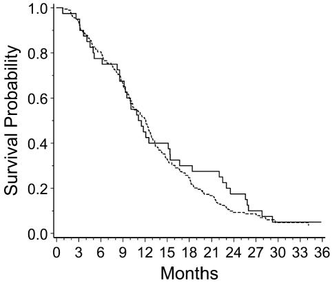
Thirty-eight of the 40 patients accrued to this study have died. The total follow-up time of all patients in this trial was 46.4 patient-years. The two surviving patients have been followed for more than 30 months. The failure rate was 0.82 (95% CI, 0.60–1.13) deaths per patient-year. Median survival was 11.3 (95% CI, 9.3–15.4) months, and median progression-free survival was 7.1 (95% CI, 6.2–8.5) months. The survival and the failure rates for patients in this study were compared to those of the NABTT reference group (Fig. 1; P = 0.52, log-rank test). At 18 months, survival in the intent-to-treat group was 30% compared to ~25% for the reference group (Table 2). Median survival for the reference group was 12.1 (95% CI, 10.3–12.9) months. The estimated hazard ratio for patients in this study in comparison to the reference group was not significant (hazard ratio = 0.89; P = 0.52; 95% CI, 0.62–1.27). Even after adjusting for age, KPS, and extent of surgery, the hazard ratio was not suggestive of survival benefit (estimated hazard ratio = 0.91, P = 0.61; Table 3).
Fig. 1.
Overall survival (Kaplan-Meier curves) for penicillamine study (solid line) compared to other NABTT trials of treatment concurrent with RT (broken line).
Table 2.
Percent survival and 95% confidence intervals
|
Penicillamine Patients |
Reference Patients |
|||
|---|---|---|---|---|
| Time (months) | Survival Rate (%) | 95% CI | Survival Rate (%) | 95% CI |
| 6 | 77.5 | 64.6–90.4 | 80.5 | 74.6–86.3 |
| 12 | 45.0 | 29.6–60.4 | 50.8 | 43.5–58.2 |
| 18 | 30.0 | 15.8–44.2 | 24.6 | 18.3–30.9 |
Table 3.
Proportional hazards regression model adjusting for prognostic factors comparing penicillamine study group to NABTT reference group
| Variable | Hazard Ratio (95% CI) | P |
|---|---|---|
| Penicillamine vs. NABTT reference | 0.91 (0.64–1.30) | 0.61 |
| Age (decade increase) | 1.32 (1.16–1.51) | 0.0001 |
| Karnofsky performance status (10 point increase) | 0.84 (0.74–0.95) | 0.007 |
| Biopsy vs. resection | 1.24 (0.86–1.79) | 0.25 |
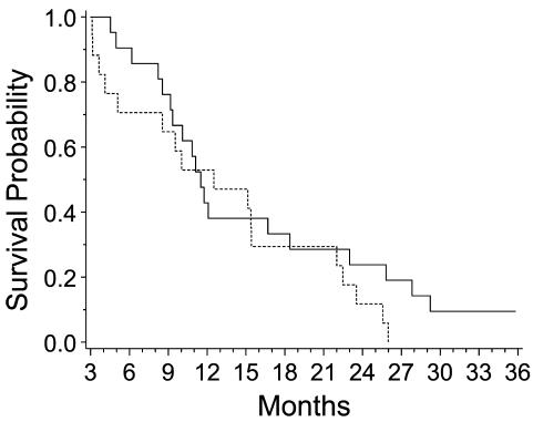
Patients surviving more than 90 days were stratified by whether or not they had attained hypocupremia (one serum copper level <50 μg/dl). Of the 38 patients alive for at least 90 days, 19 of 21 who had serum copper levels below the cut point died, whereas all 17 patients who failed to achieve hypocupremia had died. The median survival time for the hypocupremic group was 11.5 months and for the normocupremic group was 12.5 months (Fig. 2; P = 0.25, log-rank test). When this analysis was repeated using 20 μg/dl as a cutoff, there still was no significant difference in survival. One patient who had an extremely low (nondetectable) level of serum copper succumbed to a rapid recurrence of the glioblastoma.
Fig. 2.
Overall survival (Kaplan-Meier curves) for patients achieving low copper by three months (solid line) compared to those that did not (broken line).
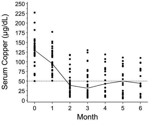
Clinically significant levels of hypocupremia were achieved after 10 weeks of penicillamine therapy (Fig. 3). The baseline levels of serum copper were either normal or elevated in all but five patients (median, 130 μg/dl; range, 50–227 μg/dl). In a few patients, the serum copper level paradoxically rose slightly after therapy was initiated, despite adherence to penicillamine and the diet, as excess copper was being mobilized from body stores. After two months, the copper levels reached the therapeutic target (median, 42 μg/dl; range, 12–118 μg/dl). Clinically significant hypocupremia was sustainable throughout the study for the majority of patients. For a few patients, the requirement of a restricted diet, including distilled water, was not tolerated, and these patients voluntarily removed themselves from the protocol. For many other patients, however, the ability to monitor and potentially control the copper level was viewed positively, and these patients enthusiastically adhered to the diet. Penicillamine compliance was confirmed by elevated levels of copper in the urine.
Fig. 3.
Hypocupremia achieved after penicillamine therapy. The dashed line represents the targeted serum copper level of 50 μg/dl. The solid line connects the median levels from each monthly visit for the patients that were sampled. At the baseline visit (month 0) data were available on 36 patients. At month 6, only 15 patients
Clinically significant hypocupremia was well tolerated. There were no reports of grade 3 or 4 anemia, sepsis, or osteoporosis attributable to hypocupremia. Hypocupremia was rapidly reversed by copper repletion in patients who developed intolerance or toxicity. There were no toxicities linked to copper repletion, such as rapid recurrence of the tumor or other “rebound” effects.
Two patients were maintained on penicillamine for more than three years without evidence of toxicity. The toxicity profile (Table 4) was consistent with known side effects of penicillamine (Singh et al., 1991) and mild copper deficiency, that is, hematological (leukopenia) and cutaneous. Potential vascular-related toxicities, such as vasogenic edema, hemorrhage, thromboembolism, neurocognitive decline, cerebral ischemia, vasculitis, nephropathy, alopecia, or impaired wound healing, were not observed. There were no neurological toxicities such as dementia, increased seizure incidence, or increased steroid dependency linked to penicillamine and copper reduction.
Table 4.
Major (grade 3/4) penicillamine-related toxicities
| Toxicity | Grade 3 | Grade 4 |
|---|---|---|
| Leukopenia | 5 | 1 |
| Granulocytopenia | 5 | __ |
| Thrombocytopenia | 3 | __ |
| Skin rash | 2 | __ |
| Hyperglycemia | 2 | __ |
| Elevated SGPT | 2 | __ |
| Elevated SGOT | 1 | __ |
| Headache | __ | 1 |
| Seizure | 1 | __ |
| Dyspnea | 1 | __ |
| Pleuritic pain | 1 | __ |
| Hypocalcemia | 1 | __ |
| Allergic reaction | 1 | __ |
| Myositis | 1 | __ |
| Insomnia | 1 | __ |
| Vomiting | 1 | __ |
| Total | 28 | 2 |
Because the study treatment was a cytostatic agent administered with radiation therapy, tumor response was not a primary end point. Significant reductions in contrast-enhancing tumor were uncommon on this protocol. Tumors treated with this therapy progressed rapidly and, as noted above, the overall survival of the patients was not improved.
Discussion
This study demonstrates that hypercupremia (Coates et al., 1989; Diez et al., 1989; Linder and Hazegh-Azam, 1996; Turecky et al., 1984), one hallmark of cancer, can be reversed by nutritional and pharmacological intervention. Because animal studies have shown that copper deficiency can suppress the malignant phenotype, including tumor growth and angiogenesis (Brem et al., 1990a, b; Khan et al., 2002; Pan et al., 2002; Yoshida et al., 1995; Yoshii et al., 2001), we evaluated copper reduction as a novel metabolic approach for the treatment of glioblastoma. An advantage of copper reduction as an antiangiogenesis therapy is that its effect can be frequently monitored through easily available serum tests of serum copper, used both as a surrogate marker of copper status and as a therapeutic target.
This study also demonstrates that both penicillamine and the low-copper diet were reasonably well tolerated. The serum copper remained low chronically, with urinary copper clearance providing further evidence of compliance. Most patients were able to cooperate and sustain hypocupremia. This study demonstrates the feasibility of nutritional and metabolic intervention for patients with brain tumors.
Until recently, there have been few angiogenesis inhibitors suitable for clinical trials (Kerbel, 2001). Penicillamine was shown previously to have antiangiogenic (Matsubara et al., 1989) and antineoplastic properties (Brem et al., 1990a, b; Hourani and Demopoulos, 1969; Jong-Hon et al., 1993; Littman et al., 1963) in laboratory investigations, but it has not previously been used in clinical trials against human cancer.
The toxicity profile of penicillamine in the current study (Table 4) is similar to that previously described for its use in rheumatoid arthritis (Singh et al., 1991), mainly hematological (leukopenia and thrombocytopenia) and correctable with dose reduction or discontinuation of penicillamine. Many of the potential toxicities associated with copper deficiency or vascular-targeted therapies were not observed in this cohort of patients. Specifically, we did not observe brain hemorrhages (Takano et al., 1994), thrombosis (DeVore et al., 2000), impaired wound healing (Brem et al., 1997), or neurocognitive decline (Strausak et al., 2001). The cutaneous and hematological toxicity of penicillamine and copper reduction was reversed by discontinuation of the drug and/or repletion of copper with copper tablets. There were no adverse events, such as tumor regrowth, associated with copper repletion. One death in this group of patients was due to Pneumocystis carinii pneumonia, previously reported in steroid-dependent patients with brain tumors (Schiff, 1996).
The effect of penicillamine on survival was estimated with reasonable precision in many ways, and none of these supported a clinically relevant benefit. The 95% CI for the adjusted hazard ratio was 0.64 to 1.3. The median survival was 11.3 months, comparable to standard therapy combining cytoreductive surgery and radiation therapy. Furthermore, when survival of patients who reached the target level of 50 μg/dl was compared with that of normocupremic patients, there was no significant difference in survival. The NABTT trials of cytostatic agents, such as antiangiogenesis compounds, are an appropriate reference group because the eligibility criteria, protocol design, and accruing institutions are similar for a broad range of trial agents. The progression-free survival and overall survival were similar to the survival data reported for the reference group.
The inability to produce a significant increase in survival in the clinical trial, in contrast to the preclinical study (Brem et al., 1990a), illustrates some of the difficulties associated with translating angiogenesis drug discovery from the laboratory to the clinic (Kerbel, 2001). Several possibilities could explain the disparity. First, the experimental animals were pretreated with penicillamine and a low-copper diet, and then the tumor cells were implanted, suggesting that hypocupremia might be more effective prior to the onset of the “switch” to the angiogenesis phenotype, as in low-grade gliomas or anaplastic astrocytomas (Brat et al., 2002). Second, genomic analysis of the human tumor endothelium indicates that the gene expression of the tumor micro-vasculature is highly variable, more closely resembling endothelial progenitor cells than the well-differentiated, mature human endothelial cells often used for antiangiogenic drug discovery (Bagley et al., 2003), including penicillamine (Matsubara et al., 1989). Third, higher doses of penicillamine with severe hypocupremia were used in the animal models (Brem et al., 1990a, b). Preclinical toxicities of anemia and increased vascular permeability were not observed in the clinical trial. Fourth, angiogenic responses to inhibitors, such as thalidomide, are species-specific (Bauer et al., 1998). In summary, the current study demonstrates the feasibility of copper reduction in patients with newly diagnosed glioblastoma. Safety was consistent with the known, mainly hematological, toxicity profile of penicillamine and copper deficiency. Survival was not significantly improved. More effective antiangiogenesis strategies could potentially improve survival in patients with newly diagnosed glioblastoma (Bello et al., 2001; Brem and Wotoczek-Obadia., 2001; Fine et al., 2003; Hurwitz et al., 2004; Jain, 2001; Kabbinavar et al., 2003; Kerbel, 2001; Redman et al., 2003).
Acknowledgment
We thank Marguerite Wotoczek-Obadia for assistance with the data analysis of the serum copper, Nagi Kumar and Karen Besterman-Dahan for development of the low-copper diet and supervision of the nutritional intervention, Steven Piantadosi for help with the statistical design and analysis, Regina Priet for the data coordination of the study, and the clinical research nurses and data managers at the participating NABTT centers.
Footnotes
This work was supported in part by grant no. UO1-CA77412 from the National Cancer Institute and by the NABTT Headquarters grant.
Preliminary results of the study were presented at the sixth International Society of Trace Elements Research in Humans, ISTERH 2002, Quebec City, Quebec, Canada, September 7–12, 2002.
Abbreviations used are as follows: CI, confidence interval; KPS, Karnofsky performance status; NABTT, New Approaches to Brain Tumor Therapy.
References
- Alpern-Elran H, Brem S. Angiogenesis in human brain tumors: Inhibition by copper depletion. Surg Forum. 1985;36:498–500. [Google Scholar]
- Bagley RG, Walter-Yohrling J, Cao X, Weber W, Simons B, Cook BP, Chartrand SD, Wang C, Madden SL, Teicher BA. Endothelial precursor cells as a model of tumor endothelium: Characterization and comparison with mature endothelial cells. Cancer Res. 2003;63:5866–5873. [PubMed] [Google Scholar]
- Barker FG, 2nd Chang, S.M. Larson, D.A. Sneed, P.K. Wara, W.M. Wilson, C.B. and, Prados MD. Age and radiation response in glioblastoma multiforme. Neurosurgery. 2001;49:1288–1298. doi: 10.1097/00006123-200112000-00002. [DOI] [PubMed] [Google Scholar]
- Bauer KS, Dixon SC, Figg WD. Inhibition of angiogenesis by thalidomide requires metabolic activation, which is species-dependent. Biochem Pharmacol. 1998;55:1827–1834. doi: 10.1016/s0006-2952(98)00046-x. [DOI] [PubMed] [Google Scholar]
- Bello L, Carrabba G, Giussani C, Lucini V, Cerutti F, Scaglione F, Landre J, Pluderi M, Tomei G, Villani R, Carroll RS, Black PM, Bikfalvi A. Low-dose chemotherapy combined with an antiangiogenic drug reduces human glioma growth in vivo. Cancer Res. 2001;61:7501–7506. [PubMed] [Google Scholar]
- Brat DJ, Castellano-Sanchez A, Kaur B, Van Meir EG. Genetic and biologic progression in astrocytomas and their relation to angiogenic dysregulation. Adv Anat Pathol. 2002;9:24–36. doi: 10.1097/00125480-200201000-00004. [DOI] [PubMed] [Google Scholar]
- Brem H, Ehrlich P, Tsakayannis D, Folkman J. Delay of wound healing by the angiogenesis inhibitor TNP-470. Surg Forum. 1997;48:714–716. [Google Scholar]
- Brem, S. (2004) Angiogenesis and brain tumors. In: Winn, H.R. (Ed.), You-mans Neurological Surgery Philadelphia: Saunders, pp. 771–789.
- Brem S, Wotoczek-Obadia M. Potential synergistic effect of copper reduction, anti-angiogenesis therapy and cytotoxic chemotherapy (BCNU) in a rat brain tumor model. Proc Am Assoc Cancer Res. 2001;42:582p. (abstract 3123) [Google Scholar]
- Brem S, Cotran R, Folkman J. Tumor angiogenesis: A quantitative method for histologic grading. J Natl Cancer Inst. 1972;48:347–356. [PubMed] [Google Scholar]
- Brem SS, Zagzag D, Tsanaclis AM, Gately S, Elkouby MP, Brien SE. Inhibition of angiogenesis and tumor growth in the brain. Suppression of endothelial cell turnover by penicillamine and the depletion of copper, an angiogenic factor. Am J Pathol. 1990a;137:1121–1142. [PMC free article] [PubMed] [Google Scholar]
- Brem S, Tsanaclis AM, Zagzag D. Anticopper treatment inhibits pseudopodial protrusion and the invasive spread of 9L gliosar-coma in the rat brain. Neurosurgery. 1990b;26:391–396. doi: 10.1097/00006123-199003000-00003. [DOI] [PubMed] [Google Scholar]
- Carmeliet P, Jain RK. Angiogenesis in cancer and other diseases. Nature. 2000;407:249–257. doi: 10.1038/35025220. [DOI] [PubMed] [Google Scholar]
- Coates RJ, Weiss NS, Daling JR, Rettmer RL, Warnick GR. Cancer risk in relation to serum copper levels. Cancer Res. 1989;49:4353–4356. [PubMed] [Google Scholar]
- Cox DR. Regression models and life tables. J R Stat Soc B. 1972;34:187–220. [Google Scholar]
- Davis FG, Freels S, Grutsch J, Barlas S, Brem S. Survival rates in patients with primary malignant brain tumors stratified by patient age and tumor histological type: An analysis based on Surveillance, Epidemiology, and End Results (SEER) data, 1973–1991. J Neuro-surg. 1998;88:1–10. doi: 10.3171/jns.1998.88.1.0001. [DOI] [PubMed] [Google Scholar]
- DeVore RF, Fehrenbacher L, Herbst RS, Langer CJ, Kelly K, Gau-dreault J, Holmgren E, Novotny WF, Kabbinavar F. A randomized phase II trial comparing rhuMAb VEGF (recombinant humanized monoclonal antibody to vascular endothelial cell growth factor) plus carboplatin/paclitaxel (CP) to CP alone in patients with stage IIIb/IV NSCLC. Proc Am Soc Clin Oncol. 2000;19:485a. (abstract 1896) [Google Scholar]
- Diez M, Cerdan FJ, Arroyo M, Balibrea JL. Use of copper/zinc ratio in the diagnosis of lung cancer. Cancer. 1989;63:726–730. doi: 10.1002/1097-0142(19890215)63:4<726::aid-cncr2820630421>3.0.co;2-p. [DOI] [PubMed] [Google Scholar]
- Fine HA, Figg WD, Jaeckle K, Wen PY, Kyritsis AP, Loeffler JS, Levin VA, Black PM, Kaplan R, Pluda JM, Yung WK. Phase II trial of the antiangiogenic agent thalidomide in patients with recurrent high-grade gliomas. J Clin Oncol. 2000;18:708–715. doi: 10.1200/JCO.2000.18.4.708. [DOI] [PubMed] [Google Scholar]
- Fine HA, Wen PY, Maher EA, Viscosi E, Batchelor T, Lakhani N, Figg WD, Purow BW, Borkowf CB. Phase II trial of thalidomide and carmustine for patients with recurrent high-grade gliomas. J Clin Oncol. 2003;21:2299–2304. doi: 10.1200/JCO.2003.08.045. [DOI] [PubMed] [Google Scholar]
- Godard S, Getz G, Delorenzi M, Farmer P, Kobayahsi H, Desbaillets I, Nozaki M, Diserens AC, Hamou MF, Dietrich PY, Regli L, Janzer RC, Bucher P, Stupp R, de Tribolet N, Domany E, Hegi ME. Classification of human astrocytic gliomas on the basis of gene expression: A correlated group of genes with angiogenic activity emerges as a strong predictor of subtypes. Cancer Res. 2003;63:6613–6625. [PubMed] [Google Scholar]
- Grossman SA, Batara J. Current management of glioblastoma multiforme. Semin Oncol. 2004;31:635–644. doi: 10.1053/j.seminoncol.2004.07.005. [DOI] [PubMed] [Google Scholar]
- Grossman, S.A., Carson, K., Piantadosi, S. and Fisher, J. (2002) Survival of adults with newly diagnosed glioblastoma multiforme (GBM) treated with experimental agents and delayed radiation therapy (RT) versus experimental agents and immediate radiation: The experience of the NABTT CNS consortium. Presented at the American Society of Clinical Oncology Annual Meeting, Orlando, Florida, May 18–21 (abstract 282). Available at: http://www.asco.org
- Hourani BT, Demopoulos HB. Inhibition of S-91 mouse melanoma metastases and growth by D-penicillamine. Lab Invest. 1969;21:434–438. [PubMed] [Google Scholar]
- Hu GF. Copper stimulates proliferation of human endothelial cells under culture. J Cell Biochem. 1998;69:326–335. doi: 10.1002/(sici)1097-4644(19980601)69:3<326::aid-jcb10>3.0.co;2-a. [DOI] [PubMed] [Google Scholar]
- Hurwitz H, Fehrenbacher L, Novotny W, Cartwright T, Hainsworth J, Heim W, Berlin J, Baron A, Griffing S, Holmgren E, Ferrara N, Fyfe G, Rogers B, Ross R, Kabbinavar F. Bevacizumab plus irinotecan, fluorouracil, and leucovorin for metastatic colorectal cancer. N Engl J Med. 2004;350:2335–2342. doi: 10.1056/NEJMoa032691. [DOI] [PubMed] [Google Scholar]
- Jain RK. Normalizing tumor vasculature with anti-angiogenic therapy: A new paradigm for combination therapy. Nat Med. 2001;7:987–989. doi: 10.1038/nm0901-987. [DOI] [PubMed] [Google Scholar]
- Jong-Hon K, Togashi Y, Kasai H, Hosokawa M, Takeichi N. Prevention of spontaneous hepatocellular carcinoma in Long-Evans cinnamon rats with hereditary hepatitis by the administration of d-penicillamine. Hepatology. 1993;18:614–620. [PubMed] [Google Scholar]
- Joyce DA. D-Penicillamine pharmacokinetics and pharmacody-namics in man. Pharmacol Ther. 1989;42:405–427. doi: 10.1016/0163-7258(89)90033-8. [DOI] [PubMed] [Google Scholar]
- Kabbinavar F, Hurwitz HI, Fehrenbacher L, Meropol NJ, Novotny WF, Lieberman G, Griffing S, Bergsland E. Phase II, randomized trial comparing bevacizumab plus fluorouracil (FU)/leu-covorin (LV) with FU/LV alone in patients with metastatic colorectal cancer. J Clin Oncol. 2003;21:60–65. doi: 10.1200/JCO.2003.10.066. [DOI] [PubMed] [Google Scholar]
- Kalbfleisch, J.D., and Prentice, R.L. (1980) The Statistical Analysis of Failure Time Data New York: John Wiley & Sons.
- Kaplan EL, Meier P. Nonparametric estimation from incomplete observations. J Am Stat Assoc. 1958;53:457–481. [Google Scholar]
- Kerbel RS. Clinical trials of antiangiogenic drugs: Opportunities, problems, and assessment of initial results. J Clin Oncol. 2001;19:45S–51S. [PubMed] [Google Scholar]
- Khan MK, Miller MW, Taylor J, Gill NK, Dick RD, Van Golen K, Brew GJ, Merajver SD. Radiotherapy and antiangiogenic TM in lung cancer. Neoplasia. 2002;4:164–170. doi: 10.1038/sj.neo.7900218. [DOI] [PMC free article] [PubMed] [Google Scholar]
- Lacroix M, Abi-Said D, Fourney DR, Gokaslan ZL, Shi W, DeMonte F, Lang FF, McCutcheon IE, Hassenbusch SJ, Holland E, Hess K, Michael C, Miller D, Sawaya R. A multivariate analysis of 416 patients with glioblastoma multiforme: Prognosis, extent of resection, and survival. J Neurosurg. 2001;95:190–198. doi: 10.3171/jns.2001.95.2.0190. [DOI] [PubMed] [Google Scholar]
- Linder MC, Hazegh-Azam M. Copper biochemistry and molecular biology. Am J Clin Nutr. 1996;63:797S–811S. doi: 10.1093/ajcn/63.5.797. [DOI] [PubMed] [Google Scholar]
- Littman ML, Taguchi T, Shimizu Y. Acceleration of growth of sarcoma 180 with pyridoxamine and retardation with penicillamine. Proc Soc Exp Biol Med. 1963;113:667–674. doi: 10.3181/00379727-113-28458. [DOI] [PubMed] [Google Scholar]
- Marsden CD. Wilson’s disease. Q J Med. 1987;248:959–966. [PubMed] [Google Scholar]
- Matsubara T, Saura R, Hirohata K, Ziff M. Inhibition of human endothelial cell proliferation in vitro and neovascularization in vivo by D-penicillamine. J Clin Invest. 1989;83:158–167. doi: 10.1172/JCI113853. [DOI] [PMC free article] [PubMed] [Google Scholar]
- Netter P, Bannwarth B, Pere P, Nicolas A. Clinical pharma-cokinetics of D-penicillamine. Clin Pharmacokinet. 1987;13:317–333. doi: 10.2165/00003088-198713050-00003. [DOI] [PubMed] [Google Scholar]
- Pan Q, Kleer CG, van Golen KL, Irani J, Bottema KM, Bias C, De Carvalho M, Mesri EA, Robins DM, Dick RD, Brewer GJ, Merajver SD. Copper deficiency induced by tetrathiomo-lybdate suppresses tumor growth and angiogenesis. Cancer Res. 2002;62:4854–4859. [PubMed] [Google Scholar]
- Puduvalli VK, Sawaya R. Antiangiogenesis—therapeutic strategies and clinical implications for brain tumors. J Neurooncol. 2000;50:189–200. doi: 10.1023/a:1006469830739. [DOI] [PubMed] [Google Scholar]
- Redman BG, Esper P, Pan Q, Dunn RL, Hussain HK, Chenevert T, Brewer GJ, Merajver SD. Phase II trial of tetrathiomo-lybdate in patients with advanced kidney cancer. Clin Cancer Res. 2003;9:1666–1672. [PubMed] [Google Scholar]
- Schiff D. Pneumocystis pneumonia in brain tumor patients: Risk factors and clinical features. J Neurooncol. 1996;27:235–240. doi: 10.1007/BF00165480. [DOI] [PubMed] [Google Scholar]
- Singh G, Fries JF, Williams CA, Zatarain E, Spitz P, Bloch DA. Toxicity profiles of disease modifying antirheumatic drugs in rheumatoid arthritis. J Rheumatol. 1991;18:188–194. [PubMed] [Google Scholar]
- Smith DB, Gallagher BB. The effect of penicillamine on seizure threshold. The role of pyridoxine. Arch Neurol. 1970;23:59–62. doi: 10.1001/archneur.1970.00480250063008. [DOI] [PubMed] [Google Scholar]
- Sproull M, Brechbiel M, Camphausen K. Antiangiogenic therapy through copper chelation. Expert Opin Ther Targets. 2003;7:405–409. doi: 10.1517/14728222.7.3.405. [DOI] [PubMed] [Google Scholar]
- Strausak D, Mercer JF, Dieter HH, Stremmel W, Multhaup G. Copper in disorders with neurological symptoms: Alzheimer’s, Menkes, and Wilson diseases. Brain Res Bull. 2001;55:175–185. doi: 10.1016/s0361-9230(01)00454-3. [DOI] [PubMed] [Google Scholar]
- Stupp R, Mason WP, van den Bent MJ, Weller M, Fisher B, Taphoorn M, Brandes AA, Cairncross G, Lacombe D, Miri-manoff RO. Concomitant and adjuvant temozolomide (TMZ) and radiotherapy (RT) for newly diagnosed glioblastoma multiforme (GBM). Conclusive results of a randomized phase III trial by the EORTC Brain & RT Groups and NCIC Clinical Trials Group. J Clin Oncol. 2004;22(14S):2. (abstract) [Google Scholar]
- Takano S, Gately S, Engelhard H, Tsanaclis AM, Brem S. Suramin inhibits glioma cell proliferation in vitro and in the brain. J Neurooncol. 1994;21:189–201. doi: 10.1007/BF01063768. [DOI] [PubMed] [Google Scholar]
- Turecky L, Kalina P, Uhlikova E, Namerova S, Krizko J. Serum ceruloplasmin and copper levels in patients with primary brain tumors. Klin Wochenschr. 1984;62:187–189. doi: 10.1007/BF01731643. [DOI] [PubMed] [Google Scholar]
- Turnlund JR, Scott KC, Peiffer GL, Jang AM, Keyes WR, Keen CL, Sakanashi TM. Copper status of young men consuming a low-copper diet. Am J Clin Nutr. 1997;65:72–78. doi: 10.1093/ajcn/65.1.72. [DOI] [PubMed] [Google Scholar]
- Weindruch R, Walford RL. Dietary restriction in mice beginning at 1 year of age: Effect on life-span and spontaneous cancer incidence. Science. 1982;215:1415–1418. doi: 10.1126/science.7063854. [DOI] [PubMed] [Google Scholar]
- Westphal M, Hilt DC, Bortey E, Delavault P, Olivares R, Warnke PC, Whittle IR, Jääskeläinen J, Ram Z. A phase 3 trial of local chemotherapy with biodegradable carmustine (BCNU) wafers (Gliadel wafers) in patients with primary malignant glioma. Neuro-Oncol. 2003;5:79–88. doi: 10.1215/S1522-8517-02-00023-6. [DOI] [PMC free article] [PubMed] [Google Scholar]
- Williams, S.R. (1993) Nutrition and Diet Therapy, 7th ed. St. Louis: Mosby-Year Book, Inc., pp. 253–268.
- Yoshida D, Ikeda Y, Nakazawa S. Copper chelation inhibits tumor angiogenesis in the experimental 9L gliosarcoma model. Neuro-surgery. 1995;37:287–293. doi: 10.1227/00006123-199508000-00014. [DOI] [PubMed] [Google Scholar]
- Yoshii J, Yoshiji H, Kuriyama S, Ikenaka Y, Noguchi R, Okuda H, Tsujinoue H, Nakatani T, Kishida H, Nakae D, Gomez DE, DeLo-renzo M.S, Tejera AM, Fukui H. The copper-chelating agent, trientine, suppresses tumor development and angiogenesis in the murine hepatocellular carcinoma cells. Int J Cancer. 2001;94:768–773. doi: 10.1002/ijc.1537. [DOI] [PubMed] [Google Scholar]
- Ziche M, Jones J, Gullino PM. Role of prostaglandin E1 and copper in angiogenesis. J Natl Cancer Inst. 1982;69:475–482. [PubMed] [Google Scholar]


