Full Text
The Full Text of this article is available as a PDF (500.2 KB).
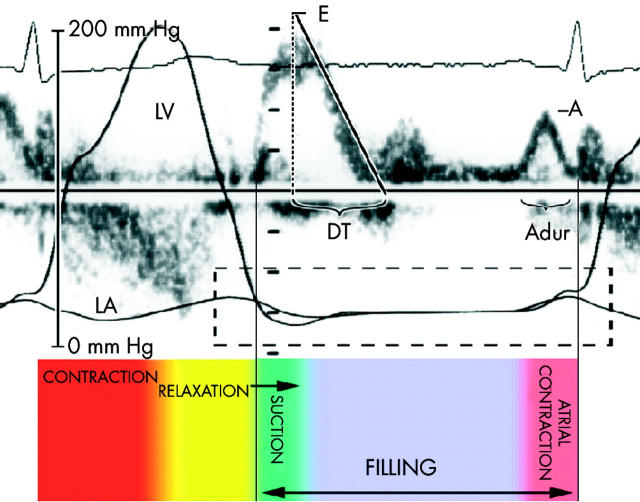
Figure 1.
The phases of left ventricular filling are relaxation, suction, filling, and atrial contraction as depicted in these simultaneous invasive pressure curves and Doppler echocardiography. A, mitral filling at atrial contraction; Adur, duration of mitral A wave; DT, mitral deceleration time; E, mitral early filling wave; LA, left atrial pressure curve; LV, left ventricular pressure curve.
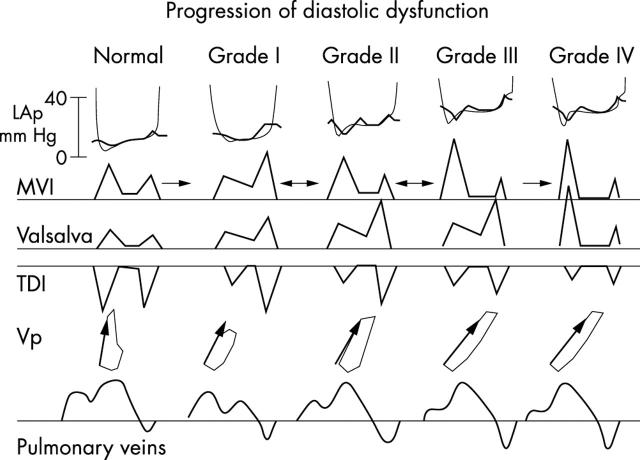
Figure 2.
The progression of left ventricular diastolic dysfunction can be readily assessed using a combination of Doppler echocardiographic variables. Each successive grade represents a worsening state of diastolic dysfunction. LAp, left atrial pressure; MVI, mitral valve inflow; TDI, tissue Doppler imaging; Valsalva, response of mitral valve inflow to Valsalva manoeuvre; Vp, mitral inflow propagation velocity.
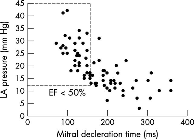
Figure 3.
Relation between mitral deceleration time and left atrial pressure as assessed from three separate simultaneous Doppler catheterisation studies. EF, left ventricular ejection fraction; LA, left atrial. Adapted from Nishimura et al,6, Ommen et al,7 and Yamamoto et al.13
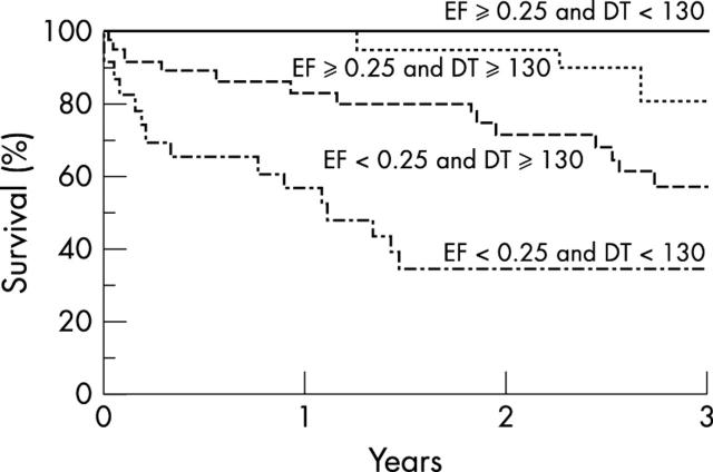
Figure 4.
Relation between systolic and diastolic function and overall survival highlights the critical role of diastolic function for the prognosis of patients with impaired left ventricular systolic function. DT, Deceleration time; EF, ejection fraction. Reproduced from Rihal et al,8 with permission.
Figure 5.
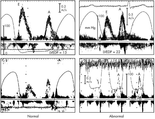
The response of the mitral inflow to preload manipulation can be useful in predicting filling pressures. On the left is a normal response to the Valsalva manoeuvre with proportional decreases in both mitral E and A waves in a patient with normal filling pressures. On the right, the Valsalva manoeuvre results in disproportionate decrease in mitral E wave in a patient with raised filling pressure. A decrease in the mitral E/A ratio of 0.5 or more is a highly specific indicator of increased filling pressures. A, mitral filling at atrial contraction; E, mitral early filling wave; LVEDP, left ventricular end diastolic pressure; m/s, metres per second.
Figure 6.
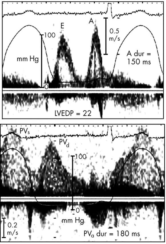
The duration of flow at atrial contraction into the left ventricle as compared to duration of flow reversal into the pulmonary veins can indicate end diastolic filling pressure. In this example, flow duration is much longer on the pulmonary vein Doppler signal (180 ms v 150 ms) indicating raised left ventricular end diastolic pressure. A, mitral filling at atrial contraction; E, mitral early filling wave; LVEDP, left ventricular end diastolic pressure; PVa dur, pulmonary vein atrial reversal duration; PVd, pulmonary vein diastolic forward flow, PVs, pulmonary vein systolic forward flow.
Figure 7.
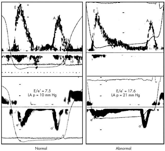
The tissue Doppler velocity of the mitral annulus can help distinguish between normal and abnormal filling. These examples are from two patients with normal systolic function and similar mitral inflow signals. On the left, a normal mitral annular velocity and E/e' ratio indicate normal filling pressures. On the right, the mitral annular e' velocity is reduced and the E/e' ratio elevated in a patient with high left atrial pressure. A, mitral filling at atrial contraction; a', late mitral annulus diastolic velocity; E, mitral early filling wave; e', early mitral annulus diastolic velocity; E/e', ratio of mitral early filling wave to early mitral annulus velocity; LA p, left atrial pressure.
Figure 8.
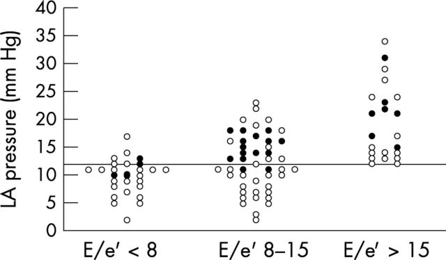
Cut-off values using the ratio of mitral early filling wave to early mitral annulus velocity (E/e') can be used to group patients according to filling pressures. Those with high ratios (> 15) have high filling pressures, while those with very low ratios tend to have normal filling pressures. The intermediate group requires additional information to correctly classify diastolic function. The closed circles represent those patients with other Doppler parameters suggesting high filling pressures. Reproduced from Ommen et al,7 with permission.
Figure 9.
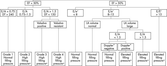
Screening assessment for diastolic function and filling pressures. Two dimensional and Doppler echocardiographic variables can be used to readily classify diastolic function. *In general, high filling pressures should be confirmed with multiple parameters (that is, E/e', E/Vp, A-dur difference, response to Valsalva manoeuvre, TR velocity, etc).
Selected References
These references are in PubMed. This may not be the complete list of references from this article.
- Appleton C. P., Galloway J. M., Gonzalez M. S., Gaballa M., Basnight M. A. Estimation of left ventricular filling pressures using two-dimensional and Doppler echocardiography in adult patients with cardiac disease. Additional value of analyzing left atrial size, left atrial ejection fraction and the difference in duration of pulmonary venous and mitral flow velocity at atrial contraction. J Am Coll Cardiol. 1993 Dec;22(7):1972–1982. doi: 10.1016/0735-1097(93)90787-2. [DOI] [PubMed] [Google Scholar]
- Bach D. S. Quantitative Doppler tissue imaging as a correlate of left ventricular contractility. Int J Card Imaging. 1996 Sep;12(3):191–195. doi: 10.1007/BF01806222. [DOI] [PubMed] [Google Scholar]
- Brunazzi M. C., Chirillo F., Pasqualini M., Gemelli M., Franceschini-Grisolia E., Longhini C., Giommi L., Barbaresi F., Stritoni P. Estimation of left ventricular diastolic pressures from precordial pulsed-Doppler analysis of pulmonary venous and mitral flow. Am Heart J. 1994 Aug;128(2):293–300. doi: 10.1016/0002-8703(94)90482-0. [DOI] [PubMed] [Google Scholar]
- Brunner-La Rocca H. P., Rickli H., Attenhofer Jost C. H., Jenni R. Left ventricular end-diastolic pressure can be estimated by either changes in transmitral inflow pattern during valsalva maneuver or analysis of pulmonary venous flow. J Am Soc Echocardiogr. 2000 Jun;13(6):599–607. doi: 10.1067/mje.2000.106077. [DOI] [PubMed] [Google Scholar]
- Dini F. L., Michelassi C., Micheli G., Rovai D. Prognostic value of pulmonary venous flow Doppler signal in left ventricular dysfunction: contribution of the difference in duration of pulmonary venous and mitral flow at atrial contraction. J Am Coll Cardiol. 2000 Oct;36(4):1295–1302. doi: 10.1016/s0735-1097(00)00821-4. [DOI] [PubMed] [Google Scholar]
- Donovan C. L., Armstrong W. F., Bach D. S. Quantitative Doppler tissue imaging of the left ventricular myocardium: validation in normal subjects. Am Heart J. 1995 Jul;130(1):100–104. doi: 10.1016/0002-8703(95)90242-2. [DOI] [PubMed] [Google Scholar]
- Dumesnil J. G., Gaudreault G., Honos G. N., Kingma J. G., Jr Use of Valsalva maneuver to unmask left ventricular diastolic function abnormalities by Doppler echocardiography in patients with coronary artery disease or systemic hypertension. Am J Cardiol. 1991 Aug 15;68(5):515–519. doi: 10.1016/0002-9149(91)90788-m. [DOI] [PubMed] [Google Scholar]
- Garcia M. J., Rodriguez L., Ares M., Griffin B. P., Klein A. L., Stewart W. J., Thomas J. D. Myocardial wall velocity assessment by pulsed Doppler tissue imaging: characteristic findings in normal subjects. Am Heart J. 1996 Sep;132(3):648–656. doi: 10.1016/s0002-8703(96)90251-3. [DOI] [PubMed] [Google Scholar]
- Garcia M. J., Smedira N. G., Greenberg N. L., Main M., Firstenberg M. S., Odabashian J., Thomas J. D. Color M-mode Doppler flow propagation velocity is a preload insensitive index of left ventricular relaxation: animal and human validation. J Am Coll Cardiol. 2000 Jan;35(1):201–208. doi: 10.1016/s0735-1097(99)00503-3. [DOI] [PubMed] [Google Scholar]
- Gulati V. K., Katz W. E., Follansbee W. P., Gorcsan J., 3rd Mitral annular descent velocity by tissue Doppler echocardiography as an index of global left ventricular function. Am J Cardiol. 1996 May 1;77(11):979–984. doi: 10.1016/s0002-9149(96)00033-1. [DOI] [PubMed] [Google Scholar]
- Hurrell D. G., Nishimura R. A., Ilstrup D. M., Appleton C. P. Utility of preload alteration in assessment of left ventricular filling pressure by Doppler echocardiography: a simultaneous catheterization and Doppler echocardiographic study. J Am Coll Cardiol. 1997 Aug;30(2):459–467. doi: 10.1016/s0735-1097(97)00184-8. [DOI] [PubMed] [Google Scholar]
- Keren G., Sherez J., Megidish R., Levitt B., Laniado S. Pulmonary venous flow pattern--its relationship to cardiac dynamics. A pulsed Doppler echocardiographic study. Circulation. 1985 Jun;71(6):1105–1112. doi: 10.1161/01.cir.71.6.1105. [DOI] [PubMed] [Google Scholar]
- Kitabatake A. Propagation velocity of left ventricular filling flow measured by color M-mode Doppler echocardiography. J Am Coll Cardiol. 1998 May;31(6):1445–1446. doi: 10.1016/s0735-1097(98)00110-7. [DOI] [PubMed] [Google Scholar]
- Kuecherer H. F., Kusumoto F., Muhiudeen I. A., Cahalan M. K., Schiller N. B. Pulmonary venous flow patterns by transesophageal pulsed Doppler echocardiography: relation to parameters of left ventricular systolic and diastolic function. Am Heart J. 1991 Dec;122(6):1683–1693. doi: 10.1016/0002-8703(91)90287-r. [DOI] [PubMed] [Google Scholar]
- Lisauskas J., Singh J., Courtois M., Kovács S. J. The relation of the peak Doppler E-wave to peak mitral annulus velocity ratio to diastolic function. Ultrasound Med Biol. 2001 Apr;27(4):499–507. doi: 10.1016/s0301-5629(00)00357-4. [DOI] [PubMed] [Google Scholar]
- Møller J. E., Poulsen S. H., Søndergaard E., Egstrup K. Preload dependence of color M-mode Doppler flow propagation velocity in controls and in patients with left ventricular dysfunction. J Am Soc Echocardiogr. 2000 Oct;13(10):902–909. doi: 10.1067/mje.2000.106572. [DOI] [PubMed] [Google Scholar]
- Møller J. E., Søndergaard E., Seward J. B., Appleton C. P., Egstrup K. Ratio of left ventricular peak E-wave velocity to flow propagation velocity assessed by color M-mode Doppler echocardiography in first myocardial infarction: prognostic and clinical implications. J Am Coll Cardiol. 2000 Feb;35(2):363–370. doi: 10.1016/s0735-1097(99)00575-6. [DOI] [PubMed] [Google Scholar]
- Nagueh S. F., Middleton K. J., Kopelen H. A., Zoghbi W. A., Quiñones M. A. Doppler tissue imaging: a noninvasive technique for evaluation of left ventricular relaxation and estimation of filling pressures. J Am Coll Cardiol. 1997 Nov 15;30(6):1527–1533. doi: 10.1016/s0735-1097(97)00344-6. [DOI] [PubMed] [Google Scholar]
- Nagueh S. F., Mikati I., Kopelen H. A., Middleton K. J., Quiñones M. A., Zoghbi W. A. Doppler estimation of left ventricular filling pressure in sinus tachycardia. A new application of tissue doppler imaging. Circulation. 1998 Oct 20;98(16):1644–1650. doi: 10.1161/01.cir.98.16.1644. [DOI] [PubMed] [Google Scholar]
- Nishimura R. A., Abel M. D., Hatle L. K., Tajik A. J. Relation of pulmonary vein to mitral flow velocities by transesophageal Doppler echocardiography. Effect of different loading conditions. Circulation. 1990 May;81(5):1488–1497. doi: 10.1161/01.cir.81.5.1488. [DOI] [PubMed] [Google Scholar]
- Nishimura R. A., Appleton C. P., Redfield M. M., Ilstrup D. M., Holmes D. R., Jr, Tajik A. J. Noninvasive doppler echocardiographic evaluation of left ventricular filling pressures in patients with cardiomyopathies: a simultaneous Doppler echocardiographic and cardiac catheterization study. J Am Coll Cardiol. 1996 Nov 1;28(5):1226–1233. doi: 10.1016/S0735-1097(96)00315-4. [DOI] [PubMed] [Google Scholar]
- Nishimura R. A., Tajik A. J. Evaluation of diastolic filling of left ventricle in health and disease: Doppler echocardiography is the clinician's Rosetta Stone. J Am Coll Cardiol. 1997 Jul;30(1):8–18. doi: 10.1016/s0735-1097(97)00144-7. [DOI] [PubMed] [Google Scholar]
- Ommen S. R., Nishimura R. A., Appleton C. P., Miller F. A., Oh J. K., Redfield M. M., Tajik A. J. Clinical utility of Doppler echocardiography and tissue Doppler imaging in the estimation of left ventricular filling pressures: A comparative simultaneous Doppler-catheterization study. Circulation. 2000 Oct 10;102(15):1788–1794. doi: 10.1161/01.cir.102.15.1788. [DOI] [PubMed] [Google Scholar]
- Redfield Margaret M., Jacobsen Steven J., Burnett John C., Jr, Mahoney Douglas W., Bailey Kent R., Rodeheffer Richard J. Burden of systolic and diastolic ventricular dysfunction in the community: appreciating the scope of the heart failure epidemic. JAMA. 2003 Jan 8;289(2):194–202. doi: 10.1001/jama.289.2.194. [DOI] [PubMed] [Google Scholar]
- Rihal C. S., Nishimura R. A., Hatle L. K., Bailey K. R., Tajik A. J. Systolic and diastolic dysfunction in patients with clinical diagnosis of dilated cardiomyopathy. Relation to symptoms and prognosis. Circulation. 1994 Dec;90(6):2772–2779. doi: 10.1161/01.cir.90.6.2772. [DOI] [PubMed] [Google Scholar]
- Rodriguez L., Garcia M., Ares M., Griffin B. P., Nakatani S., Thomas J. D. Assessment of mitral annular dynamics during diastole by Doppler tissue imaging: comparison with mitral Doppler inflow in subjects without heart disease and in patients with left ventricular hypertrophy. Am Heart J. 1996 May;131(5):982–987. doi: 10.1016/s0002-8703(96)90183-0. [DOI] [PubMed] [Google Scholar]
- Rossvoll O., Hatle L. K. Pulmonary venous flow velocities recorded by transthoracic Doppler ultrasound: relation to left ventricular diastolic pressures. J Am Coll Cardiol. 1993 Jun;21(7):1687–1696. doi: 10.1016/0735-1097(93)90388-h. [DOI] [PubMed] [Google Scholar]
- Schwammenthal E., Popescu B. A., Popescu A. C., Di Segni E., Kaplinsky E., Rabinowitz B., Guetta V., Rath S., Feinberg M. S. Noninvasive assessment of left ventricular end-diastolic pressure by the response of the transmitral a-wave velocity to a standardized Valsalva maneuver. Am J Cardiol. 2000 Jul 15;86(2):169–174. doi: 10.1016/s0002-9149(00)00855-9. [DOI] [PubMed] [Google Scholar]
- Senni M., Tribouilloy C. M., Rodeheffer R. J., Jacobsen S. J., Evans J. M., Bailey K. R., Redfield M. M. Congestive heart failure in the community: a study of all incident cases in Olmsted County, Minnesota, in 1991. Circulation. 1998 Nov 24;98(21):2282–2289. doi: 10.1161/01.cir.98.21.2282. [DOI] [PubMed] [Google Scholar]
- Sohn D. W., Chai I. H., Lee D. J., Kim H. C., Kim H. S., Oh B. H., Lee M. M., Park Y. B., Choi Y. S., Seo J. D. Assessment of mitral annulus velocity by Doppler tissue imaging in the evaluation of left ventricular diastolic function. J Am Coll Cardiol. 1997 Aug;30(2):474–480. doi: 10.1016/s0735-1097(97)88335-0. [DOI] [PubMed] [Google Scholar]
- Temporelli P. L., Corrà U., Imparato A., Bosimini E., Scapellato F., Giannuzzi P. Reversible restrictive left ventricular diastolic filling with optimized oral therapy predicts a more favorable prognosis in patients with chronic heart failure. J Am Coll Cardiol. 1998 Jun;31(7):1591–1597. doi: 10.1016/s0735-1097(98)00165-x. [DOI] [PubMed] [Google Scholar]
- Thomas J. D., Garcia M. J., Greenberg N. L. Application of color Doppler M-mode echocardiography in the assessment of ventricular diastolic function: potential for quantitative analysis. Heart Vessels. 1997;Suppl 12:135–137. [PubMed] [Google Scholar]
- Vasan R. S., Benjamin E. J., Levy D. Prevalence, clinical features and prognosis of diastolic heart failure: an epidemiologic perspective. J Am Coll Cardiol. 1995 Dec;26(7):1565–1574. doi: 10.1016/0735-1097(95)00381-9. [DOI] [PubMed] [Google Scholar]
- Vasan R. S., Levy D. Defining diastolic heart failure: a call for standardized diagnostic criteria. Circulation. 2000 May 2;101(17):2118–2121. doi: 10.1161/01.cir.101.17.2118. [DOI] [PubMed] [Google Scholar]
- Xie G. Y., Berk M. R., Smith M. D., Gurley J. C., DeMaria A. N. Prognostic value of Doppler transmitral flow patterns in patients with congestive heart failure. J Am Coll Cardiol. 1994 Jul;24(1):132–139. doi: 10.1016/0735-1097(94)90553-3. [DOI] [PubMed] [Google Scholar]
- Yamamoto K., Nishimura R. A., Chaliki H. P., Appleton C. P., Holmes D. R., Jr, Redfield M. M. Determination of left ventricular filling pressure by Doppler echocardiography in patients with coronary artery disease: critical role of left ventricular systolic function. J Am Coll Cardiol. 1997 Dec;30(7):1819–1826. doi: 10.1016/s0735-1097(97)00390-2. [DOI] [PubMed] [Google Scholar]


