Full Text
The Full Text of this article is available as a PDF (11.7 MB).
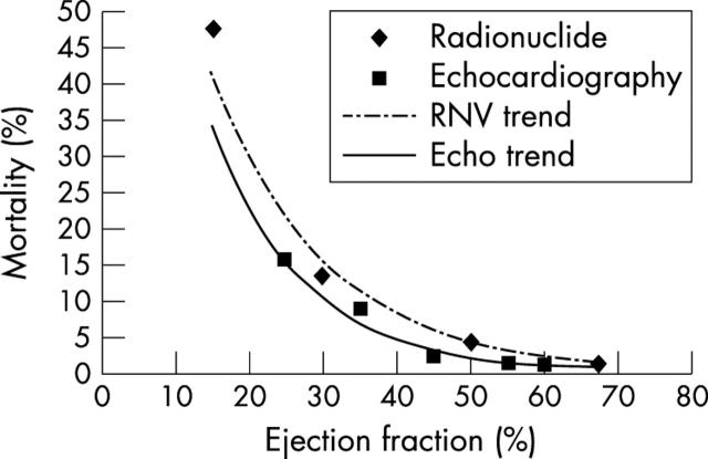
Figure 1.
Mortality after measurement of ejection fraction by radionuclide ventriculography (one year follow up, Multicentre Post-infarction Research Group, 1983) and after 2D echo measurement of ejection fraction (six month follow up, GISSI study 1993). Note the close correlation of the curves, allowing for the difference in follow up between the two cohorts.
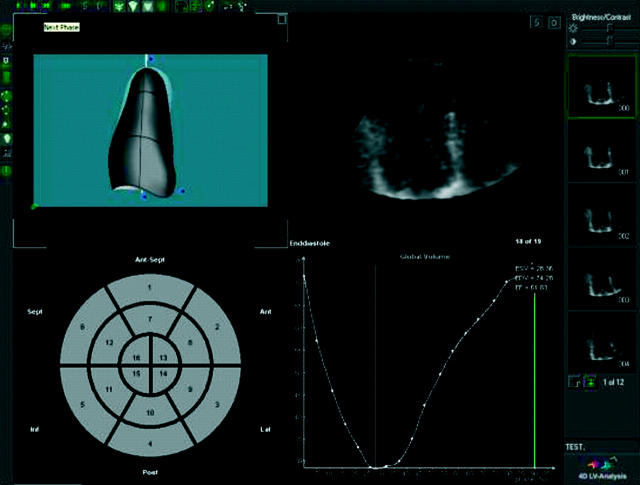
Figure 2.
Three dimensional (3D) echocardiographic evaluation of left ventricular (LV) volumes and ejection fraction. Images from a volumetric dataset are reconstructed (upper centre and right column) and semi-automated edge detection is used to trace the endocardial border and construct a 3D model in systole and diastole (upper left). Expression of volumes in each image yields a time–volume curve (lower left) from which ejection fraction is calculated.
Figure 3.
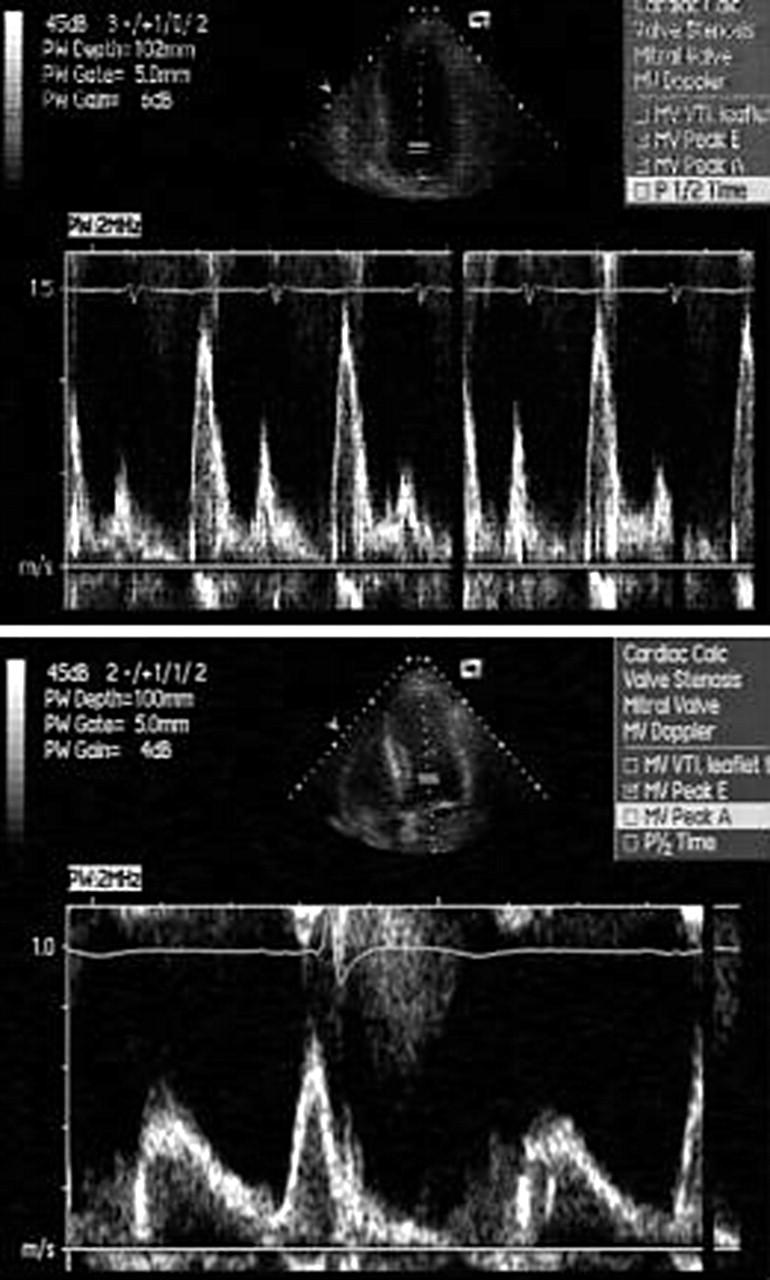
Dependence of LV volumes and standard Doppler indices of diastolic function on loading conditions. This patient with renal failure was studied before (above) and after dialysis (below). LV size and LV filling patterns are altered by loading.
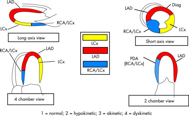
Figure 4.
The 16 segment American Society of Echocardiography model for characterisation of regional LV function, and usual coronary artery distribution of the segments. Reproduced from: Marwick TH. Stress echocardiography – its role in the diagnosis and evaluation of coronary artery disease. Boston: Kluwer Academic Publishers, 2003, with permission of the publisher.
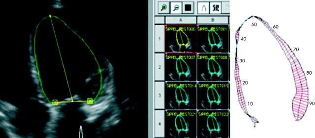
Figure 5.
Centre line approach to quantification of regional LV function. Automated detection of the endocardial border is applied in each view (left), repeated in each frame (thumbprint images, centre), and regional excursion between end diastole and end systole is measured using the centre line method. Reproduced from: Marwick TH. Stress echocardiography – its role in the diagnosis and evaluation of coronary artery disease. Boston: Kluwer Academic Publishers, 2003, with permission of the publisher.
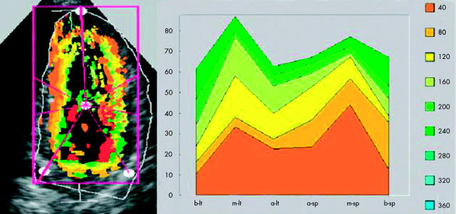
Figure 6.
Quantitation of radial function using colour kinesis. Each successive ultrasound frame on the image (left) is coded with a different colour. The histogram (right) shows the fractional area change within each segment (x axis). Reproduced from: Marwick TH. Stress echocardiography – its role in the diagnosis and evaluation of coronary artery disease. Boston: Kluwer Academic Publishers, 2003, with permission of the publisher.
Selected References
These references are in PubMed. This may not be the complete list of references from this article.
- Armstrong G. P., Carlier S. G., Fukamachi K., Thomas J. D., Marwick T. H. Estimation of cardiac reserve by peak power: validation and initial application of a simplified index. Heart. 1999 Sep;82(3):357–364. doi: 10.1136/hrt.82.3.357. [DOI] [PMC free article] [PubMed] [Google Scholar]
- Aurigemma G. P., Gaasch W. H., Villegas B., Meyer T. E. Noninvasive assessment of left ventricular mass, chamber volume, and contractile function. Curr Probl Cardiol. 1995 Jun;20(6):361–440. [PubMed] [Google Scholar]
- Baik H. K., Budoff M. J., Lane K. L., Bakhsheshi H., Brundage B. H. Accurate measures of left ventricular ejection fraction using electron beam tomography: a comparison with radionuclide angiography, and cine angiography. Int J Card Imaging. 2000 Oct;16(5):391–398. doi: 10.1023/a:1026536510821. [DOI] [PubMed] [Google Scholar]
- Bednarz James, Vignon Philippe, Mor-Avi Victor, V, Weinert Lynn, Koch Rick, Spencer Kirk, Lang Roberto M. Color Kinesis: Principles of Operation and Technical Guidelines. Echocardiography. 1998 Jan;15(1):21–34. doi: 10.1111/j.1540-8175.1998.tb00574.x. [DOI] [PubMed] [Google Scholar]
- Bottini P. B., Carr A. A., Prisant L. M., Flickinger F. W., Allison J. D., Gottdiener J. S. Magnetic resonance imaging compared to echocardiography to assess left ventricular mass in the hypertensive patient. Am J Hypertens. 1995 Mar;8(3):221–228. doi: 10.1016/0895-7061(94)00178-E. [DOI] [PubMed] [Google Scholar]
- Broderick T., Sawada S., Armstrong W. F., Ryan T., Dillon J. C., Bourdillon P. D., Feigenbaum H. Improvement in rest and exercise-induced wall motion abnormalities after coronary angioplasty: an exercise echocardiographic study. J Am Coll Cardiol. 1990 Mar 1;15(3):591–599. doi: 10.1016/0735-1097(90)90632-y. [DOI] [PubMed] [Google Scholar]
- Calnon D. A., Kastner R. J., Smith W. H., Segalla D., Beller G. A., Watson D. D. Validation of a new counts-based gated single photon emission computed tomography method for quantifying left ventricular systolic function: comparison with equilibrium radionuclide angiography. J Nucl Cardiol. 1997 Nov-Dec;4(6):464–471. doi: 10.1016/s1071-3581(97)90003-9. [DOI] [PubMed] [Google Scholar]
- Chan J., Wahi S., Cain P., Marwick T. H. Anatomical M-mode: A novel technique for the quantitative evaluation of regional wall motion analysis during dobutamine echocardiography. Int J Card Imaging. 2000 Aug;16(4):247–255. doi: 10.1023/a:1026539708034. [DOI] [PubMed] [Google Scholar]
- Chuang M. L., Hibberd M. G., Salton C. J., Beaudin R. A., Riley M. F., Parker R. A., Douglas P. S., Manning W. J. Importance of imaging method over imaging modality in noninvasive determination of left ventricular volumes and ejection fraction: assessment by two- and three-dimensional echocardiography and magnetic resonance imaging. J Am Coll Cardiol. 2000 Feb;35(2):477–484. doi: 10.1016/s0735-1097(99)00551-3. [DOI] [PubMed] [Google Scholar]
- Davie A. P., Francis C. M., Love M. P., Caruana L., Starkey I. R., Shaw T. R., Sutherland G. R., McMurray J. J. Value of the electrocardiogram in identifying heart failure due to left ventricular systolic dysfunction. BMJ. 1996 Jan 27;312(7025):222–222. doi: 10.1136/bmj.312.7025.222. [DOI] [PMC free article] [PubMed] [Google Scholar]
- Denault A. Y., Gorcsan J., 3rd, Mandarino W. A., Kancel M. J., Pinsky M. R. Left ventricular performance assessed by echocardiographic automated border detection and arterial pressure. Am J Physiol. 1997 Jan;272(1 Pt 2):H138–H147. doi: 10.1152/ajpheart.1997.272.1.H138. [DOI] [PubMed] [Google Scholar]
- Foster E., Cahalan M. K. The search for intelligent quantitation in echocardiography: "eyeball," "trackball" and beyond. J Am Coll Cardiol. 1993 Sep;22(3):848–850. doi: 10.1016/0735-1097(93)90201-b. [DOI] [PubMed] [Google Scholar]
- Gaasch W. H., Zile M. R., Hoshino P. K., Apstein C. S., Blaustein A. S. Stress-shortening relations and myocardial blood flow in compensated and failing canine hearts with pressure-overload hypertrophy. Circulation. 1989 Apr;79(4):872–883. doi: 10.1161/01.cir.79.4.872. [DOI] [PubMed] [Google Scholar]
- Gaudio C., Tanzilli G., Mazzarotto P., Motolese M., Romeo F., Marino B., Reale A. Comparison of left ventricular ejection fraction by magnetic resonance imaging and radionuclide ventriculography in idiopathic dilated cardiomyopathy. Am J Cardiol. 1991 Feb 15;67(5):411–415. doi: 10.1016/0002-9149(91)90051-l. [DOI] [PubMed] [Google Scholar]
- Ginzton L. E., Laks M. M., Brizendine M., Conant R., Mena I. Noninvasive measurement of the rest and exercise peak systolic pressure/end-systolic volume ratio: a sensitive two-dimensional echocardiographic indicator of left ventricular function. J Am Coll Cardiol. 1984 Sep;4(3):509–516. doi: 10.1016/s0735-1097(84)80094-7. [DOI] [PubMed] [Google Scholar]
- Glower D. D., Spratt J. A., Snow N. D., Kabas J. S., Davis J. W., Olsen C. O., Tyson G. S., Sabiston D. C., Jr, Rankin J. S. Linearity of the Frank-Starling relationship in the intact heart: the concept of preload recruitable stroke work. Circulation. 1985 May;71(5):994–1009. doi: 10.1161/01.cir.71.5.994. [DOI] [PubMed] [Google Scholar]
- Gordon E. P., Schnittger I., Fitzgerald P. J., Williams P., Popp R. L. Reproducibility of left ventricular volumes by two-dimensional echocardiography. J Am Coll Cardiol. 1983 Sep;2(3):506–513. doi: 10.1016/s0735-1097(83)80278-2. [DOI] [PubMed] [Google Scholar]
- Griffin B. P., Shah P. K., Diamond G. A., Berman D. S., Ferguson J. G. Incremental prognostic accuracy of clinical, radionuclide and hemodynamic data in acute myocardial infarction. Am J Cardiol. 1991 Sep 15;68(8):707–712. doi: 10.1016/0002-9149(91)90640-7. [DOI] [PubMed] [Google Scholar]
- Haley J. H., Miller T. D., Christian T. F., Hodge D. O., Lerman A., Gibbons R. J. Twelve-year outcome of patients with an abnormal exercise radionuclide left ventricular angiogram and angiographically insignificant coronary artery disease. Am J Cardiol. 1998 Aug 15;82(4):418–422. doi: 10.1016/s0002-9149(98)00352-x. [DOI] [PubMed] [Google Scholar]
- Haluska Brian A., Short Leanne, Marwick Thomas H. Relationship of ventricular longitudinal function to contractile reserve in patients with mitral regurgitation. Am Heart J. 2003 Jul;146(1):183–188. doi: 10.1016/S0002-8703(03)00173-X. [DOI] [PubMed] [Google Scholar]
- He Z. X., Cwajg E., Preslar J. S., Mahmarian J. J., Verani M. S. Accuracy of left ventricular ejection fraction determined by gated myocardial perfusion SPECT with Tl-201 and Tc-99m sestamibi: comparison with first-pass radionuclide angiography. J Nucl Cardiol. 1999 Jul-Aug;6(4):412–417. doi: 10.1016/s1071-3581(99)90007-7. [DOI] [PubMed] [Google Scholar]
- Himelman R. B., Cassidy M. M., Landzberg J. S., Schiller N. B. Reproducibility of quantitative two-dimensional echocardiography. Am Heart J. 1988 Feb;115(2):425–431. doi: 10.1016/0002-8703(88)90491-7. [DOI] [PubMed] [Google Scholar]
- Hoffmann R., Lethen H., Marwick T., Arnese M., Fioretti P., Pingitore A., Picano E., Buck T., Erbel R., Flachskampf F. A. Analysis of interinstitutional observer agreement in interpretation of dobutamine stress echocardiograms. J Am Coll Cardiol. 1996 Feb;27(2):330–336. doi: 10.1016/0735-1097(95)00483-1. [DOI] [PubMed] [Google Scholar]
- Hoffmann R., Lethen H., Marwick T., Rambaldi R., Fioretti P., Pingitore A., Picano E., Buck T., Erbel R., Flachskampf F. A. Standardized guidelines for the interpretation of dobutamine echocardiography reduce interinstitutional variance in interpretation. Am J Cardiol. 1998 Dec 15;82(12):1520–1524. doi: 10.1016/s0002-9149(98)00697-3. [DOI] [PubMed] [Google Scholar]
- Hoffmann R., Marwick T. H., Poldermans D., Lethen H., Ciani R., van der Meer P., Tries H-P, Gianfagna P., Fioretti P., Bax J. J. Refinements in stress echocardiographic techniques improve inter-institutional agreement in interpretation of dobutamine stress echocardiograms. Eur Heart J. 2002 May;23(10):821–829. doi: 10.1053/euhj.2001.2968. [DOI] [PubMed] [Google Scholar]
- King D. L., Harrison M. R., King D. L., Jr, Gopal A. S., Martin R. P., DeMaria A. N. Improved reproducibility of left atrial and left ventricular measurements by guided three-dimensional echocardiography. J Am Coll Cardiol. 1992 Nov 1;20(5):1238–1245. doi: 10.1016/0735-1097(92)90383-x. [DOI] [PubMed] [Google Scholar]
- Koch R., Lang R. M., Garcia M. J., Weinert L., Bednarz J., Korcarz C., Coughlan B., Spiegel A., Kaji E., Spencer K. T. Objective evaluation of regional left ventricular wall motion during dobutamine stress echocardiographic studies using segmental analysis of color kinesis images. J Am Coll Cardiol. 1999 Aug;34(2):409–419. doi: 10.1016/s0735-1097(99)00233-8. [DOI] [PubMed] [Google Scholar]
- Lawson M. A., Blackwell G. G., Davis N. D., Roney M., Dell'Italia L. J., Pohost G. M. Accuracy of biplane long-axis left ventricular volume determined by cine magnetic resonance imaging in patients with regional and global dysfunction. Am J Cardiol. 1996 May 15;77(12):1098–1104. doi: 10.1016/s0002-9149(96)00140-3. [DOI] [PubMed] [Google Scholar]
- Leung D. Y., Griffin B. P., Stewart W. J., Cosgrove D. M., 3rd, Thomas J. D., Marwick T. H. Left ventricular function after valve repair for chronic mitral regurgitation: predictive value of preoperative assessment of contractile reserve by exercise echocardiography. J Am Coll Cardiol. 1996 Nov 1;28(5):1198–1205. doi: 10.1016/S0735-1097(96)00281-1. [DOI] [PubMed] [Google Scholar]
- Mandarino W. A., Pinsky M. R., Gorcsan J., 3rd Assessment of left ventricular contractile state by preload-adjusted maximal power using echocardiographic automated border detection. J Am Coll Cardiol. 1998 Mar 15;31(4):861–868. doi: 10.1016/s0735-1097(98)00005-9. [DOI] [PubMed] [Google Scholar]
- Marwick T. H. Quantitative techniques for stress echocardiography: dream or reality? Eur J Echocardiogr. 2002 Sep;3(3):171–176. doi: 10.1053/euje.2002.0160. [DOI] [PubMed] [Google Scholar]
- McCullough Peter A., Nowak Richard M., McCord James, Hollander Judd E., Herrmann Howard C., Steg Philippe G., Duc Philippe, Westheim Arne, Omland Torbjørn, Knudsen Cathrine Wold. B-type natriuretic peptide and clinical judgment in emergency diagnosis of heart failure: analysis from Breathing Not Properly (BNP) Multinational Study. Circulation. 2002 Jul 23;106(4):416–422. doi: 10.1161/01.cir.0000025242.79963.4c. [DOI] [PubMed] [Google Scholar]
- Murkofsky R. L., Dangas G., Diamond J. A., Mehta D., Schaffer A., Ambrose J. A. A prolonged QRS duration on surface electrocardiogram is a specific indicator of left ventricular dysfunction [see comment]. J Am Coll Cardiol. 1998 Aug;32(2):476–482. doi: 10.1016/s0735-1097(98)00242-3. [DOI] [PubMed] [Google Scholar]
- Nishimura R. A., Reeder G. S., Miller F. A., Jr, Ilstrup D. M., Shub C., Seward J. B., Tajik A. J. Prognostic value of predischarge 2-dimensional echocardiogram after acute myocardial infarction. Am J Cardiol. 1984 Feb 1;53(4):429–432. doi: 10.1016/0002-9149(84)90007-9. [DOI] [PubMed] [Google Scholar]
- O'Keefe J. H., Jr, Zinsmeister A. R., Gibbons R. J. Value of normal electrocardiographic findings in predicting resting left ventricular function in patients with chest pain and suspected coronary artery disease. Am J Med. 1989 Jun;86(6 Pt 1):658–662. doi: 10.1016/0002-9343(89)90439-7. [DOI] [PubMed] [Google Scholar]
- Oberman A., Fan P. H., Nanda N. C., Lee J. Y., Huster W. J., Sulentic J. A., Storey O. F. Reproducibility of two-dimensional exercise echocardiography. J Am Coll Cardiol. 1989 Oct;14(4):923–928. doi: 10.1016/0735-1097(89)90467-1. [DOI] [PubMed] [Google Scholar]
- Pan C., Hoffmann R., Kühl H., Severin E., Franke A., Hanrath P. Tissue tracking allows rapid and accurate visual evaluation of left ventricular function. Eur J Echocardiogr. 2001 Sep;2(3):197–202. doi: 10.1053/euje.2001.0098. [DOI] [PubMed] [Google Scholar]
- Pearlman A. S. Measurement of left ventricular volume by three-dimensional echocardiography--present promise and potential problems. J Am Coll Cardiol. 1993 Nov 1;22(5):1538–1540. doi: 10.1016/0735-1097(93)90568-l. [DOI] [PubMed] [Google Scholar]
- Pryor D. B., Harrell F. E., Jr, Lee K. L., Rosati R. A., Coleman R. E., Cobb F. R., Califf R. M., Jones R. H. Prognostic indicators from radionuclide angiography in medically treated patients with coronary artery disease. Am J Cardiol. 1984 Jan 1;53(1):18–22. doi: 10.1016/0002-9149(84)90677-5. [DOI] [PubMed] [Google Scholar]
- Reichek N., Wilson J., St John Sutton M., Plappert T. A., Goldberg S., Hirshfeld J. W. Noninvasive determination of left ventricular end-systolic stress: validation of the method and initial application. Circulation. 1982 Jan;65(1):99–108. doi: 10.1161/01.cir.65.1.99. [DOI] [PubMed] [Google Scholar]
- Rich S., Chomka E. V., Stagl R., Shanes J. G., Kondos G. T., Brundage B. H. Determination of left ventricular ejection fraction using ultrafast computed tomography. Am Heart J. 1986 Aug;112(2):392–396. doi: 10.1016/0002-8703(86)90280-2. [DOI] [PubMed] [Google Scholar]
- Rihal C. S., Davis K. B., Kennedy J. W., Gersh B. J. The utility of clinical, electrocardiographic, and roentgenographic variables in the prediction of left ventricular function. Am J Cardiol. 1995 Feb 1;75(4):220–223. doi: 10.1016/0002-9149(95)80023-l. [DOI] [PubMed] [Google Scholar]
- Schalla S., Nagel E., Lehmkuhl H., Klein C., Bornstedt A., Schnackenburg B., Schneider U., Fleck E. Comparison of magnetic resonance real-time imaging of left ventricular function with conventional magnetic resonance imaging and echocardiography. Am J Cardiol. 2001 Jan 1;87(1):95–99. doi: 10.1016/s0002-9149(00)01279-0. [DOI] [PubMed] [Google Scholar]
- Sharir T., Germano G., Kavanagh P. B., Lai S., Cohen I., Lewin H. C., Friedman J. D., Zellweger M. J., Berman D. S. Incremental prognostic value of post-stress left ventricular ejection fraction and volume by gated myocardial perfusion single photon emission computed tomography. Circulation. 1999 Sep 7;100(10):1035–1042. doi: 10.1161/01.cir.100.10.1035. [DOI] [PubMed] [Google Scholar]
- Spencer Kirk T., Bednarz Jim, Mor-Avi Victor, DeCara Jeanne, Lang Roberto M. Automated endocardial border detection and evaluation of left ventricular function from contrast-enhanced images using modified acoustic quantification. J Am Soc Echocardiogr. 2002 Aug;15(8):777–781. doi: 10.1067/mje.2002.120505. [DOI] [PubMed] [Google Scholar]
- Stamm R. B., Carabello B. A., Mayers D. L., Martin R. P. Two-dimensional echocardiographic measurement of left ventricular ejection fraction: prospective analysis of what constitutes an adequate determination. Am Heart J. 1982 Jul;104(1):136–144. doi: 10.1016/0002-8703(82)90651-2. [DOI] [PubMed] [Google Scholar]
- Strotmann J. M., Kvitting J. P., Wilkenshoff U. M., Wranne B., Hatle L., Sutherland G. R. Anatomic M-mode echocardiography: A new approach to assess regional myocardial function--A comparative in vivo and in vitro study of both fundamental and second harmonic imaging modes. J Am Soc Echocardiogr. 1999 May;12(5):300–307. doi: 10.1016/s0894-7317(99)70050-7. [DOI] [PubMed] [Google Scholar]
- Talreja D., Gruver C., Sklenar J., Dent J., Kaul S. Efficient utilization of echocardiography for the assessment of left ventricular systolic function. Am Heart J. 2000 Mar;139(3):394–398. [PubMed] [Google Scholar]
- Tan L. B. Evaluation of cardiac dysfunction, cardiac reserve and inotropic response. Postgrad Med J. 1991;67 (Suppl 1):S10–S20. [PubMed] [Google Scholar]
- Tei C., Nishimura R. A., Seward J. B., Tajik A. J. Noninvasive Doppler-derived myocardial performance index: correlation with simultaneous measurements of cardiac catheterization measurements. J Am Soc Echocardiogr. 1997 Mar;10(2):169–178. doi: 10.1016/s0894-7317(97)70090-7. [DOI] [PubMed] [Google Scholar]
- Vasan Ramachandran S., Benjamin Emelia J., Larson Martin G., Leip Eric P., Wang Thomas J., Wilson Peter W. F., Levy Daniel. Plasma natriuretic peptides for community screening for left ventricular hypertrophy and systolic dysfunction: the Framingham heart study. JAMA. 2002 Sep 11;288(10):1252–1259. doi: 10.1001/jama.288.10.1252. [DOI] [PubMed] [Google Scholar]
- Wyatt H. L., Heng M. K., Meerbaum S., Gueret P., Hestenes J., Dula E., Corday E. Cross-sectional echocardiography. II. Analysis of mathematic models for quantifying volume of the formalin-fixed left ventricle. Circulation. 1980 Jun;61(6):1119–1125. doi: 10.1161/01.cir.61.6.1119. [DOI] [PubMed] [Google Scholar]
- Zaret B. L., Wackers F. J., Terrin M. L., Forman S. A., Williams D. O., Knatterud G. L., Braunwald E. Value of radionuclide rest and exercise left ventricular ejection fraction in assessing survival of patients after thrombolytic therapy for acute myocardial infarction: results of Thrombolysis in Myocardial Infarction (TIMI) phase II study. The TIMI Study Group. J Am Coll Cardiol. 1995 Jul;26(1):73–79. doi: 10.1016/0735-1097(95)00146-q. [DOI] [PubMed] [Google Scholar]


