Full Text
The Full Text of this article is available as a PDF (22.6 MB).
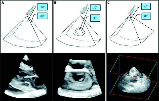
Figure 1.
Different modes of data acquisition using the matrix array transducer: (A) narrow angled scan, (B) zoom mode, and (C) wide angled acquisition. See text for further explanation.
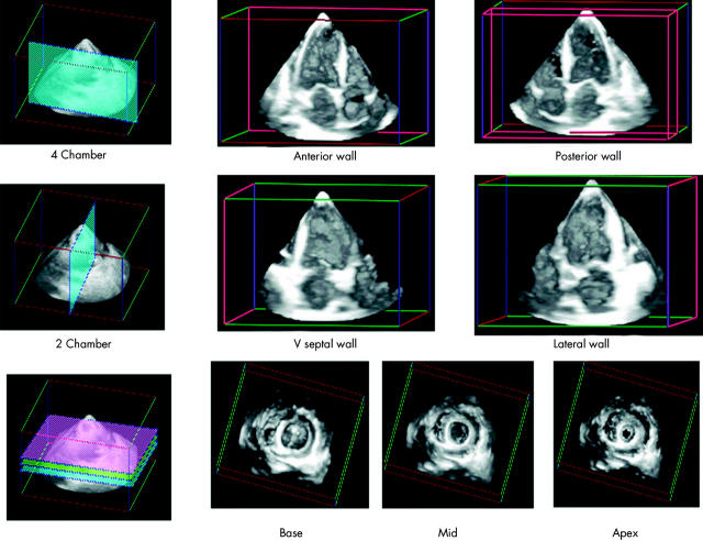
Figure 2.
Wide angled scan of the left ventricle sliced using multiple cut planes. From a typical apical four chamber view using a longitudinal cut (top row), the septal and lateral walls are seen along with the surface of the anterior and posterior wall. From the two chamber view (middle row), the anterior and inferior walls are visualised together with the surface of the septal and lateral wall. Multiple cuts of the short axis from base to apex may be also derived from this scan, as seen on the bottom row.
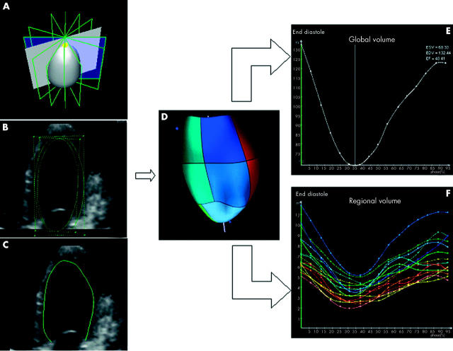
Figure 3.
Panel A depicts the pyramidal volume of data divided automatically into eight equi-angled longitudinal slices through the apex. Panels B and C depict the automated border detection algorithm used to track endocardial borders throughout the cardiac cycle. A dynamic ventricular cast is automatically displayed as a result of the endocardial borders and surface reconstruction (panel D). Global volumes and ejection fraction is displayed in panel E. Regional volumes of all 16 segments are shown in panel F.
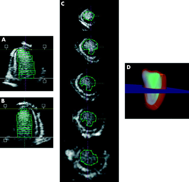
Figure 4.
The disc summation method is an alternative method to calculate left ventricular volume and ejection fraction. The left ventricle is placed in a longitudinal position (panels A and B). With predefined distance intervals, multiple short axis cut planes are derived and endocardial borders traced in all end systolic and end diastolic frames (panel C). The summation of the volumes of each slice results in left ventricular volumes demonstrated in panel D.
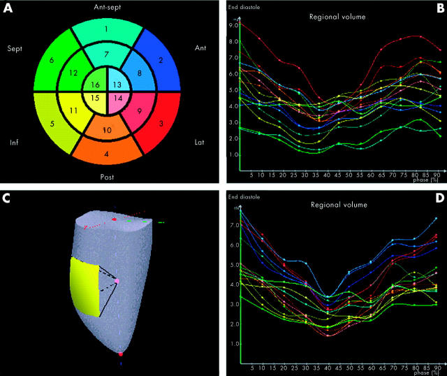
Figure 5.
Bulls-eye demonstrating the left ventricle divided into 16 segments, corresponding to the regional volume of each segment (panel A). For each segment a regional volume curve is displayed (panel B). An example of a regional volume is depicted in panel C. Three dimensional echocardiography may help depict the more uniform mechanism of contraction during biventricular pacing. Preliminary observations demonstrate more synchronised regional volume curves, as seen in panel D, compared to panel B.
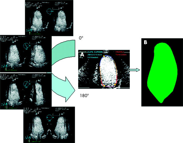
Figure 6.
Biplane imaging from the apical windows provides an alternative method of data acquisition which may be useful in patients with dilated ventricles. Continuous contrast infusion enhances the endocardial border facilitating automated tracking (panel A). Multiple images are obtained at 10° increments over 180° rotation without respiratory gating shown on the left. A sphere is then fitted over the set of contours to calculate volume and ejection fraction (panel B).
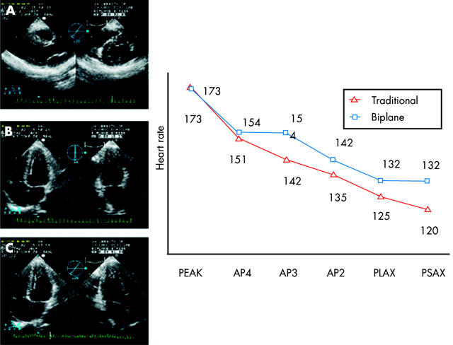
Figure 7.
Comparison of the decline in heart rate at the time of data acquisition between the traditional 2D probe and the x 4 transducer as demonstrated by the graph.63 Biplane views are displayed on the left in panels A–C.
Selected References
These references are in PubMed. This may not be the complete list of references from this article.
- Acar P., Maunoury C., Antonietti T., Bonnet D., Sidi D., Kachaner J. Left ventricular ejection fraction in children measured by three-dimensional echocardiography using a new transthoracic integrated 3D-probe. A comparison with equilibrium radionuclide angiography. Eur Heart J. 1998 Oct;19(10):1583–1588. doi: 10.1053/euhj.1998.1091. [DOI] [PubMed] [Google Scholar]
- Ahmad M., Xie T., McCulloch M., Abreo G., Runge M. Real-time three-dimensional dobutamine stress echocardiography in assessment stress echocardiography in assessment of ischemia: comparison with two-dimensional dobutamine stress echocardiography. J Am Coll Cardiol. 2001 Apr;37(5):1303–1309. doi: 10.1016/s0735-1097(01)01159-7. [DOI] [PubMed] [Google Scholar]
- Altmann K., Shen Z., Boxt L. M., King D. L., Gersony W. M., Allan L. D., Apfel H. D. Comparison of three-dimensional echocardiographic assessment of volume, mass, and function in children with functionally single left ventricles with two-dimensional echocardiography and magnetic resonance imaging. Am J Cardiol. 1997 Oct 15;80(8):1060–1065. doi: 10.1016/s0002-9149(97)00603-6. [DOI] [PubMed] [Google Scholar]
- Califf R. M., White H. D., Van de Werf F., Sadowski Z., Armstrong P. W., Vahanian A., Simoons M. L., Simes R. J., Lee K. L., Topol E. J. One-year results from the Global Utilization of Streptokinase and TPA for Occluded Coronary Arteries (GUSTO-I) trial. GUSTO-I Investigators. Circulation. 1996 Sep 15;94(6):1233–1238. doi: 10.1161/01.cir.94.6.1233. [DOI] [PubMed] [Google Scholar]
- Collins M., Hsieh A., Ohazama C. J., Ota T., Stetten G., Donovan C. L., Kisslo J., Ryan T. Assessment of regional wall motion abnormalities with real-time 3-dimensional echocardiography. J Am Soc Echocardiogr. 1999 Jan;12(1):7–14. doi: 10.1016/s0894-7317(99)70167-7. [DOI] [PubMed] [Google Scholar]
- Erbel R., Schweizer P., Lambertz H., Henn G., Meyer J., Krebs W., Effert S. Echoventriculography -- a simultaneous analysis of two-dimensional echocardiography and cineventriculography. Circulation. 1983 Jan;67(1):205–215. doi: 10.1161/01.cir.67.1.205. [DOI] [PubMed] [Google Scholar]
- Folland E. D., Parisi A. F., Moynihan P. F., Jones D. R., Feldman C. L., Tow D. E. Assessment of left ventricular ejection fraction and volumes by real-time, two-dimensional echocardiography. A comparison of cineangiographic and radionuclide techniques. Circulation. 1979 Oct;60(4):760–766. doi: 10.1161/01.cir.60.4.760. [DOI] [PubMed] [Google Scholar]
- Franke A., Flachskampf F. A., Kühl H. P., Klues H. G., Job F. P., Merx M., Krebs W., Hanrath P. Dreidimensionale Rekonstruktion multiplaner transösophagealer echokardiographischer Schnittbilder: Ein Methodenbericht mit Fallbeispielen. Z Kardiol. 1995 Aug;84(8):633–642. [PubMed] [Google Scholar]
- Fulton D. R., Marx G. R., Pandian N. G., Romero B. A., Mumm B., Krauss M., Wollschläger H., Ludomirsky A., Cao Q. L. Dynamic three-dimensional echocardiographic imaging of congenital heart defects in infants and children by computer-controlled tomographic parallel slicing using a single integrated ultrasound instrument. Echocardiography. 1994 Mar;11(2):155–164. doi: 10.1111/j.1540-8175.1994.tb01061.x. [DOI] [PubMed] [Google Scholar]
- Geiser E. A., Lupkiewicz S. M., Christie L. G., Ariet M., Conetta D. A., Conti C. R. A framework for three-dimensional time-varying reconstruction of the human left ventricle: sources of error and estimation of their magnitude. Comput Biomed Res. 1980 Jun;13(3):225–241. doi: 10.1016/0010-4809(80)90018-x. [DOI] [PubMed] [Google Scholar]
- Gopal A. S., Keller A. M., Rigling R., King D. L., Jr, King D. L. Left ventricular volume and endocardial surface area by three-dimensional echocardiography: comparison with two-dimensional echocardiography and nuclear magnetic resonance imaging in normal subjects. J Am Coll Cardiol. 1993 Jul;22(1):258–270. doi: 10.1016/0735-1097(93)90842-o. [DOI] [PubMed] [Google Scholar]
- Gopal A. S., Keller A. M., Shen Z., Sapin P. M., Schroeder K. M., King D. L., Jr, King D. L. Three-dimensional echocardiography: in vitro and in vivo validation of left ventricular mass and comparison with conventional echocardiographic methods. J Am Coll Cardiol. 1994 Aug;24(2):504–513. doi: 10.1016/0735-1097(94)90310-7. [DOI] [PubMed] [Google Scholar]
- Gopal A. S., King D. L., Katz J., Boxt L. M., King D. L., Jr, Shao M. Y. Three-dimensional echocardiographic volume computation by polyhedral surface reconstruction: in vitro validation and comparison to magnetic resonance imaging. J Am Soc Echocardiogr. 1992 Mar-Apr;5(2):115–124. doi: 10.1016/s0894-7317(14)80541-5. [DOI] [PubMed] [Google Scholar]
- Gopal A. S., Schnellbaecher M. J., Shen Z., Akinboboye O. O., Sapin P. M., King D. L. Freehand three-dimensional echocardiography for measurement of left ventricular mass: in vivo anatomic validation using explanted human hearts. J Am Coll Cardiol. 1997 Sep;30(3):802–810. doi: 10.1016/s0735-1097(97)00198-8. [DOI] [PubMed] [Google Scholar]
- Gopal A. S., Schnellbaecher M. J., Shen Z., Boxt L. M., Katz J., King D. L. Freehand three-dimensional echocardiography for determination of left ventricular volume and mass in patients with abnormal ventricles: comparison with magnetic resonance imaging. J Am Soc Echocardiogr. 1997 Oct;10(8):853–861. doi: 10.1016/s0894-7317(97)70045-2. [DOI] [PubMed] [Google Scholar]
- Handschumacher M. D., Lethor J. P., Siu S. C., Mele D., Rivera J. M., Picard M. H., Weyman A. E., Levine R. A. A new integrated system for three-dimensional echocardiographic reconstruction: development and validation for ventricular volume with application in human subjects. J Am Coll Cardiol. 1993 Mar 1;21(3):743–753. doi: 10.1016/0735-1097(93)90108-d. [DOI] [PubMed] [Google Scholar]
- Heusch A., Rübo J., Krogmann O. N., Bourgeois M. Volumetric analysis of the right ventricle in children with congenital heart defects: comparison of biplane angiography and transthoracic 3-dimensional echocardiography. Cardiol Young. 1999 Nov;9(6):577–584. doi: 10.1017/s1047951100005618. [DOI] [PubMed] [Google Scholar]
- Hozumi T., Yoshikawa J., Yoshida K., Akasaka T., Takagi T., Yamamuro A. Three-dimensional echocardiographic measurement of left ventricular volumes and ejection fraction using a multiplane transesophageal probe in patients. Am J Cardiol. 1996 Nov 1;78(9):1077–1080. doi: 10.1016/s0002-9149(96)00591-7. [DOI] [PubMed] [Google Scholar]
- Jiang Leng, Levine Robert A., Weyman Arthur E. Echocardiographic Assessment of Right Ventricular Volume and Function. Echocardiography. 1997 Mar;14(2):189–206. doi: 10.1111/j.1540-8175.1997.tb00711.x. [DOI] [PubMed] [Google Scholar]
- Kasprzak J. D., Vletter W. B., Roelandt J. R., van Meegen J. R., Johnson R., Ten Cate F. J. Visualization and quantification of myocardial mass at risk using three-dimensional contrast echocardiography. Cardiovasc Res. 1998 Nov;40(2):314–321. doi: 10.1016/s0008-6363(98)00178-3. [DOI] [PubMed] [Google Scholar]
- Kasprzak J. D., Vletter W. B., van Meegen J. R., Nosir Y. F., Johnson R., Ten Cate F. J., Roelandt J. R. Improved quantification of myocardial mass by three-dimensional echocardiography using a deposit contrast agent. Ultrasound Med Biol. 1998 Jun;24(5):647–653. doi: 10.1016/s0301-5629(98)00035-0. [DOI] [PubMed] [Google Scholar]
- Kawai Junichi, Tanabe Kazuaki, Morioka Shigefumi, Shiotani Hideyuki. Rapid freehand scanning three-dimensional echocardiography: accurate measurement of left ventricular volumes and ejection fraction compared with quantitative gated scintigraphy. J Am Soc Echocardiogr. 2003 Feb;16(2):110–115. doi: 10.1067/mje.2003.4. [DOI] [PubMed] [Google Scholar]
- King D. L., Harrison M. R., King D. L., Jr, Gopal A. S., Martin R. P., DeMaria A. N. Improved reproducibility of left atrial and left ventricular measurements by guided three-dimensional echocardiography. J Am Coll Cardiol. 1992 Nov 1;20(5):1238–1245. doi: 10.1016/0735-1097(92)90383-x. [DOI] [PubMed] [Google Scholar]
- Kupferwasser I., Mohr-Kahaly S., Stähr P., Rupprecht H. J., Nixdorff U., Fenster M., Voigtländer T., Erbel R., Meyer J. Transthoracic three-dimensional echocardiographic volumetry of distorted left ventricles using rotational scanning. J Am Soc Echocardiogr. 1997 Oct;10(8):840–852. doi: 10.1016/s0894-7317(97)70044-0. [DOI] [PubMed] [Google Scholar]
- Kühl H. P., Franke A., Janssens U., Merx M., Graf J., Krebs W., Reul H., Rau G., Hoffmann R., Klues H. G. Three-dimensional echocardiographic determination of left ventricular volumes and function by multiplane transesophageal transducer: dynamic in vitro validation and in vivo comparison with angiography and thermodilution. J Am Soc Echocardiogr. 1998 Dec;11(12):1113–1124. doi: 10.1016/s0894-7317(98)80006-0. [DOI] [PubMed] [Google Scholar]
- Lehmann M. H., Timothy K. W., Frankovich D., Fromm B. S., Keating M., Locati E. H., Taggart R. T., Towbin J. A., Moss A. J., Schwartz P. J. Age-gender influence on the rate-corrected QT interval and the QT-heart rate relation in families with genotypically characterized long QT syndrome. J Am Coll Cardiol. 1997 Jan;29(1):93–99. doi: 10.1016/s0735-1097(96)00454-8. [DOI] [PubMed] [Google Scholar]
- Leotta D. F., Munt B., Bolson E. L., Kraft C., Martin R. W., Otto C. M., Sheehan F. H. Quantitative three-dimensional echocardiography by rapid imaging from multiple transthoracic windows: in vitro validation and initial in vivo studies. J Am Soc Echocardiogr. 1997 Oct;10(8):830–839. doi: 10.1016/s0894-7317(97)70043-9. [DOI] [PubMed] [Google Scholar]
- Ludomirsky A., Vermilion R., Nesser J., Marx G., Vogel M., Derman R., Pandian N. Transthoracic real-time three-dimensional echocardiography using the rotational scanning approach for data acquisition. Echocardiography. 1994 Nov;11(6):599–606. doi: 10.1111/j.1540-8175.1994.tb01104.x. [DOI] [PubMed] [Google Scholar]
- Marx G. R., Fulton D. R., Pandian N. G., Vogel M., Cao Q. L., Ludomirsky A., Delabays A., Sugeng L., Klas B. Delineation of site, relative size and dynamic geometry of atrial septal defects by real-time three-dimensional echocardiography. J Am Coll Cardiol. 1995 Feb;25(2):482–490. doi: 10.1016/0735-1097(94)00372-w. [DOI] [PubMed] [Google Scholar]
- Mele D., Levine R. A. Quantitation of ventricular size and function: principles and accuracy of transthoracic rotational scanning. Echocardiography. 2000 Nov;17(8):749–755. doi: 10.1111/j.1540-8175.2000.tb01233.x. [DOI] [PubMed] [Google Scholar]
- Moore N. R., Parry A. J., Trottman-Dickenson B., Pillai R., Westaby S. Fate of the native aorta after repair of acute type A dissection: a magnetic resonance imaging study. Heart. 1996 Jan;75(1):62–66. doi: 10.1136/hrt.75.1.62. [DOI] [PMC free article] [PubMed] [Google Scholar]
- Nixon J. V., Saffer S. I., Lipscomb K., Blomqvist C. G. Three-dimensional echoventriculography. Am Heart J. 1983 Sep;106(3):435–443. doi: 10.1016/0002-8703(83)90683-x. [DOI] [PubMed] [Google Scholar]
- Nosir Y. F., Vletter W. B., Kasprzak J. D., Boersma E., Lequin M. H., Elhendy A. A., Yao J., Stoker J., Ten Cate F. J., Roelandt J. R. Optimal rotational interval for 3-dimensional echocardiography data acquisition for rapid and accurate measurement of left ventricular function. J Am Soc Echocardiogr. 2000 Aug;13(8):715–722. doi: 10.1067/mje.2000.104899. [DOI] [PubMed] [Google Scholar]
- Papavassiliou D. P., Parks W. J., Hopkins K. L., Fyfe D. A. Three-dimensional echocardiographic measurement of right ventricular volume in children with congenital heart disease validated by magnetic resonance imaging. J Am Soc Echocardiogr. 1998 Aug;11(8):770–777. doi: 10.1016/s0894-7317(98)70051-3. [DOI] [PubMed] [Google Scholar]
- Raichlen J. S., Trivedi S. S., Herman G. T., St John Sutton M. G., Reichek N. Dynamic three-dimensional reconstruction of the left ventricle from two-dimensional echocardiograms. J Am Coll Cardiol. 1986 Aug;8(2):364–370. doi: 10.1016/s0735-1097(86)80052-3. [DOI] [PubMed] [Google Scholar]
- Salustri A., Spitaels S., McGhie J., Vletter W., Roelandt J. R. Transthoracic three-dimensional echocardiography in adult patients with congenital heart disease. J Am Coll Cardiol. 1995 Sep;26(3):759–767. doi: 10.1016/0735-1097(95)00245-Y. [DOI] [PubMed] [Google Scholar]
- Sapin P. M., Schroeder K. D., Smith M. D., DeMaria A. N., King D. L. Three-dimensional echocardiographic measurement of left ventricular volume in vitro: comparison with two-dimensional echocardiography and cineventriculography. J Am Coll Cardiol. 1993 Nov 1;22(5):1530–1537. doi: 10.1016/0735-1097(93)90567-k. [DOI] [PubMed] [Google Scholar]
- Sapin P. M., Schröder K. M., Gopal A. S., Smith M. D., DeMaria A. N., King D. L. Comparison of two- and three-dimensional echocardiography with cineventriculography for measurement of left ventricular volume in patients. J Am Coll Cardiol. 1994 Oct;24(4):1054–1063. doi: 10.1016/0735-1097(94)90869-9. [DOI] [PubMed] [Google Scholar]
- Schnittger I., Fitzgerald P. J., Daughters G. T., Ingels N. B., Kantrowitz N. E., Schwarzkopf A., Mead C. W., Popp R. L. Limitations of comparing left ventricular volumes by two dimensional echocardiography, myocardial markers and cineangiography. Am J Cardiol. 1982 Sep;50(3):512–519. doi: 10.1016/0002-9149(82)90317-4. [DOI] [PubMed] [Google Scholar]
- Sheikh K., Smith S. W., von Ramm O., Kisslo J. Real-time, three-dimensional echocardiography: feasibility and initial use. Echocardiography. 1991 Jan;8(1):119–125. doi: 10.1111/j.1540-8175.1991.tb01409.x. [DOI] [PubMed] [Google Scholar]
- Shiota T., Jones M., Chikada M., Fleishman C. E., Castellucci J. B., Cotter B., DeMaria A. N., von Ramm O. T., Kisslo J., Ryan T. Real-time three-dimensional echocardiography for determining right ventricular stroke volume in an animal model of chronic right ventricular volume overload. Circulation. 1998 May 19;97(19):1897–1900. doi: 10.1161/01.cir.97.19.1897. [DOI] [PubMed] [Google Scholar]
- Shiota T., McCarthy P. M., White R. D., Qin J. X., Greenberg N. L., Flamm S. D., Wong J., Thomas J. D. Initial clinical experience of real-time three-dimensional echocardiography in patients with ischemic and idiopathic dilated cardiomyopathy. Am J Cardiol. 1999 Nov 1;84(9):1068–1073. doi: 10.1016/s0002-9149(99)00500-7. [DOI] [PubMed] [Google Scholar]
- Siu S. C., Rivera J. M., Guerrero J. L., Handschumacher M. D., Lethor J. P., Weyman A. E., Levine R. A., Picard M. H. Three-dimensional echocardiography. In vivo validation for left ventricular volume and function. Circulation. 1993 Oct;88(4 Pt 1):1715–1723. doi: 10.1161/01.cir.88.4.1715. [DOI] [PubMed] [Google Scholar]
- Snyder J. E., Kisslo J., von Ramm O. Real-time orthogonal mode scanning of the heart. I. System design. J Am Coll Cardiol. 1986 Jun;7(6):1279–1285. doi: 10.1016/s0735-1097(86)80147-4. [DOI] [PubMed] [Google Scholar]
- Starling M. R., Crawford M. H., Sorensen S. G., Levi B., Richards K. L., O'Rourke R. A. Comparative accuracy of apical biplane cross-sectional echocardiography and gated equilibrium radionuclide angiography for estimating left ventricular size and performance. Circulation. 1981 May;63(5):1075–1084. doi: 10.1161/01.cir.63.5.1075. [DOI] [PubMed] [Google Scholar]
- Teupe C., Takeuchi M., Ram S. P., Pandian N. G. Three-dimensional echocardiography: in-vitro validation of a new, voxel-based method for rapid quantification of ventricular volume in normal and aneurysmal left ventricles. Int J Cardiovasc Imaging. 2001 Apr;17(2):99–105. doi: 10.1023/a:1010671305700. [DOI] [PubMed] [Google Scholar]
- Teupe Claudius, Takeuchi Masaaki, Yao Jiefen, Pandian Natesa. Determination of left ventricular mass by three-dimensional echocardiography: in vitro validation of a novel quantification method using multiple equi-angular rotational planes for rapid measurements. Int J Cardiovasc Imaging. 2002 Jun;18(3):161–167. doi: 10.1023/a:1014665408355. [DOI] [PubMed] [Google Scholar]
- Vogel M., Gutberlet M., Dittrich S., Hosten N., Lange P. E. Comparison of transthoracic three dimensional echocardiography with magnetic resonance imaging in the assessment of right ventricular volume and mass. Heart. 1997 Aug;78(2):127–130. doi: 10.1136/hrt.78.2.127. [DOI] [PMC free article] [PubMed] [Google Scholar]
- Vogel M., White P. A., Redington A. N. In vitro validation of right ventricular volume measurement by three dimensional echocardiography. Br Heart J. 1995 Oct;74(4):460–463. doi: 10.1136/hrt.74.4.460. [DOI] [PMC free article] [PubMed] [Google Scholar]
- Yao J., Cao Q. L., Masani N., Delabays A., Magni G., Acar P., Laskari C., Pandian N. G. Three-dimensional echocardiographic estimation of infarct mass based on quantification of dysfunctional left ventricular mass. Circulation. 1997 Sep 2;96(5):1660–1666. doi: 10.1161/01.cir.96.5.1660. [DOI] [PubMed] [Google Scholar]
- Zwas D. R., Takuma S., Mullis-Jansson S., Fard A., Chaudhry H., Wu H., Di Tullio M. R., Homma S. Feasibility of real-time 3-dimensional treadmill stress echocardiography. J Am Soc Echocardiogr. 1999 May;12(5):285–289. doi: 10.1016/s0894-7317(99)70048-9. [DOI] [PubMed] [Google Scholar]


