Abstract
Activation of endothelial cells, fibrin deposition, and coagulation within the tumor vasculature has been shown in vivo to correlate with the occurrence of tumor necrosis factor (TNF)-induced tumor necrosis in mice. In the present study we investigated which target cells mediate the TNF-induced necrosis in fibrosarcomas grown in wild type (wt), TNF receptor type 1-deficient (TNFRp55−/−), and TNF receptor type 2-deficient (TNFRp75−/−) mice. TNF administration resulted in tumor necrosis exclusively in wt and TNFRp75−/−, but not in TNFRp55−/− mice, indicating a dependence of TNF-mediated tumor necrosis on the expression of TNF receptor type 1. However, using wt and TNFRp55−/− fibrosarcomas in wt mice, we found that TNF-mediated tumor necrosis was completely independent of TNF receptor type 1 expression in tumor cells. Thus we could exclude any direct tumoricidal effect of TNF in this model. Soluble TNF induced leukostasis in wt and TNFRp75−/− mice but not in TNFRp55−/− mice. TNF-induced leukostasis in TNFRp55−/− mice was restored by adoptive bone marrow transplantation of wt hematopoietic cells, but TNF failed to induce tumor necrosis in these chimeric mice. Because TNF administration resulted in both activation and focal damage of tumor endothelium, TNF receptor type 1-expressing cells of the tumor vasculature, likely to be endothelial cells, appear to be target cells for mediating TNF-induced tumor necrosis.
Bacterial endotoxin (lipopolysaccharide, LPS) has long been shown to have antitumor activity in vivo by induction of tumor necrosis or tumor regression in mouse tumor models. 1-3 This finding led to the identification of tumor necrosis factor (TNF) as the major mediator for the necrotizing effect of LPS. 4 TNF is a pleiotropic cytokine, which is mainly produced by activated monocytes/macrophages, neutrophils, and T cells. 5 TNF exerts its activity via two distinct receptors with an apparent molecular mass of 55 kd (TNF receptor type 1, TNFRp55) and 75 kd (TNF receptor type 2, TNFRp75), respectively. These TNF receptors are expressed on almost every cell type such as leukocytes, endothelial cells, and tumor cells, but a number of differences in TNFRp55- and TNFRp75-mediated signals are known. 6 For activation of blood-borne as well as endothelial cells, recombinant human TNF (rhTNF), which only binds to the TNFRp55 in the mouse system, is sufficient. Furthermore, the ability to induce tumor necrosis in mice with rhTNF indicated an important role for the TNF receptor type 1 in mediating the necrotizing effects.
Earlier studies suggested that TNF acts on the tumor-associated endogenic cells rather than directly on the tumor cells because methylcholanthrene-induced fibrosarcomas, known to be relatively insensitive to TNF-mediated cytotoxicity in vitro, are extremely sensitive to the necrotizing effect in vivo. 7 Until now a TNF-mediated tumor cytotoxicity, however, could not be fully excluded. In addition, it was demonstrated that TNF-induced coagulation in the tumor vasculature may be one potential mechanism for tumor necrosis. 8 This was stressed by the fact that necrosis was achieved through selective induction of thrombosis within the tumor vessels by targeting procoagulant tissue factor (TF), by means of a bispecific antibody to an experimentally induced marker on tumor vascular endothelial cells. 9 In a less artificial system we could show that coagulation within the vascular bed of the tumor correlated with the expression of tissue factor (TF) on endothelial cells, which is known to be induced by TNF in vivo. 10 In addition, when TF expression was inhibited by somatic gene transfer of IκB in this model, TNF-mediated fibrin deposition was decreased and free blood flow could be restored. 11 Besides a direct endothelial cell stimulation by LPS, TNF, or other cytokines, adhesion of lymphocytes to endothelium or coculture of monocytes with endothelial cells can also induce TF expression on endothelium. 12-14 This, together with the observation of TF expression on endothelial cells after TNFRp75 stimulation 15 and the fact that activated monocytes also express TF, 16 leaves room for a potential role for both leukocytes and endothelial cells in initiating tumor necrosis. However, it is still unclear whether coagulation as a result of induction of TF expression is the cause or rather a consequence of the disruption of the tumor vasculature. Ruegg and co-workers demonstrated that TNF reduces αvβ3 integrin-mediated endothelial cell adhesion in vitro, resulting in detachment and apoptotic cell death. 17 Such endothelial cell destruction has been suggested to be the effector mechanism in the only antitumoral TNF therapy of isolated limp perfusion currently used, 18 which points to the endothelium as a target for TNF-induced tumor necrosis.
Although in TNF-induced tumor necrosis a role of endothelial cells was suggested by a number of previous studies, neither a direct tumor cell cytotoxic activity nor a leukocyte-mediated event could be fully excluded until now. By using mice and tumor cells deficient for TNFRs and by generating bone marrow chimeric mice, we investigated the mechanism by which recombinant soluble mouse TNF, which, in contrast to human TNF, interacts with both the TNFRp55 and the TNFRp75, initiates tumor necrosis.
Materials and Methods
Animals
Female wild-type (wt) mice of strain C57BL/6 and C57BL/6 × 129/Sv were obtained from Charles River, Germany, and from RCC, Füllinsdorf, Switzerland, respectively. TNFRp55-deficient mice (TNFRp55−/−), 19 (kindly provided by K. Pfeffer, Munich) and TNFRp75-deficient mice (TNFRp75−/−) 20 have been back-crossed six times to C57BL/6. Mice deficient for both TNF receptors were obtained by crossing single TNFR-deficient mice and were of hybrid C57BL/6 × 129/Sv background. Mice used for tumor experiments were age- and sex-matched. All mice were fed with a standard diet, received water ad libitum, and were kept in the animal facility of the University of Regensburg according to institutional guidelines and in accordance with the German law for animal experimentation.
Reagents
Recombinant mouse (rm)TNF was expressed in Escherichia coli and purified according to standard procedures. The specific activity was 9 × 10 8 U/mg, as tested in the L929 TNF bioassay in the presence of 2 μg/ml actinomycin D. LPS contamination of the purified material was <0.16 μg/mg, as determined in a Limulus lysate turbidity assay. 21 LPS from Salmonella minnesota (LPSW S. minnesota 9700) was purchased from Difco (Detroit, MI).
Tumors
The wtBFS-1 MethA-induced fibrosarcoma was generated in a female C57BL/6 mouse and the p55−/−BFS-1 MethA-induced fibrosarcoma in a female TNFRp55−/− mouse by injection of 1 mg of 3-methylcholanthrene (Sigma, Deisenhofen, Germany) dissolved in 200 μl tricaprylin (Sigma) i.d. in the back of a mouse essentially as described earlier. 5 To adapt wtBFS-1 cells for growth in C57BL/6 × 129/Sv mice, wtBFS-1 cells were passaged three times in C57BL/6 × 129/Sv mice and named BFS-2c. The tumor cells were maintained in vitro in RPMI 1640 supplemented with 5% heat-inactivated fetal calf serum (all from Life Technologies, Eggenstein, Germany).
Tumor Experiments
Mice received 1.5 × 10 6 BFS-1, p55−/−BFS-1, or BFS-2c cells in 50 μl medium i.d. in the back, and tumors were allowed to grow for 9–11 days to reach a size approximately 8 mm in diameter before i.p. injection of rmTNF (5 μg) in 100 μl PBS or 100 μl PBS alone as a control. After 12 hours or 2 days, mice were sacrificed and the tumors were removed for immunohistochemistry or histology, respectively.
Histology
The tumors were excised, fixed overnight in 4% PBS-buffered formalin, and embedded in paraffin. For immunohistochemistry, dewaxed sections were treated with 0.05% pronase (Dako, Hamburg, Germany) (15 minutes, 37°C) before incubation with anti-von Willebrand factor (vWF) antibody (Dako) (serum dilution 1:400, 1 hour, 37°C) to look for vascular endothelial damage as described earlier. 22 Central vertical sections (4 μm) were stained with hematoxylin and eosin and examined with a Leitz Axioplan microscope (×40 magnification). The percentage of necrotic area versus total tumor area of two sections per tumor was quantified using the image analysis software analySIS (Soft-Imaging Software GmbH, Münster, Germany). Tumors with >20% necrotic area were considered positive for tumor necrosis.
Leukocyte Adhesion in Vivo
Mice received rmTNF (5 μg) i.p. in 100 μl PBS or PBS alone as a control. After 4 hours, mice were sacrificed and the lungs were removed and processed like the tumors. The sections were examined with a Leitz Axioplan microscope (×160 magnification). Adherent leukocytes in vessels with an inner diameter of 50–150 μm were counted, and the inner circumference of the vessels was measured using the image analysis software analySIS (Soft-Imaging Software GmbH). To quantify leukostasis, two sections per lung and at least five vessels per section were examined and expressed as adherent leukocytes per mm endothelium ± SE.
Adoptive Transfer of Bone Marrow Cells
Donor mice were sacrificed and the femurs were removed. The ends of the femurs were cut off, gently ground in medium, and filtered through a sieve (0.2-μm mesh) to remove bone fragments. The bone marrow cells were flushed out of the femurs with ice-cold RPMI 1640 containing 5% fetal calf serum, using a syringe. Bone marrow cells were pooled, washed twice, and resuspended in medium at a concentration of 108/ml. After lethal x-irradiation (10 Gy) recipient mice received 10 7 donor bone marrow cells i.v. in 100 μl medium into the tail vein. For the following 2 weeks drinking water was supplemented with 0.1 g/l neomycin sulfate (Sigma) and 10 mg/l polymycin (Sigma). Four weeks after reconstitution, mice were used for tumor experiments.
PCR
To determine the quality of the adoptive cell transfer, PCR of genomic DNA derived from leukocytes from reconstituted mice was performed to test for the presence or absence of either TNFR allele. Reconstituted mice were anesthetized and bled through the retroorbital plexus into 0.1 mol/L citrate buffer (pH 8). Genomic DNA was isolated from approximately 200 μl whole blood, using the Quiamp Blood Kit (Quiagen, Hilden, Germany), following the supplier’s instructions. The following primers were used: gT6E515 (AGAAATGTCCCAGGTGATCTC), gT6IP2 (TTGCCAGACGTTTGCAAGCG), and HSVTK-AS (ATTCGGCAATGACAAGACGCTCC). PCR was carried out following the instructions from Perkin-Elmer (Applied Biosystems, Weiterstadt, Germany). By use of the primers gT6H515 and gT6IP2, a 600-bp fragment of the wt TNFRp55 allele was amplified. By use of the primers HSVTK-AS and gT6IP2, a 800-bp fragment of the mutated TNFRp55 allele was amplified. The annealing temperature for the primers used was 63°C (0.5 minutes) and 72°C (1.5 minutes) for the elongation reaction.
Statistics
P values were determined using Student’s t-test.
Results and Discussion
Generally, presence or deficiency of the TNFRp55 on the tumor or on endogenic cells does not seem to affect tumor growth per se. Wild-type (wtBFS-1 and BFS-2c) and TNFRp55-deficient fibrosarcoma cells (p55−/−BFS-1) grew with similar growth characteristics in C57BL/6 wt, TNFRp55−/−, and TNFRp75−/− mice. In addition, tumors grew in a comparable way in mice deficient for both TNF receptors (data not shown). Tumor-bearing wt or TNFR-deficient mice were treated with rmTNF, and the incidence of tumor necrosis was determined 2 days after treatment. Using these models, we found that soluble rmTNF, which interacts with both TNFRp55 and TNFRp75, did not induce necrosis in fibrosarcomas of TNFRp55−/− mice (Table 1) ▶ . On the other hand, rmTNF treatment caused necrosis in TNFRp75−/− mice, although to a somewhat reduced rate as compared to wt mice (60% in TNFRp75−/− versus 84% in wt mice), which could be due to the enhancer function of the TNFRp75 lacking in these mice. Although it was recently stated that a TNF mutein with a specificity to TNFRp75 is capable of induction of some tumor necrosis, 23 our results from TNF-deficient mice are clearly in line with the finding that rhTNF, which binds only to the mouse TNFRp55 and does not interact with the mouse TNFRp75, could also induce necrosis ( ref. 7 and our own data). Interestingly, LPS-induced tumor necrosis seems to be a more complex process, inasmuch as LPS treatment, in contrast to that with rmTNF, still causes tumor necrosis in TNFRp55−/− mice (data not shown). One reason for this finding could be that LPS-induced, endogenously produced membrane-bound TNF, before maturation to soluble TNF, preferentially activates cells via TNFRp75.
Table 1.
Incidence of Tumor Necrosis after rmTNF Administration in Wild-Type and TNFR-Deficient Mice
| Mouse strain | Incidence of tumor necrosis | ||
|---|---|---|---|
| PBS | rmTNF | P value | |
| C57BL/6 | 1 /30 | 38 /43 | <10−7 |
| TNFRp55−/− | 1 /7 | 0 /26 | |
| TNFRp75−/− | 0 /5 | 9 /15 | <0.05 |
Tumor (wt BFS-1)-bearing C57BL/6 wt or TNFR-deficient mice were treated with rmTNF (5 μg in 100 μl PBS) or PBS (100 μl) i.p., and the incidence of tumor necrosis in histological sections was scored 2 days after treatment. Results are pooled from at least three experiments per group. P values for the comparison of the PBS-treated versus rmTNF-treated groups are given.
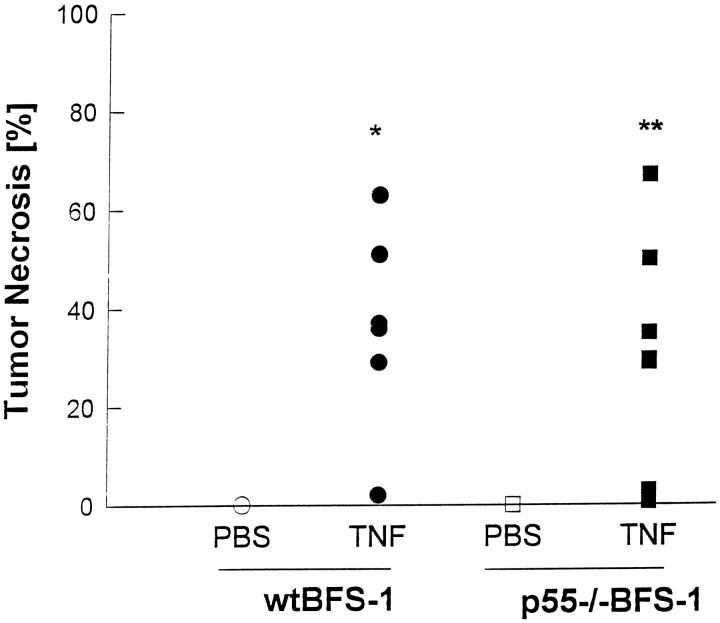
When C57BL/6 wt mice bearing solid tumors derived from wt BFS-1 or p55−/−BFS-1 cells were treated with rmTNF, we found that necrosis was induced by rmTNF in p55BFS-1 tumors as efficiently as in wt BFS-1 tumors (Figure 1) ▶ . The induction of necrosis with rmTNF was obviously independent of the TNFRp55 expression on the tumor cells, thus excluding any direct tumoricidal effect of TNF. Therefore, we concluded that the presence of TNFRp55 on tumor-associated, endogenic cells beside the tumor cells has to be necessary and sufficient to mediate TNF-induced tumor necrosis.
Figure 1.
TNF-induced tumor necrosis independent of the TNFR on tumor cells. C57BL/6 wt mice bearing wt BFS-1 (○, •) or p55−/−BFS-1 (□, ▪) tumors were treated with rmTNF (5 μg in 100 μl PBS, n = 13, •, ▪) or PBS (100 μl, n = 8, ○, □) i.p., and tumor necrosis was determined 2 days after treatment. Results from two experiments with at least three mice per group are shown. P values for the comparison of the PBS versus rmTNF-treated groups: *P < 0.05; **P < 0.005.
Activation of endothelial cells, fibrin deposition, and coagulation within the tumor vasculature has been shown in vivo to be correlated with the occurrence of TNF-induced tumor necrosis in other and very similar tumor models. 8-11,24 Accordingly we found in the TNF-treated tumors intravascular fibrin formation (not shown). In addition, 12 hours after TNF administration we observed increased amounts of vWF in the tumor endothelium by immunohistochemistry (not shown), indicating an injury and/or regeneration of endothelial cells. 22,25 Therefore, one potential mechanism for TNF-mediated tumor necrosis might be the induction of a procoagulatory state within the tumor vasculature by direct stimulation of TF expression on the tumor endothelium and/or via activated blood leukocytes (ie, monocytes). 10,12,13,26 In addition, inflammatory mediators like LPS and TNF are known to induce leukocyte sequestration. 26,27 Four to twelve hours after rmTNF treatment of tumor-bearing mice we observed leukostasis in the vessels of different organ systems such as lung and kidney, but also in the tumor vasculature (data not shown). Detection of leukostasis in the tumor vessels, however, was difficult because of the structure (diameter, wall thickness) of the tumor vasculature. Because rmTNF-activated leukocytes release inflammatory cytokines (which, furthermore, should be able to stimulate TF expression on endothelial cells), rmTNF-induced adherence of activated leukocytes may contribute to the tumor necrosis in our mouse model.
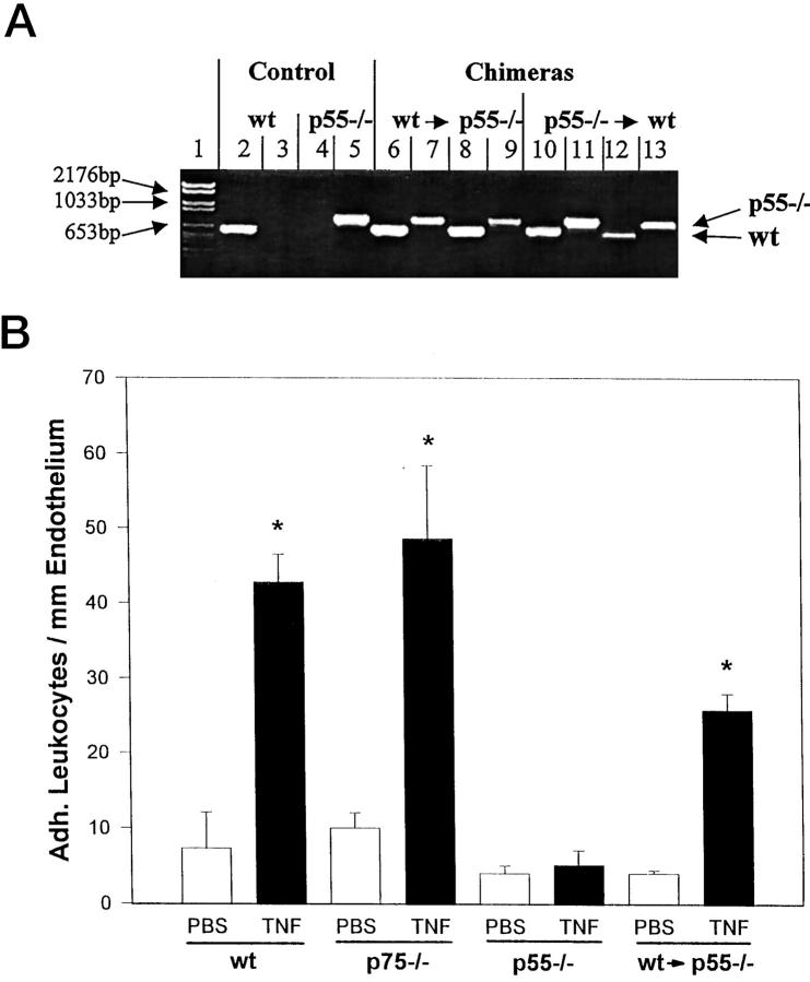
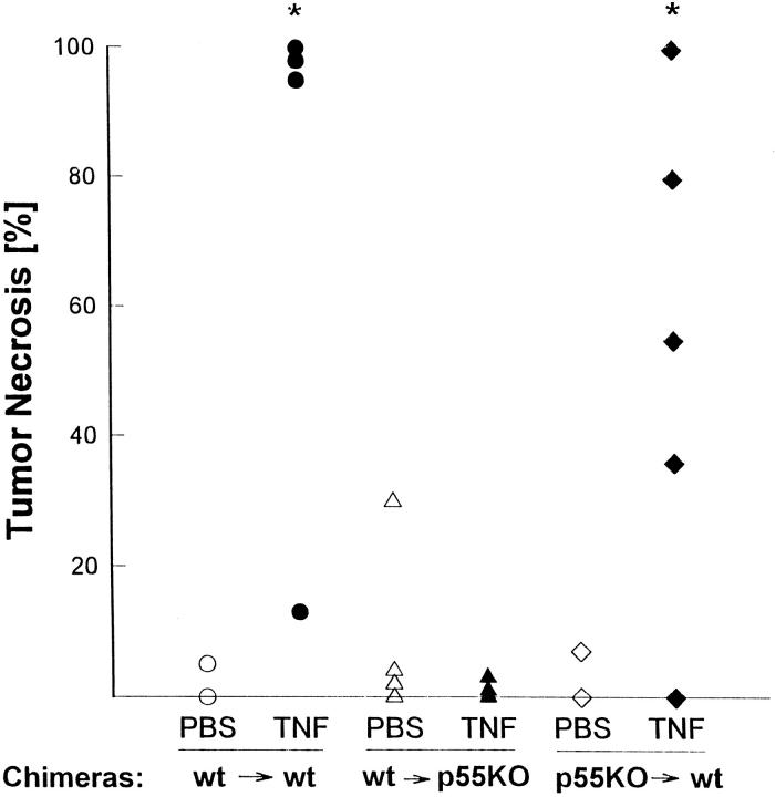
To analyze the TNF receptor-dependent effect of systemic rmTNF treatment on the sequestration of peripheral blood leukocytes, the occurrence of pulmonary leukostasis was quantified in wt and TNFR-deficient mice 4 hours after TNF application. Administration of rmTNF resulted in adhesion of leukocytes to lung vessels in wt and TNFRp75−/− mice. However, as in tumor necrosis, soluble rmTNF was not able to induce leukostasis in TNFRp55−/− mice (Figure 2B) ▶ . To determine the role of hematopoietic cells for mediating these rmTNF-induced effects, chimeric mice were generated by adoptive bone marrow transplantation. Either TNFRp55−/− mice received wt bone marrow cells or wt mice received bone marrow cells from TNFRp55−/− donors. To test the successful adoptive bone marrow cell transfer, the genotype of blood leukocytes in chimeric mice was determined 4 weeks after reconstitution. In all chimeric mice, specific bands of the donor genotype and the recipient genotype were detectable, which was likely due to incomplete reconstitution (Figure 2A) ▶ . Nevertheless, adoptive transfer of TNFRp55-positive bone marrow cells into TNFRp55−/− mice was sufficient to restore rmTNF-induced sequestration of leukocytes in TNFRp55−/− mice (Figure 2B) ▶ , indicating that rmTNF was acting on the hematopoietic cells for this function. In contrast, we could not induce tumor necrosis with rmTNF in chimeric TNFRp55−/− mice, even after reconstitution with wt bone marrow cells (Figure 3) ▶ , whereas rmTNF caused tumor necrosis in chimeric wt mice reconstituted with TNFRp55−/− bone marrow cells as well as it did in wt mice reconstituted for control purposes with wt bone marrow cells. LPS, in the same experiment, induced tumor necrosis in TNFRp55−/− mice reconstituted with bone marrow from wt as well as TNFRp55−/− mice, documenting the general ability for tumor necrosis in such mice (data not shown).
Figure 2.
TNF-induced leukostasis in TNFR-deficient and bone marrow chimeric mice. A: Genomic DNA was isolated from leukocytes of C57BL/6 wt (lanes 2 and 3) and TNFRp55−/− mice (lanes 4 and 5), or TNFRp55−/− mice reconstituted with wt bone marrow (lanes 6–9), or wt mice reconstituted with TNFRp55−/− bone marrow (lanes 10–13). The presence of the wt (lanes 2, 4, 6, 8, 10, 12) or mutated (lanes 3, 5, 7, 9, 11, 13) TNFRp55 allele was determined by PCR. A DNA length standard is shown in lane 1. B: C57BL/6 mice (wt, n = 6), TNFRp75−/− mice (p75−/−, n = 6), TNFRp55−/− mice (p55−/−, n = 6), or TNFRp55−/− mice reconstituted with wt bone marrow cells (wt → p55−/−, n = 5) were treated i.p. with rmTNF (5 μg in 100 μl PBS) or PBS (100 μl). Bars represent the mean value ± SE of adherent leukocytes per mm endothelium. *P < 0.001.
Figure 3.
TNF-induced tumor necrosis in bone marrow chimeric mice. Tumor (wt BFS-1)-bearing C57BL/6 wt mice reconstituted with wt bone marrow (wt→wt, n = 7, ○, •), TNFRp55−/− mice reconstituted with wt bone marrow (p55−/− →wt, n = 11, ⋄, ♦) or wt mice reconstituted with TNFRp55−/− bone marrow (wt→p55−/−, n = 16, ▵, ▴) were treated with PBS (100 μl, ○, □, ⋄) or rmTNF (5 μg in 100 μl PBS, •, ▪, ♦) i.p. and tumor necrosis was determined 2 days after treatment. Pooled results from two experiments with at least three mice per group are shown. P values for the comparison of the PBS versus rmTNF-treated groups: *P < 0.05.
Taken together, even though leukocyte adhesion after rmTNF treatment was dependent on the presence of the TNFRp55 in the host cells, an essential contribution of leukocytes to rmTNF-induced tumor necrosis is unlikely, because in chimeric mice the occurrence of tumor necrosis did not correlate with leukostasis. The hypothesis of TNF action on endothelial cells for tumor necrosis induction, 8,10 however, is strengthened by the finding that wt bone marrow transplantation did not change the phenotype of the tumor-bearing TNFRp55−/− mice, which are not able to mediate rmTNF-induced tumor necrosis. In these mice the endothelial cells lining the vascular system do not carry a functional TNFRp55 but only the adoptively transferred hematopoietic cells. This demonstrated clearly that rmTNF-induced tumor necrosis was not mediated by hematopoietic cells like granulocytes, T cells, or monocytes/macrophages, but likely by tumor vascular endothelium. This is in line with the observed toxic effect on endothelial cells after therapeutic administration of TNF and IFNγ in isolated limp perfusion of melanoma patients. 17 From these studies, disruption of the tumor vasculature is discussed as result of anoikis due to decreased αvβ3-dependent endothelial cell adhesion and ensuing detachment of these cells. 17
In conclusion, we found that 1) rmTNF does not directly exert a cytotoxic effect on the tumor cells, 2) the TNFRp55 expression on hematopoietic cells was essential for TNF-induced leukocyte sequestration, 3) but not sufficient to mediate tumor necrosis. TNFR type 1-expressing endothelial cells lining the tumor vasculature are most likely the target cells for mediation of TNF-induced tumor necrosis.
Footnotes
Address reprint requests to Dr. Daniela N. Männel, Department of Pathology, University of Regensburg, D-93042 Regensburg, Germany. E-mail: daniela.maennel@klinik.uni-regensburg.de.
References
- 1.O’Malley WE, Achinstein B, Shear MJ: Journal of the National Cancer Institute, Vol. 29, 1962, Action of bacterial polysaccharide on tumors. II. Damage of sarcoma 37 by serum of mice treated with Serratia marcescens polysaccharide, and induced tolerance (classical article). Nutr Rev 1988, 46:389–391 [DOI] [PubMed]
- 2.Parr I, Wheeler E, Alexander P: Similarities of the anti-tumour actions of endotoxin, lipid A, and double-stranded RNA. Br J Cancer 1973, 27:370-389 [DOI] [PMC free article] [PubMed] [Google Scholar]
- 3.Rietschel ET, Brade H, Holst O, Brade L, Müller Loennies S, Mamat U, Zähringer U, Beckmann F, Seydel U, Brandenburg K, Ulmer AJ, Mattern T, Heine H, Schletter J, Loppnow H, Schönbeck U, Flad HD, Hauschildt S, Schade UF, Di Padova F, Kusumoto S, Schumann RR: Bacterial endotoxin: chemical constitution, biological recognition, host response, and immunological detoxification. Curr Top Microbiol Immunol 1996, 216:39–81 [DOI] [PubMed]
- 4.Carswell EA, Old LJ, Kassel RL, Green S, Fiore N, Williamson B: An endotoxin-induced serum factor that causes necrosis of tumor. Proc Natl Acad Sci USA 1975, 72:3666-3670 [DOI] [PMC free article] [PubMed] [Google Scholar]
- 5.Männel DN, Rosenstreich DL, Mergenhagen SE: Mechanism of lipopolysaccharide-induced tumor necrosis: requirement for lipopolysaccharide-sensitive lymphoreticular cells. Infect Immun 1979, 24:573-576 [DOI] [PMC free article] [PubMed] [Google Scholar]
- 6.Tartaglia LA, Goeddel DV: Two TNF receptors. Immunol Today 1992, 13:151-153 [DOI] [PubMed] [Google Scholar]
- 7.Palladino MA, Jr, Shalaby MR, Kramer SM, Ferraiolo BL, Baughman RA, Deleo AB, Crase D, Marafino B, Aggarwal BB, Figari IS: Characterization of the antitumor activities of human tumor necrosis factor-alpha and the comparison with other cytokines: induction of tumor-specific immunity. J Immunol 1987, 138:4023-4032 [PubMed] [Google Scholar]
- 8.Shimomura K, Manda T, Mukumoto S, Kobayashi K, Nakano K, Mori J: Recombinant human tumor necrosis factor-alpha: thrombus formation is a cause of anti-tumor activity. Int J Cancer 1988, 41:243-247 [DOI] [PubMed] [Google Scholar]
- 9.Huang X, Molema G, King S, Watkins L, Edgington TS, Thorpe PE: Tumor infarction in mice by antibody-directed targeting of tissue factor to tumor vasculature. Science 1997, 275:547-550 [DOI] [PubMed] [Google Scholar]
- 10.Zhang Y, Deng Y, Wendt T, Liliensiek B, Bierhaus A, Greten J, He W, Chen B, Hach Wunderle V, Waldherr R, Ziegler R, Männel D, Stern DM, Nawroth PP: Intravenous somatic gene transfer with antisense tissue factor restores blood flow by reducing tumor necrosis factor-induced tissue factor expression and fibrin deposition in mouse meth-A sarcoma. J Clin Invest 1996, 97:2213–2224 [DOI] [PMC free article] [PubMed]
- 11.Bierhaus A, Zhang Y, Deng Y, Mackman N, Quehenberger P, Haase M, Luther T, Müller M, Bohrer H, Greten J, Martin E, Bäuerle P, Waldherr R, Kisiel W, Ziegler R, Stern DM, Nawroth PP: Mechanism of the tumor necrosis factor alpha-mediated induction of endothelial tissue factor. J Biol Chem 1995, 270:26419-26432 [DOI] [PubMed] [Google Scholar]
- 12.Schmid E, Müller TH, Budzinski RM, Pfizenmaier K, Binder K: Lymphocyte adhesion to human endothelial cells induces tissue factor expression via a juxtacrine pathway. Thromb Haemost 1995, 73:421-428 [PubMed] [Google Scholar]
- 13.Napoleone E, Di Santo A, Lorenzet R: Monocytes upregulate endothelial cell expression of tissue factor: a role for cell-cell contact and cross-talk. Blood 1997, 89:541-549 [PubMed] [Google Scholar]
- 14.Lewis JC, Jones NL, Hermanns MI, Rohrig O, Klein CL, Kirkpatrick CJ: Tissue factor expression during coculture of endothelial cells and monocytes. Exp Mol Pathol 1995, 62:207-218 [DOI] [PubMed] [Google Scholar]
- 15.Vandenabeele P, Declercq W, Vercammen D, Van de Craen M, Grooten J, Loetscher H, Brockhaus M, Lesslauer W, Fiers W: Functional characterization of the human tumor necrosis factor receptor p75 in a transfected rat/mouse T cell hybridoma. J Exp Med 1992, 176:1015-1024 [DOI] [PMC free article] [PubMed] [Google Scholar]
- 16.Gregory SA, Morrissey JH, Edgington TS: Regulation of tissue factor gene expression in the monocyte procoagulant response to endotoxin. Mol Cell Biol 1989, 9:2752-2755 [DOI] [PMC free article] [PubMed] [Google Scholar]
- 17.Ruegg C, Yilmaz A, Bieler G, Bamat J, Chaubert P, Lejeune FJ: Evidence for the involvement of endothelial cell integrin alphaVbeta3 in the disruption of the tumor vasculature induced by TNF and IFN-gamma. Nature Med 1998, 4:408-414 [DOI] [PubMed] [Google Scholar]
- 18.Lejeune FJ, Ruegg C, Lienard D: Clinical applications of TNF-alpha in cancer. Curr Opin Immunol 1998, 10:573-580 [DOI] [PubMed] [Google Scholar]
- 19.Pfeffer K, Matsuyama T, Kundig TM, Wakeham A, Kishihara K, Shahinian A, Wiegmann K, Ohashi PS, Krönke M, Mak TW: Mice deficient for the 55 kd tumor necrosis factor receptor are resistant to endotoxic shock, yet succumb to L. monocytogenes infection. Cell 1993, 73:457-467 [DOI] [PubMed] [Google Scholar]
- 20.Erickson SL, de Sauvage FJ, Kikly K, Carver Moore K, Pitts Meek S, Gillett N, Sheehan KC, Schreiber RD, Goeddel DV, Moore MW: Decreased sensitivity to tumour-necrosis factor but normal T-cell development in TNF receptor-2-deficient mice. Nature 1994, 372:560–563 [DOI] [PubMed]
- 21.Ditter B, Becker KP, Urbaschek R, Urbaschek B: Detection of endotoxin in blood and other specimens by evaluation of photometrically registered LAL-reaction-kinetics in microtiter plates. Prog Clin Biol Res 1982, 93:385-392 [PubMed] [Google Scholar]
- 22.Kasper M, Schobl R, Haroske G, Fischer R, Neubert F, Dimmer V, Muller M: Distribution of von Willebrand factor in capillary endothelial cells of rat lungs with pulmonary fibrosis. Exp Toxicol Pathol 1996, 48:283-288 [DOI] [PubMed] [Google Scholar]
- 23.Marr RA, Hitt M, Gauldie J, Muller WJ, Graham FL: A p75 tumor necrosis factor receptor-specific mutant of murine tumor necrosis factor α expressed from an adenovirus vector induces an antitumor response with reduced toxicity. Cancer Gene Ther 1999, 6:465-474 [DOI] [PubMed] [Google Scholar]
- 24.Nawroth P, Handley D, Matsueda G, de Waal R, Gerlach H, Blohm D, Stern D: Tumor necrosis factor/cachectin-induced intravascular fibrin formation in meth A fibrosarcomas. J Exp Med 1988, 168:637-647 [DOI] [PMC free article] [PubMed] [Google Scholar]
- 25.Reidy MA, Chopek M, Chao S, McDonald T, Schwartz SM: Injury induces increase of von Willebrand factor in rat endothelial cells. Am J Pathol 1989, 134:857-864 [PMC free article] [PubMed] [Google Scholar]
- 26.Luther T, Bierhaus A, Kasper M, Kotzsch M, Nawroth PP, Müller M: Increased tissue factor expression on monocytes in response to endotoxin. Immunology 1995, 194:A82 [Google Scholar]
- 27.Chang SW: Endotoxin-induced pulmonary leukostasis in the rat: role of platelet-activating factor and tumor necrosis factor. J Lab Clin Med 1994, 123:65-72 [PubMed] [Google Scholar]


