

Abstract
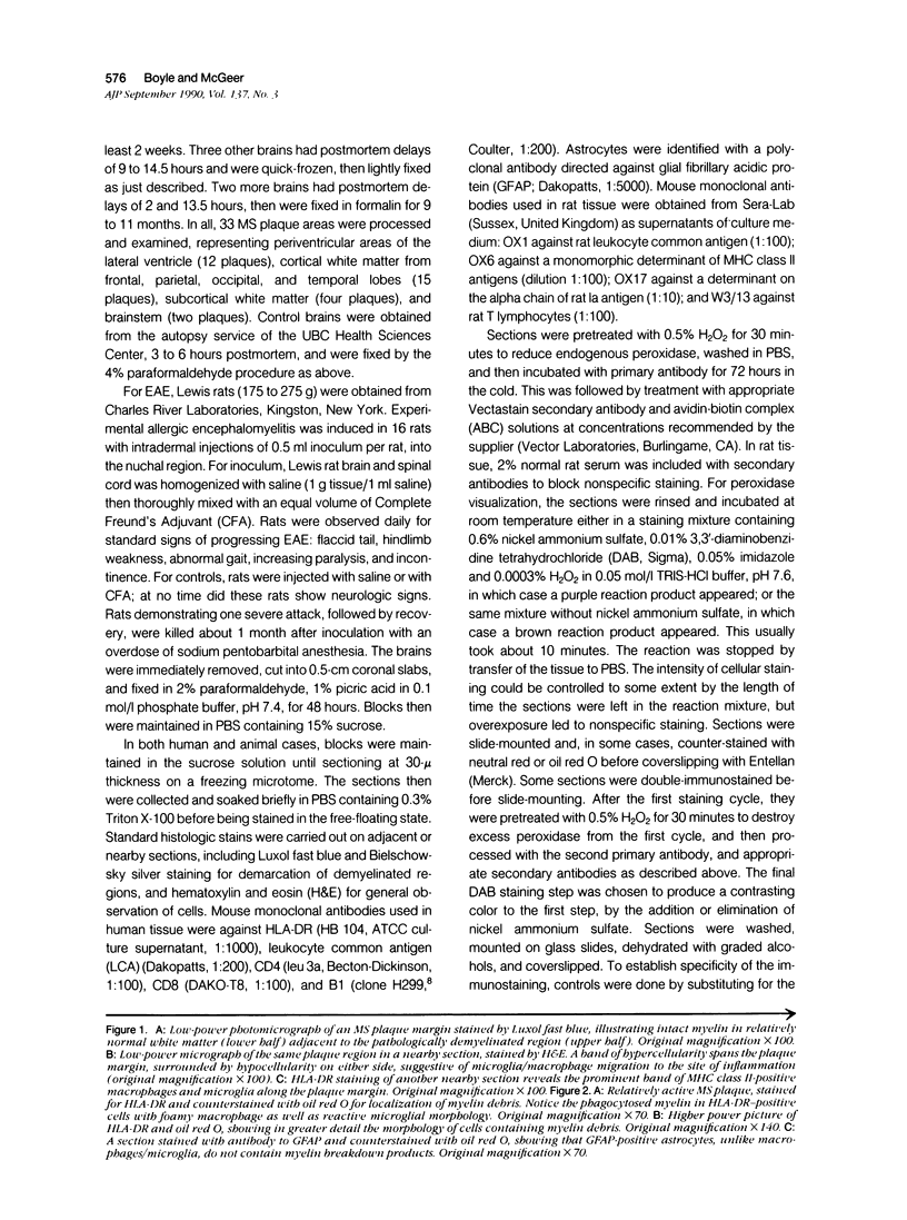
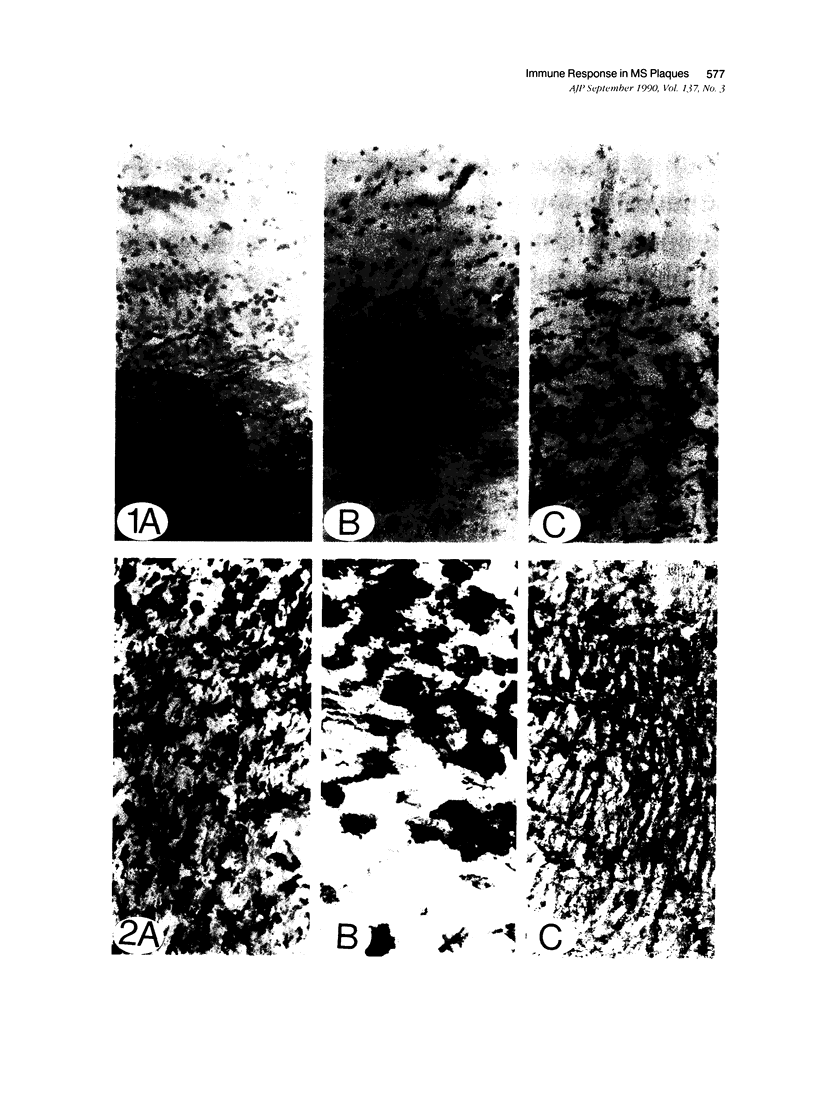
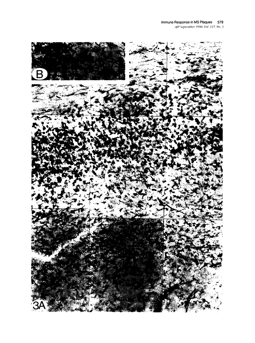
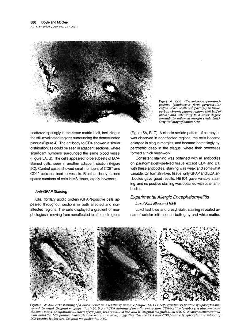
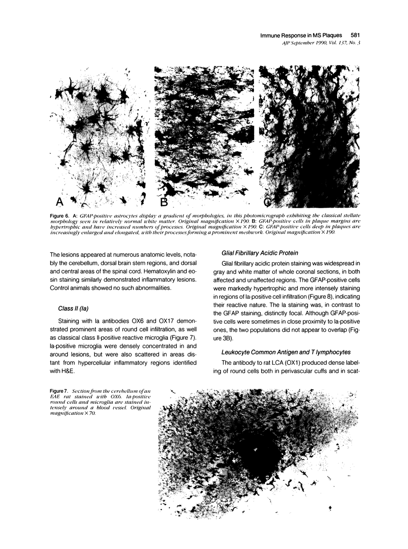
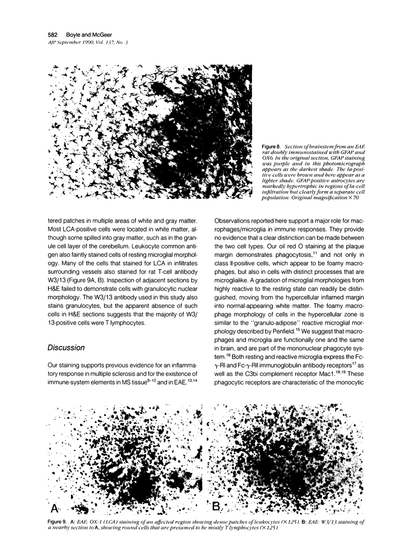
Multiple sclerosis plaques were immunohistochemically stained to exhibit cells expressing immune-system antigens. Human leukocyte antigen (HLA)-DR-positive cells formed dense rings around all plaque regions. The majority were reactive microglia/macrophages. Counterstaining with oil red O revealed heavy myelin debris within these cells. They were distinct from astrocytes, which were identified with an antibody to glial fibrillary acidic protein (GFAP) and which did not contain oil red O myelin debris. Numerous leukocytes and microglia were stained with antibody to leukocyte common antigen (LCA). Lymphocytes in cuffs around vessels, along the margins of capillary walls, and, sparingly, in the tissue matrix of affected areas, were stained with antibodies to CD4 (T-helper/inducer) and CD8 (T-cytotoxic/suppressor). In experimental allergic encephalomyelitis (EAE) induced in Lewis rats, a similar proliferation of Ia-positive (OX6, OX17) cells displaying reactive microglia/macrophage morphology was observed. These Ia-positive cells also were easily distinguished from GFAP-positive astrocytes. The results suggest that macrophages/reactive microglia, and not astrocytes, express class II MHC antigens in multiple sclerosis and EAE plaques.
Full text
PDF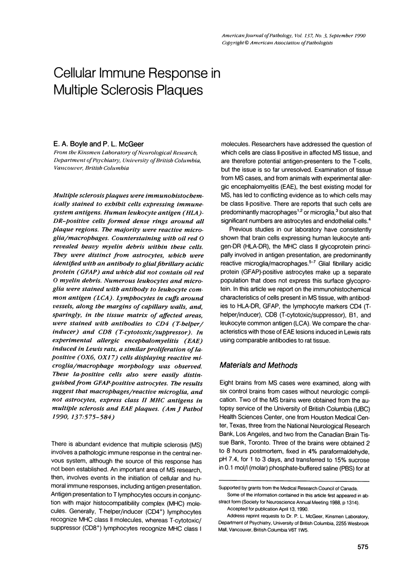
Images in this article

Selected References
These references are in PubMed. This may not be the complete list of references from this article.
- Booss J., Esiri M. M., Tourtellotte W. W., Mason D. Y. Immunohistological analysis of T lymphocyte subsets in the central nervous system in chronic progressive multiple sclerosis. J Neurol Sci. 1983 Dec;62(1-3):219–232. doi: 10.1016/0022-510x(83)90201-0. [DOI] [PubMed] [Google Scholar]
- Butter C., Healey D. G., Agha N., Turk J. L. An immunoelectron microscopical study of the expression of class II MHC and a T lymphocyte surface marker during chronic relapsing experimental allergic encephalomyelitis. J Neuroimmunol. 1988 Nov;20(1):45–51. doi: 10.1016/0165-5728(88)90113-0. [DOI] [PubMed] [Google Scholar]
- Craggs R. I., Webster H. D. Ia antigens in the normal rat nervous system and in lesions of experimental allergic encephalomyelitis. Acta Neuropathol. 1985;68(4):263–272. doi: 10.1007/BF00690828. [DOI] [PubMed] [Google Scholar]
- Cuzner M. L., Hayes G. M., Newcombe J., Woodroofe M. N. The nature of inflammatory components during demyelination in multiple sclerosis. J Neuroimmunol. 1988 Dec;20(2-3):203–209. doi: 10.1016/0165-5728(88)90161-0. [DOI] [PubMed] [Google Scholar]
- Erb P., Kennedy M., Wassmer P., Huegli G. Antigen-presenting cells and T cell activation. Agents Actions. 1986 Dec;19(5-6):266–268. doi: 10.1007/BF01971224. [DOI] [PubMed] [Google Scholar]
- Esiri M. M., Reading M. C. Macrophage populations associated with multiple sclerosis plaques. Neuropathol Appl Neurobiol. 1987 Nov-Dec;13(6):451–465. doi: 10.1111/j.1365-2990.1987.tb00074.x. [DOI] [PubMed] [Google Scholar]
- Fontana A., Fierz W., Wekerle H. Astrocytes present myelin basic protein to encephalitogenic T-cell lines. Nature. 1984 Jan 19;307(5948):273–276. doi: 10.1038/307273a0. [DOI] [PubMed] [Google Scholar]
- Frei K., Siepl C., Groscurth P., Bodmer S., Fontana A. Immunobiology of microglial cells. Ann N Y Acad Sci. 1988;540:218–227. doi: 10.1111/j.1749-6632.1988.tb27064.x. [DOI] [PubMed] [Google Scholar]
- Giulian D., Baker T. J. Characterization of ameboid microglia isolated from developing mammalian brain. J Neurosci. 1986 Aug;6(8):2163–2178. doi: 10.1523/JNEUROSCI.06-08-02163.1986. [DOI] [PMC free article] [PubMed] [Google Scholar]
- Hauser S. L., Bhan A. K., Gilles F., Kemp M., Kerr C., Weiner H. L. Immunohistochemical analysis of the cellular infiltrate in multiple sclerosis lesions. Ann Neurol. 1986 Jun;19(6):578–587. doi: 10.1002/ana.410190610. [DOI] [PubMed] [Google Scholar]
- Hayashi T., Morimoto C., Burks J. S., Kerr C., Hauser S. L. Dual-label immunocytochemistry of the active multiple sclerosis lesion: major histocompatibility complex and activation antigens. Ann Neurol. 1988 Oct;24(4):523–531. doi: 10.1002/ana.410240408. [DOI] [PubMed] [Google Scholar]
- Hirsch M. R., Wietzerbin J., Pierres M., Goridis C. Expression of Ia antigens by cultured astrocytes treated with gamma-interferon. Neurosci Lett. 1983 Oct 31;41(1-2):199–204. doi: 10.1016/0304-3940(83)90247-1. [DOI] [PubMed] [Google Scholar]
- Itagaki S., McGeer P. L., Akiyama H. Presence of T-cytotoxic suppressor and leucocyte common antigen positive cells in Alzheimer's disease brain tissue. Neurosci Lett. 1988 Sep 12;91(3):259–264. doi: 10.1016/0304-3940(88)90690-8. [DOI] [PubMed] [Google Scholar]
- Matsumoto Y., Hara N., Tanaka R., Fujiwara M. Immunohistochemical analysis of the rat central nervous system during experimental allergic encephalomyelitis, with special reference to Ia-positive cells with dendritic morphology. J Immunol. 1986 May 15;136(10):3668–3676. [PubMed] [Google Scholar]
- McGeer P. L., Akiyama H., Itagaki S., McGeer E. G. Immune system response in Alzheimer's disease. Can J Neurol Sci. 1989 Nov;16(4 Suppl):516–527. doi: 10.1017/s0317167100029863. [DOI] [PubMed] [Google Scholar]
- McGeer P. L., Itagaki S., McGeer E. G. Expression of the histocompatibility glycoprotein HLA-DR in neurological disease. Acta Neuropathol. 1988;76(6):550–557. doi: 10.1007/BF00689592. [DOI] [PubMed] [Google Scholar]
- Pardridge W. M., Yang J., Buciak J., Tourtellotte W. W. Human brain microvascular DR-antigen. J Neurosci Res. 1989 Jul;23(3):337–341. doi: 10.1002/jnr.490230314. [DOI] [PubMed] [Google Scholar]
- Penfield W. Microglia and the Process of Phagocytosis in Gliomas. Am J Pathol. 1925 Jan;1(1):77–90.15. [PMC free article] [PubMed] [Google Scholar]
- Poltorak M., Freed W. J. Immunological reactions induced by intracerebral transplantation: evidence that host microglia but not astroglia are the antigen-presenting cells. Exp Neurol. 1989 Mar;103(3):222–233. doi: 10.1016/0014-4886(89)90046-0. [DOI] [PubMed] [Google Scholar]
- Prineas J. W., Wright R. G. Macrophages, lymphocytes, and plasma cells in the perivascular compartment in chronic multiple sclerosis. Lab Invest. 1978 Apr;38(4):409–421. [PubMed] [Google Scholar]
- Rozemuller J. M., Eikelenboom P., Pals S. T., Stam F. C. Microglial cells around amyloid plaques in Alzheimer's disease express leucocyte adhesion molecules of the LFA-1 family. Neurosci Lett. 1989 Jul 3;101(3):288–292. doi: 10.1016/0304-3940(89)90547-8. [DOI] [PubMed] [Google Scholar]
- Sobel R. A., Blanchette B. W., Bhan A. K., Colvin R. B. The immunopathology of experimental allergic encephalomyelitis. II. Endothelial cell Ia increases prior to inflammatory cell infiltration. J Immunol. 1984 May;132(5):2402–2407. [PubMed] [Google Scholar]
- Stashenko P., Nadler L. M., Hardy R., Schlossman S. F. Characterization of a human B lymphocyte-specific antigen. J Immunol. 1980 Oct;125(4):1678–1685. [PubMed] [Google Scholar]
- Streit W. J., Graeber M. B., Kreutzberg G. W. Functional plasticity of microglia: a review. Glia. 1988;1(5):301–307. doi: 10.1002/glia.440010502. [DOI] [PubMed] [Google Scholar]
- Traugott U. Multiple sclerosis: relevance of class I and class II MHC-expressing cells to lesion development. J Neuroimmunol. 1987 Oct;16(2):283–302. doi: 10.1016/0165-5728(87)90082-8. [DOI] [PubMed] [Google Scholar]
- Traugott U., Raine C. S. Multiple sclerosis. Evidence for antigen presentation in situ by endothelial cells and astrocytes. J Neurol Sci. 1985 Jul;69(3):365–370. doi: 10.1016/0022-510x(85)90147-9. [DOI] [PubMed] [Google Scholar]
- Vass K., Lassmann H., Wekerle H., Wisniewski H. M. The distribution of Ia antigen in the lesions of rat acute experimental allergic encephalomyelitis. Acta Neuropathol. 1986;70(2):149–160. doi: 10.1007/BF00691433. [DOI] [PubMed] [Google Scholar]
- Woodroofe M. N., Bellamy A. S., Feldmann M., Davison A. N., Cuzner M. L. Immunocytochemical characterisation of the immune reaction in the central nervous system in multiple sclerosis. Possible role for microglia in lesion growth. J Neurol Sci. 1986 Jul;74(2-3):135–152. doi: 10.1016/0022-510x(86)90100-0. [DOI] [PubMed] [Google Scholar]
- van Furth R. Current view on the mononuclear phagocyte system. Immunobiology. 1982 Apr;161(3-4):178–185. doi: 10.1016/S0171-2985(82)80072-7. [DOI] [PubMed] [Google Scholar]