

Abstract
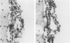
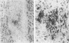
Cytomegalovirus (CMV) infection of the brain is common in AIDS; however, little is known of the host defense response to CMV in the central nervous system (CNS). A guinea pig model was developed to study this problem. In the present studies the percentages of T cells and monocytes invading the leptomeninges during the course of acute CMV infection were compared. In addition, qualitative observations on parenchymal infiltrates were made. Such studies have not been performed previously in CMV infection of the CNS. Monocytes, defined cytochemically, predominated in the leptomeninges and in parenchymal foci. In contrast, T cells, defined immunohistologically, were found in a low percentage in the leptomeningeal reaction and only rarely in the parenchyma. These novel results differ significantly from other viral infections in which the T cell predominates in the leptomeningeal response and plays a major role in the parenchyma.
Full text
PDF


Images in this article


Selected References
These references are in PubMed. This may not be the complete list of references from this article.
- Bixler G. S., Jr, Booss J. Adherent spleen cells from mice acutely infected with cytomegalovirus suppress the primary antibody response in vitro. J Immunol. 1981 Oct;127(4):1294–1299. [PubMed] [Google Scholar]
- Booss J., Dann P. R., Griffith B. P., Kim J. H. Glial nodule encephalitis in the guinea pig: serial observations following cytomegalovirus infection. Acta Neuropathol. 1988;75(5):465–473. doi: 10.1007/BF00687133. [DOI] [PubMed] [Google Scholar]
- Booss J. Establishment of cytomegaloviral infection in mice: role of a macrophage-enriched subpopulation. J Infect Dis. 1980 Apr;141(4):466–472. doi: 10.1093/infdis/141.4.466. [DOI] [PubMed] [Google Scholar]
- Booss J., Wheelock E. F. Progressive inhibition of T-cell function preceding clinical signs of cytomegalovirus infection in mice. J Infect Dis. 1977 Mar;135(3):478–481. doi: 10.1093/infdis/135.3.478. [DOI] [PubMed] [Google Scholar]
- Bozdech M. J., Bainton D. F. Identification of alpha-naphthyl butyrate esterase as a plasma membrane ectoenzyme of monocytes and as a discrete intracellular membrane-bounded organelle in lymphocytes. J Exp Med. 1981 Jan 1;153(1):182–195. doi: 10.1084/jem.153.1.182. [DOI] [PMC free article] [PubMed] [Google Scholar]
- Chiba J., Chused T. M., Leiserson W. M., Zweig S. E., Shevach E. M. Production and characterization of monoclonal antibodies to guinea pig lymphoid differentiation antigens. J Immunol Methods. 1983 Oct 14;63(2):247–261. doi: 10.1016/0022-1759(83)90429-5. [DOI] [PubMed] [Google Scholar]
- Elias J. M., Chiba J., Shevach E. M., Godfrey H. P. Guinea pig T lymphocyte development analyzed by enzyme histocytochemistry, monoclonal antibodies, and flow cytometry. Lab Invest. 1985 Mar;52(3):270–277. [PubMed] [Google Scholar]
- Esiri M. M., Booss J. Comparison of methods to identify microglial cells and macrophages in the human central nervous system. J Clin Pathol. 1984 Feb;37(2):150–156. doi: 10.1136/jcp.37.2.150. [DOI] [PMC free article] [PubMed] [Google Scholar]
- Esiri M. M., McGee J. O. Monoclonal antibody to macrophages (EMB/11) labels macrophages and microglial cells in human brain. J Clin Pathol. 1986 Jun;39(6):615–621. doi: 10.1136/jcp.39.6.615. [DOI] [PMC free article] [PubMed] [Google Scholar]
- Goff E., Griffith B. P., Booss J. Delayed amplification of cytomegalovirus infection in the placenta and maternal tissues during late gestation. Am J Obstet Gynecol. 1987 May;156(5):1265–1270. doi: 10.1016/0002-9378(87)90159-1. [DOI] [PubMed] [Google Scholar]
- Griffith B. P., Lavallee J. T., Booss J., Hsiung G. D. Asynchronous depression of responses to T- and B-cell mitogens during acute infection with cytomegalovirus in the guinea pig. Cell Immunol. 1984 Sep;87(2):727–733. doi: 10.1016/0008-8749(84)90043-1. [DOI] [PubMed] [Google Scholar]
- Griffith B. P., Lucia H. L., Bia F. J., Hsiung G. D. Cytomegalovirus-induced mononucleosis in guinea pigs. Infect Immun. 1981 May;32(2):857–863. doi: 10.1128/iai.32.2.857-863.1981. [DOI] [PMC free article] [PubMed] [Google Scholar]
- Johnson R. T., Burke D. S., Elwell M., Leake C. J., Nisalak A., Hoke C. H., Lorsomrudee W. Japanese encephalitis: immunocytochemical studies of viral antigen and inflammatory cells in fatal cases. Ann Neurol. 1985 Nov;18(5):567–573. doi: 10.1002/ana.410180510. [DOI] [PubMed] [Google Scholar]
- Kam-Hansen S., Frydén A., Link H. B and T lymphocytes in cerebrospinal fluid and blood in multiple sclerosis, optic neuritis and mumps meningitis. Acta Neurol Scand. 1978 Aug;58(2):95–103. doi: 10.1111/j.1600-0404.1978.tb02866.x. [DOI] [PubMed] [Google Scholar]
- Koenig S., Gendelman H. E., Orenstein J. M., Dal Canto M. C., Pezeshkpour G. H., Yungbluth M., Janotta F., Aksamit A., Martin M. A., Fauci A. S. Detection of AIDS virus in macrophages in brain tissue from AIDS patients with encephalopathy. Science. 1986 Sep 5;233(4768):1089–1093. doi: 10.1126/science.3016903. [DOI] [PubMed] [Google Scholar]
- Moench T. R., Griffin D. E. Immunocytochemical identification and quantitation of the mononuclear cells in the cerebrospinal fluid, meninges, and brain during acute viral meningoencephalitis. J Exp Med. 1984 Jan 1;159(1):77–88. doi: 10.1084/jem.159.1.77. [DOI] [PMC free article] [PubMed] [Google Scholar]
- Morgello S., Cho E. S., Nielsen S., Devinsky O., Petito C. K. Cytomegalovirus encephalitis in patients with acquired immunodeficiency syndrome: an autopsy study of 30 cases and a review of the literature. Hum Pathol. 1987 Mar;18(3):289–297. doi: 10.1016/s0046-8177(87)80012-6. [DOI] [PubMed] [Google Scholar]
- Sobel R. A., Blanchette B. W., Bhan A. K., Colvin R. B. The immunopathology of experimental allergic encephalomyelitis. I. Quantitative analysis of inflammatory cells in situ. J Immunol. 1984 May;132(5):2393–2401. [PubMed] [Google Scholar]