Abstract
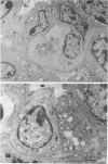
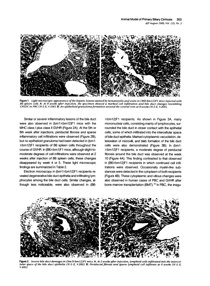
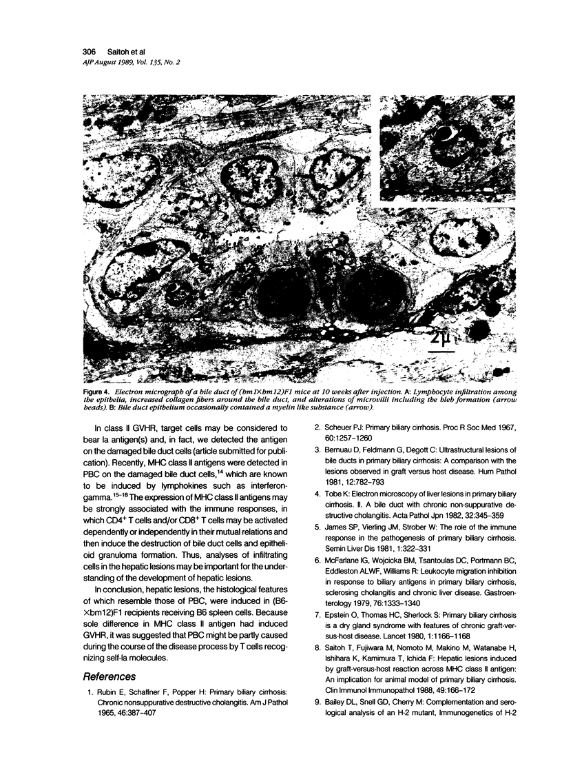
By light and electron microscopic examinations, histologic changes in the liver of mice with graft-versus-host reaction (GVHR) were analyzed. To induce GVHR, C57BL/6 (B6) spleen cells were injected into (B6Xbm1)F1, (B6Xbm12)F1, and (bm1Xbm12)F1 mice. In (B6Xbm12)F1 recipient mice, bile duct changes resembling chronic non-suppurative destructive cholangitis (CNSDC) and a formation of epithelioid granulomas were observed during the course of GVHR. An epithelioid granuloma in the liver of (B6Xbm1)F1 or (bm1.Xbm12)F1 recipients was not detected. By electron microscopy, the bile duct epithelia were seen to be in close contact with infiltrating cells, and marked alterations of their cytoplasm and microvilli were demonstrated; ie, vacuolation of the cytoplasm, deterioration of microvilli, and bleb formation were frequently observed in the liver of class II-disparate hosts. Concerning the basement membrane, no marked changes characteristic of primary biliary cirrhosis (PBC), such as many-layered basement membranes containing osmium positive substance, were detected. Because the major histocompatibility complex (MHC) class II-disparate system was used in our experimental system in the GVHR, the antigen expressed on the bile duct might be a target and be associated with the formation of the initial hepatic lesions in PBC such as CNSDC and epithelioid granuloma formation. Thus, GVHR across the MHC class II antigen is believed to play an important role in the development of PBC.
Full text
PDF


Images in this article
Selected References
These references are in PubMed. This may not be the complete list of references from this article.
- Bernuau D., Feldmann G., Degott C., Gisselbrecht C. Ultrastructural lesions of bile ducts in primary biliary cirrhosis. A comparison with the lesions observed in graft versus host disease. Hum Pathol. 1981 Sep;12(9):782–793. doi: 10.1016/s0046-8177(81)80081-0. [DOI] [PubMed] [Google Scholar]
- Colucci G., Schaffner F., Paronetto F. In situ characterization of the cell-surface antigens of the mononuclear cell infiltrate and bile duct epithelium in primary biliary cirrhosis. Clin Immunol Immunopathol. 1986 Oct;41(1):35–42. doi: 10.1016/0090-1229(86)90049-8. [DOI] [PubMed] [Google Scholar]
- Epstein O., Thomas H. C., Sherlock S. Primary biliary cirrhosis is a dry gland syndrome with features of chronic graft-versus-host disease. Lancet. 1980 May 31;1(8179):1166–1168. doi: 10.1016/s0140-6736(80)91621-9. [DOI] [PubMed] [Google Scholar]
- James S. P., Vierling J. M., Strober W. The role of the immune response in the pathogenesis of primary biliary cirrhosis. Semin Liver Dis. 1981 Nov;1(4):322–337. doi: 10.1055/s-2008-1040735. [DOI] [PubMed] [Google Scholar]
- McFarlane I. G., Wojcicka B. M., Tsantoulas D. C., Portmann B. C., Eddleston A. L., Williams R. Leukocyte migration inhibition in response to biliary antigens in primary biliary cirrhosis, sclerosing cholangitis, and other chronic liver diseases. Gastroenterology. 1979 Jun;76(6):1333–1340. [PubMed] [Google Scholar]
- McKenzie I. F., Morgan G. M., Sandrin M. S., Michaelides M. M., Melvold R. W., Kohn H. I. B6.C-H-2bm12. A new H-2 mutation in the I region in the mouse. J Exp Med. 1979 Dec 1;150(6):1323–1338. doi: 10.1084/jem.150.6.1323. [DOI] [PMC free article] [PubMed] [Google Scholar]
- RUBIN E., SCHAFFNER F., POPPER H. PRIMARY BILIARY CIRRHOSIS. CHRONIC NON-SUPPURATIVE DESTRUCTIVE CHOLANGITIS. Am J Pathol. 1965 Mar;46:387–407. [PMC free article] [PubMed] [Google Scholar]
- Rolink A. G., Gleichmann H., Gleichmann E. Diseases caused by reactions of T lymphocytes to incompatible structures of the major histocompatibility complex. VII. Immune-complex glomerulonephritis. J Immunol. 1983 Jan;130(1):209–215. [PubMed] [Google Scholar]
- Rolink A. G., Pals S. T., Gleichmann E. Allosuppressor and allohelper T cells in acute and chronic graft-vs.-host disease. II. F1 recipients carrying mutations at H-2K and/or I-A. J Exp Med. 1983 Feb 1;157(2):755–771. doi: 10.1084/jem.157.2.755. [DOI] [PMC free article] [PubMed] [Google Scholar]
- Saitoh T., Fujiwara M., Nomoto M., Makino M., Watanabe H., Ishihara K., Kamimura T., Ichida F. Hepatic lesions induced by graft-versus-host reaction across MHC class II antigens: an implication for animal model of primary biliary cirrhosis. Clin Immunol Immunopathol. 1988 Oct;49(1):166–172. doi: 10.1016/0090-1229(88)90106-7. [DOI] [PubMed] [Google Scholar]
- Sale G. E. Ileal predilection of graft-versus-host disease. Lancet. 1986 Nov 22;2(8517):1221–1221. doi: 10.1016/s0140-6736(86)92230-0. [DOI] [PubMed] [Google Scholar]
- Scheuer P. Primary biliary cirrhosis. Proc R Soc Med. 1967 Dec;60(12):1257–1260. [PMC free article] [PubMed] [Google Scholar]
- Tobe K. Electron microscopy of liver lesions in primary biliary cirrhosis. II. A bile duct with chronic non-suppurative destructive cholangitis. Acta Pathol Jpn. 1982 Mar;32(2):345–357. [PubMed] [Google Scholar]
- Todd I., Pujol-Borrell R., Hammond L. J., Bottazzo G. F., Feldmann M. Interferon-gamma induces HLA-DR expression by thyroid epithelium. Clin Exp Immunol. 1985 Aug;61(2):265–273. [PMC free article] [PubMed] [Google Scholar]
- Wadgymar A., Urmson J., Baumal R., Halloran P. F. Changes in Ia expression in mouse kidney during acute graft-vs-host disease. J Immunol. 1984 Apr;132(4):1826–1832. [PubMed] [Google Scholar]
- Yamada G., Hyodo I., Tobe K., Mizuno M., Nishihara T., Kobayashi T., Nagashima H. Ultrastructural immunocytochemical analysis of lymphocytes infiltrating bile duct epithelia in primary biliary cirrhosis. Hepatology. 1986 May-Jun;6(3):385–391. doi: 10.1002/hep.1840060309. [DOI] [PubMed] [Google Scholar]


