

Abstract
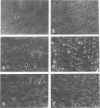
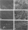
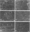
The authors probed the vascular endothelial cell cytoskeleton in strains of pigeons that are atherosclerosis-susceptible and disease-resistant, namely, the White Carneau and Show Racer pigeons. Endothelial cell actin and myosin were localized with the use of affinity-purified antibodies in conjunction with indirect immunofluorescence microscopy. The endothelial cell cytoskeleton was characterized in a site-specific and time-dependent manner by examination of arterial segments from each strain of pigeons. Anti-actin and anti-myosin fluorescence staining patterns of endothelial cells lining the ascending aorta, aortic arch, and thoracic aorta from the White Carneau and Show Racer pigeons sacrificed at 1 and 12 months of age were compared and analyzed. In the Show Racer, irrespective of arterial site or chronologic age, endothelial cell cytoskeletal organization is similar. Actin and myosin fluorescence is brightest at the cortex, where endothelial cells meet their neighbors. There is also an amorphous (diffuse) fluorescence throughout the cytoplasm. In addition to the diffuse and cortical cytoskeletal fluorescence in the endothelial cells of the Show Racers, the White Carneau also possess a unique cytoskeletal array of linear fluorescence, ie, the endothelial cell ridge. At 1 month of age, anti-actin staining of endothelial cell ridges averages 28.5 mu in length in the ascending aorta, 28.0 mu in the aortic arch, and 40.0 mu in the thoracic aorta. At the same time, anti-myosin fluorescence extends past both ends of the anti-actin-stained endothelial cell ridge fluorescence. In the ascending aorta, anti-myosin labeling of endothelial cell ridges is 3.5 times longer than anti-actin staining. This staining is absent in the aortic arch, whereas the thoracic aorta possesses endothelial cell ridges that extend over the entire length of the vessel segment. At 12 months of age, actin-stained endothelial cell ridges increase 1.6- and 1.4-fold in the ascending aorta and aortic arch, respectively. The thoracic aorta possesses endothelial cell ridges that cover its entire length. At 12 months of age, the length of myosin-stained endothelial cell ridges does not increase in the ascending or thoracic aorta. In contrast, the aortic arch expresses endothelial cell ridges that exceed 150 mu in length. It is proposed that the endothelial cell ridge assembles from cytoskeletal components as a focal endothelial cell response to injury, perhaps promoting endothelial cell adhesion to the underlying basal lamina through a transmembrane linkage.
Full text
PDF


Images in this article

Selected References
These references are in PubMed. This may not be the complete list of references from this article.
- Alavi M., Dunnett C. W., Moore S. Lipid composition of rabbit aortic wall following removal of endothelium by balloon catheter. Arteriosclerosis. 1983 Sep-Oct;3(5):413–419. doi: 10.1161/01.atv.3.5.413. [DOI] [PubMed] [Google Scholar]
- Bell F. P., Day A. J., Gent M., Schwartz C. J. Differing patterns of cholesterol accumulation and 3-H-cholesterol influx in areas of the cholesterol-fed pig aorta identified by evans blue dye. Exp Mol Pathol. 1975 Jun;22(3):336–375. doi: 10.1016/0014-4800(75)90081-7. [DOI] [PubMed] [Google Scholar]
- Curwen K. D., Smith S. C. Aortic glycosaminoglycans in atherosclerosis-susceptible and -resistant pigeons. Exp Mol Pathol. 1977 Aug;27(1):121–133. doi: 10.1016/0014-4800(77)90024-7. [DOI] [PubMed] [Google Scholar]
- Fishman J. A., Ryan G. B., Karnovsky M. J. Endothelial regeneration in the rat carotid artery and the significance of endothelial denudation in the pathogenesis of myointimal thickening. Lab Invest. 1975 Mar;32(3):339–351. [PubMed] [Google Scholar]
- Fox J. A., Hugh A. E. Localization of atheroma: a theory based on boundary layer separation. Br Heart J. 1966 May;28(3):388–399. doi: 10.1136/hrt.28.3.388. [DOI] [PMC free article] [PubMed] [Google Scholar]
- Gabbiani G., Gabbiani F., Heimark R. L., Schwartz S. M. Organization of actin cytoskeleton during early endothelial regeneration in vitro. J Cell Sci. 1984 Mar;66:39–50. doi: 10.1242/jcs.66.1.39. [DOI] [PubMed] [Google Scholar]
- Gabbiani G., Gabbiani F., Lombardi D., Schwartz S. M. Organization of actin cytoskeleton in normal and regenerating arterial endothelial cells. Proc Natl Acad Sci U S A. 1983 Apr;80(8):2361–2364. doi: 10.1073/pnas.80.8.2361. [DOI] [PMC free article] [PubMed] [Google Scholar]
- Gerrity R. G., Naito H. K., Richardson M., Schwartz C. J. Dietary induced atherogenesis in swine. Morphology of the intima in prelesion stages. Am J Pathol. 1979 Jun;95(3):775–792. [PMC free article] [PubMed] [Google Scholar]
- Gerrity R. G. The role of the monocyte in atherogenesis: I. Transition of blood-borne monocytes into foam cells in fatty lesions. Am J Pathol. 1981 May;103(2):181–190. [PMC free article] [PubMed] [Google Scholar]
- Gerrity R. G. The role of the monocyte in atherogenesis: II. Migration of foam cells from atherosclerotic lesions. Am J Pathol. 1981 May;103(2):191–200. [PMC free article] [PubMed] [Google Scholar]
- Gospodarowicz D., Ill C. Extracellular matrix and control of proliferation of vascular endothelial cells. J Clin Invest. 1980 Jun;65(6):1351–1364. doi: 10.1172/JCI109799. [DOI] [PMC free article] [PubMed] [Google Scholar]
- Gotlieb A. I., Spector W. Migration into an in vitro experimental wound: a comparison of porcine aortic endothelial and smooth muscle cells and the effect of culture irradiation. Am J Pathol. 1981 May;103(2):271–282. [PMC free article] [PubMed] [Google Scholar]
- Gotlieb A. I., Spector W., Wong M. K., Lacey C. In vitro reendothelialization. Microfilament bundle reorganization in migrating porcine endothelial cells. Arteriosclerosis. 1984 Mar-Apr;4(2):91–96. doi: 10.1161/01.atv.4.2.91. [DOI] [PubMed] [Google Scholar]
- Gotlieb A. I., Subrahmanyan L., Kalnins V. I. Microtubule-organizing centers and cell migration: effect of inhibition of migration and microtubule disruption in endothelial cells. J Cell Biol. 1983 May;96(5):1266–1272. doi: 10.1083/jcb.96.5.1266. [DOI] [PMC free article] [PubMed] [Google Scholar]
- Hajjar D. P., Falcone D. J., Fowler S., Minick C. R. Endothelium modifies the altered metabolism of the injured aortic wall. Am J Pathol. 1981 Jan;102(1):28–39. [PMC free article] [PubMed] [Google Scholar]
- Haudenschild C. C., Schwartz S. M. Endothelial regeneration. II. Restitution of endothelial continuity. Lab Invest. 1979 Nov;41(5):407–418. [PubMed] [Google Scholar]
- Haudenschild C. C., Van Sickle W., Chobanian A. V. Response of the aorta of the obese Zucker rat to injury. Arteriosclerosis. 1981 May-Jun;1(3):186–191. doi: 10.1161/01.atv.1.3.186. [DOI] [PubMed] [Google Scholar]
- Herman I. M., Brant A. M., Warty V. S., Bonaccorso J., Klein E. C., Kormos R. L., Borovetz H. S. Hemodynamics and the vascular endothelial cytoskeleton. J Cell Biol. 1987 Jul;105(1):291–302. doi: 10.1083/jcb.105.1.291. [DOI] [PMC free article] [PubMed] [Google Scholar]
- Herman I. M., Castellot J. J., Jr Regulation of vascular smooth muscle cell growth by endothelial-synthesized extracellular matrices. Arteriosclerosis. 1987 Sep-Oct;7(5):463–469. doi: 10.1161/01.atv.7.5.463. [DOI] [PubMed] [Google Scholar]
- Herman I. M., Crisona N. J., Pollard T. D. Relation between cell activity and the distribution of cytoplasmic actin and myosin. J Cell Biol. 1981 Jul;90(1):84–91. doi: 10.1083/jcb.90.1.84. [DOI] [PMC free article] [PubMed] [Google Scholar]
- Herman I. M. Extracellular matrix-cytoskeletal interactions in vascular cells. Tissue Cell. 1987;19(1):1–19. doi: 10.1016/0040-8166(87)90052-8. [DOI] [PubMed] [Google Scholar]
- Herman I. M., Pollard T. D., Wong A. J. Contractile proteins in endothelial cells. Ann N Y Acad Sci. 1982;401:50–60. doi: 10.1111/j.1749-6632.1982.tb25706.x. [DOI] [PubMed] [Google Scholar]
- Hirsch E. Z., Robertson A. L., Jr Selective acute arterial endothelial injury and repair. I. Methodology and surface characteristics. Atherosclerosis. 1977 Nov;28(3):271–287. doi: 10.1016/0021-9150(77)90176-9. [DOI] [PubMed] [Google Scholar]
- Hormia M., Badley R. A., Lehto V. P., Virtanen I. Actomyosin organization in stationary and migrating sheets of cultured human endothelial cells. Exp Cell Res. 1985 Mar;157(1):116–126. doi: 10.1016/0014-4827(85)90156-9. [DOI] [PubMed] [Google Scholar]
- Kellie S., Patel B., Mitchell A., Critchley D. R., Wigglesworth N. M., Wyke J. A. Comparison of the relative importance of tyrosine-specific vinculin phosphorylation and the loss of surface-associated fibronectin in the morphology of cells transformed by Rous sarcoma virus. J Cell Sci. 1986 Jun;82:129–142. doi: 10.1242/jcs.82.1.129. [DOI] [PubMed] [Google Scholar]
- Kinsella M. G., Wight T. N. Modulation of sulfated proteoglycan synthesis by bovine aortic endothelial cells during migration. J Cell Biol. 1986 Mar;102(3):679–687. doi: 10.1083/jcb.102.3.679. [DOI] [PMC free article] [PubMed] [Google Scholar]
- Lauper N. T., Unni K. K., Kottke B. A., Titus J. L. Anatomy and histology of aorta of White Carneau pigeon. Lab Invest. 1975 Apr;32(4):536–551. [PubMed] [Google Scholar]
- Liepsch D. W. Flow in tubes and arteries--a comparison. Biorheology. 1986;23(4):395–433. doi: 10.3233/bir-1986-23408. [DOI] [PubMed] [Google Scholar]
- Lutz R. J., Cannon J. N., Bischoff K. B., Dedrick R. L., Stiles R. K., Fry D. L. Wall shear stress distribution in a model canine artery during steady flow. Circ Res. 1977 Sep;41(3):391–399. doi: 10.1161/01.res.41.3.391. [DOI] [PubMed] [Google Scholar]
- O'Halloran T., Beckerle M. C., Burridge K. Identification of talin as a major cytoplasmic protein implicated in platelet activation. Nature. 1985 Oct 3;317(6036):449–451. doi: 10.1038/317449a0. [DOI] [PubMed] [Google Scholar]
- PRICHARD R. W., CLARKSON T. B., GOODMAN H. O., LOFLAND H. B. AORTIC ATHEROSCLEROSIS IN PIGEONS AND ITS COMPLICATIONS. Arch Pathol. 1964 Mar;77:244–257. [PubMed] [Google Scholar]
- Pratt B. M., Harris A. S., Morrow J. S., Madri J. A. Mechanisms of cytoskeletal regulation. Modulation of aortic endothelial cell spectrin by the extracellular matrix. Am J Pathol. 1984 Dec;117(3):349–354. [PMC free article] [PubMed] [Google Scholar]
- Ramsay M. M., Walker L. N., Bowyer D. E. Narrow superficial injury to rabbit aortic endothelium. The healing process as observed by scanning electron microscopy. Atherosclerosis. 1982 Jun;43(2-3):233–243. doi: 10.1016/0021-9150(82)90025-9. [DOI] [PubMed] [Google Scholar]
- Reidy M. A., Schwartz S. M. Endothelial regeneration. III. Time course of intimal changes after small defined injury to rat aortic endothelium. Lab Invest. 1981 Apr;44(4):301–308. [PubMed] [Google Scholar]
- Rodkiewicz C. M. Localization of early atherosclerotic lesions in the aortic arch in the light of fluid flow. J Biomech. 1975 Mar;8(2):149–156. doi: 10.1016/0021-9290(75)90096-2. [DOI] [PubMed] [Google Scholar]
- Ross R., Glomset J. A. The pathogenesis of atherosclerosis (second of two parts). N Engl J Med. 1976 Aug 19;295(8):420–425. doi: 10.1056/NEJM197608192950805. [DOI] [PubMed] [Google Scholar]
- Ross R. The pathogenesis of atherosclerosis--an update. N Engl J Med. 1986 Feb 20;314(8):488–500. doi: 10.1056/NEJM198602203140806. [DOI] [PubMed] [Google Scholar]
- Rowe H. A., Wagner W. D. Arterial dermatan sulfate proteoglycan structure in pigeons susceptible to atherosclerosis. Arteriosclerosis. 1985 Jan-Feb;5(1):101–109. doi: 10.1161/01.atv.5.1.101. [DOI] [PubMed] [Google Scholar]
- Schwartz S. M., Benditt E. P. Cell replication in the aortic endothelium: a new method for study of the problem. Lab Invest. 1973 Jun;28(6):699–707. [PubMed] [Google Scholar]
- Schwartz S. M., Haudenschild C. C., Eddy E. M. Endothelial regneration. I. Quantitative analysis of initial stages of endothelial regeneration in rat aortic intima. Lab Invest. 1978 May;38(5):568–580. [PubMed] [Google Scholar]
- Selden S. C., 3rd, Rabinovitch P. S., Schwartz S. M. Effects of cytoskeletal disrupting agents on replication of bovine endothelium. J Cell Physiol. 1981 Aug;108(2):195–211. doi: 10.1002/jcp.1041080210. [DOI] [PubMed] [Google Scholar]
- Selden S. C., 3rd, Schwartz S. M. Cytochalasin B inhibition of endothelial proliferation at wound edges in vitro. J Cell Biol. 1979 May;81(2):348–354. doi: 10.1083/jcb.81.2.348. [DOI] [PMC free article] [PubMed] [Google Scholar]
- St Clair R. W., Leight M. A., Barakat H. A. Metabolism of low density lipoproteins by pigeon skin fibroblasts and aortic smooth muscle cells. Comparison of cells from atherosclerosis-susceptible and atherosclerosis-resistant pigeons. Arteriosclerosis. 1986 Mar-Apr;6(2):170–177. doi: 10.1161/01.atv.6.2.170. [DOI] [PubMed] [Google Scholar]
- WILLIS G. C. Localizing factors in atherosclerosis. Can Med Assoc J. 1954 Jan;70(1):1–9. [PMC free article] [PubMed] [Google Scholar]
- Wagner W. D., Clarkson T. B., Feldner M. A., Prichard R. W. The development of pigeon strains with selected atherosclerosis characteristics. Exp Mol Pathol. 1973 Dec;19(3):304–319. doi: 10.1016/0014-4800(73)90062-2. [DOI] [PubMed] [Google Scholar]
- Wagner W. D., Nohlgren S. R. Aortic glycosaminoglycans in genetically selected WC-2 pigeons with increased atherosclerosis susceptibility. Arteriosclerosis. 1981 May-Jun;1(3):192–201. doi: 10.1161/01.atv.1.3.192. [DOI] [PubMed] [Google Scholar]
- Wagner W. D. Proteoglycan structure and function as related to atherosclerosis. Ann N Y Acad Sci. 1985;454:52–68. doi: 10.1111/j.1749-6632.1985.tb11844.x. [DOI] [PubMed] [Google Scholar]
- Wagner W. D. Risk factors in pigeons genetically selected for increased atherosclerosis susceptibility. Atherosclerosis. 1978 Dec;31(4):453–463. doi: 10.1016/0021-9150(78)90141-7. [DOI] [PubMed] [Google Scholar]
- Werth D. K., Pastan I. Vinculin phosphorylation in response to calcium and phorbol esters in intact cells. J Biol Chem. 1984 Apr 25;259(8):5264–5270. [PubMed] [Google Scholar]
- White G. E., Gimbrone M. A., Jr, Fujiwara K. Factors influencing the expression of stress fibers in vascular endothelial cells in situ. J Cell Biol. 1983 Aug;97(2):416–424. doi: 10.1083/jcb.97.2.416. [DOI] [PMC free article] [PubMed] [Google Scholar]
- Wong A. J., Pollard T. D., Herman I. M. Actin filament stress fibers in vascular endothelial cells in vivo. Science. 1983 Feb 18;219(4586):867–869. doi: 10.1126/science.6681677. [DOI] [PubMed] [Google Scholar]
- Wong M. K., Gotlieb A. I. In vitro reendothelialization of a single-cell wound. Role of microfilament bundles in rapid lamellipodia-mediated wound closure. Lab Invest. 1984 Jul;51(1):75–81. [PubMed] [Google Scholar]
- Young W. C., Herman I. M. Extracellular matrix modulation of endothelial cell shape and motility following injury in vitro. J Cell Sci. 1985 Feb;73:19–32. doi: 10.1242/jcs.73.1.19. [DOI] [PubMed] [Google Scholar]