

Abstract
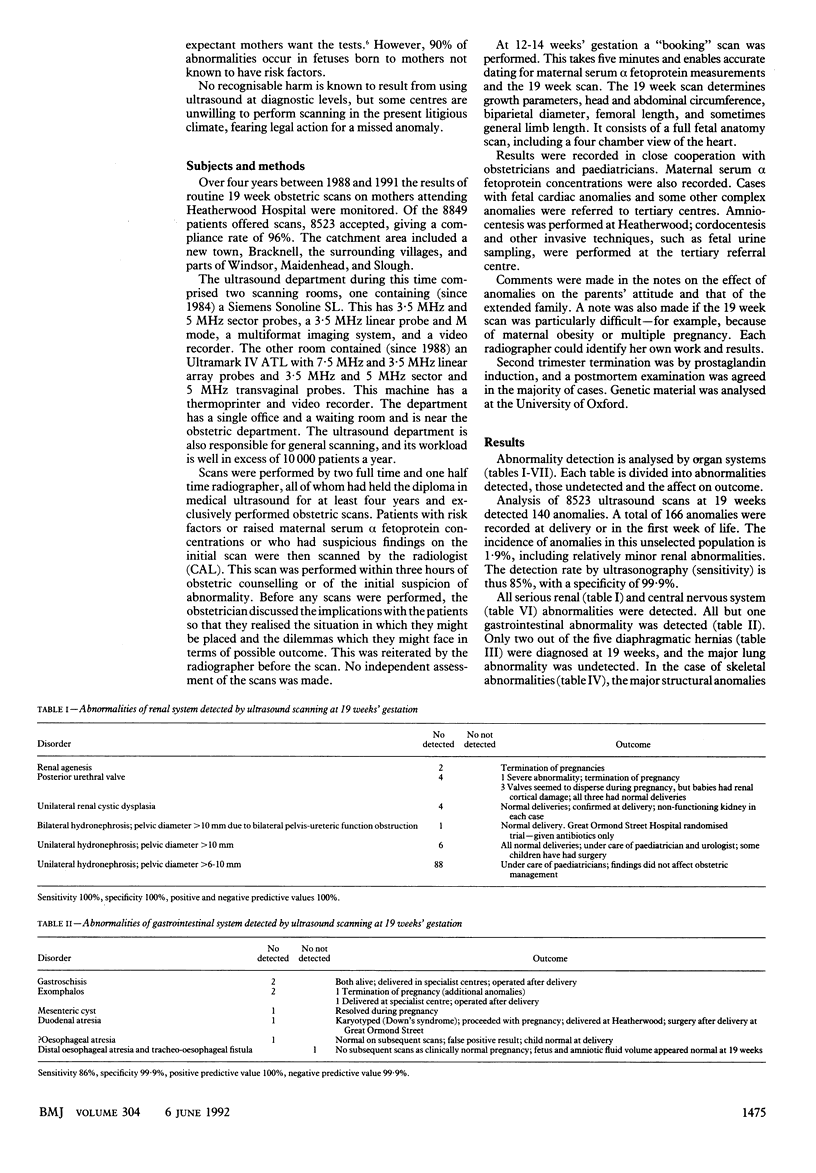
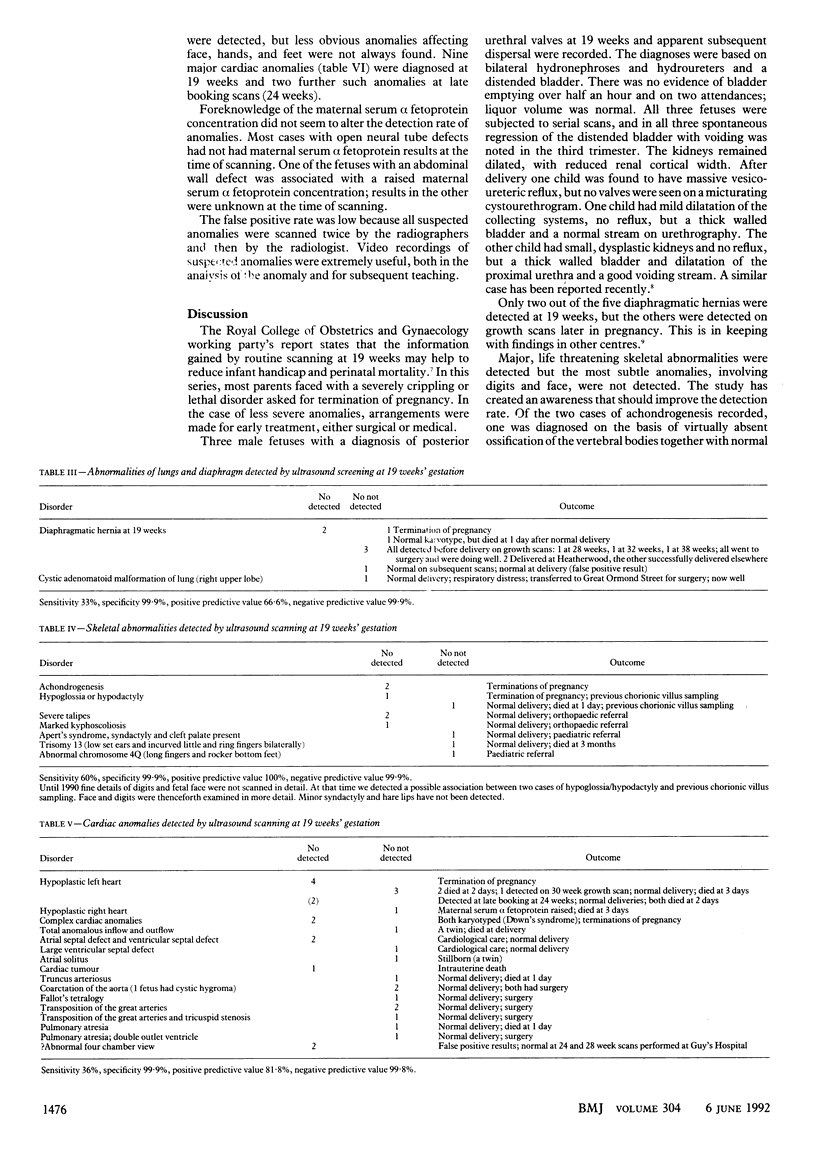
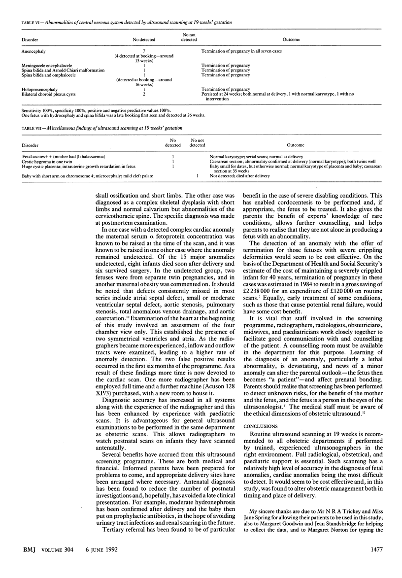
OBJECTIVE--To evaluate the effectiveness of routine ultrasound scanning at 19 weeks' gestation in an unselected population in terms of accuracy of detection of fetal structural abnormality and the effect on obstetric and neonatal care. DESIGN--Prospective study over four years. Scans performed by radiographers with overall supervision by a radiologist. SETTING--Ultrasound department of district general hospital. SUBJECTS--All pregnant women were offered scans; 8523 of 8849 (96%) accepted. MAIN OUTCOME MEASURES--Information obtained from hospital records, genetic analysis, and post-mortem findings. RESULTS--166 fetal anomalies occurred; 140 were detected at 19 weeks (sensitivity 85%; specificity 99.9%). In 27 cases fetuses were shown to have severely crippling or lethal abnormalities; termination of pregnancy was requested in 25. Early diagnosis influenced timing and place of delivery in babies with severe cardiac or gastrointestinal anomalies. CONCLUSION--Scanning at 19 weeks with availability of termination can reduce perinatal morbidity and mortality. Scanning can be performed in a general ultrasound department with adequate counselling facilities and close cooperation between radiographers, midwives, obstetricians, paediatricians, and the radiologist.
Full text
PDF
Selected References
These references are in PubMed. This may not be the complete list of references from this article.
- Benacerraf B. R., Adzick N. S. Fetal diaphragmatic hernia: ultrasound diagnosis and clinical outcome in 19 cases. Am J Obstet Gynecol. 1987 Mar;156(3):573–576. doi: 10.1016/0002-9378(87)90053-6. [DOI] [PubMed] [Google Scholar]
- Campbell S., Pearce J. M. The prenatal diagnosis of fetal structural anomalies by ultrasound. Clin Obstet Gynaecol. 1983 Dec;10(3):475–506. [PubMed] [Google Scholar]
- Chervenak F. A., McCullough L. B. Ethics, an emerging subdiscipline of obstetric ultrasound, and its relevance to the routine obstetric scan. Ultrasound Obstet Gynecol. 1991 Jan 1;1(1):18–20. doi: 10.1046/j.1469-0705.1991.01010018.x. [DOI] [PubMed] [Google Scholar]
- Chitty L. S., Hunt G. H., Moore J., Lobb M. O. Effectiveness of routine ultrasonography in detecting fetal structural abnormalities in a low risk population. BMJ. 1991 Nov 9;303(6811):1165–1169. doi: 10.1136/bmj.303.6811.1165. [DOI] [PMC free article] [PubMed] [Google Scholar]
- Crawford D. C., Chita S. K., Allan L. D. Prenatal detection of congenital heart disease: factors affecting obstetric management and survival. Am J Obstet Gynecol. 1988 Aug;159(2):352–356. doi: 10.1016/s0002-9378(88)80083-8. [DOI] [PubMed] [Google Scholar]
- Hecher K., Henning K., Spernol R., Szalay S. Spontaneous remission of urinary tract obstruction and ascites in a fetus with posterior urethral valves. Ultrasound Obstet Gynecol. 1991 Nov 1;1(6):426–430. doi: 10.1046/j.1469-0705.1991.01060426.x. [DOI] [PubMed] [Google Scholar]