Abstract
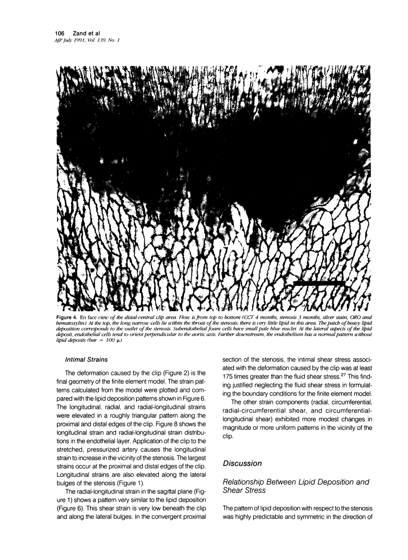
These experiments were designed to study the topography of lipid deposition in the stenotic aorta of hypercholesterolemic rats, and to correlate it with flow conditions and intimal stresses and strains studied in a scale biophysical model and in a computer model. A 69% +/- 5% stenosis was produced with a U-shaped metal clip. One month to 8 months later, the aorta was studied en face by light microscopy after fixation and lipid staining. The intima in the throat of the stenosis was almost completely free of lipid, whereas symmetric lipid deposits occurred as bands just above and especially just below the stenosis; elsewhere lipid deposits appeared to be random. The flow data obtained from the scale model showed that the intima in the throat of the stenosis was subjected to an increase of as much as 20 times in shear stress, whereas the lipid deposits just above and just below the stenosis were associated with asymmetric flow conditions: the proximal area corresponded to a region of rapidly increasing shear stress, the distal area to a region of low to normal shear stress and separated flow. A finite element computer model based on the aortic deformations indicated that the endothelium at the inlet and outlet of the stenosis is subjected to a symmetric pattern of elevated stresses and strains. These results indicate that 1) the pattern of lipid deposition can not be adequately explained by a hypothesis based solely on flow conditions, and 2) lipid deposits can develop in areas of increased fluid shear stress, decreased fluid shear stress, and increased intimal strains.
Full text
PDF


Images in this article
Selected References
These references are in PubMed. This may not be the complete list of references from this article.
- Bomberger R. A., Zarins C. K., Glagov S. Resident research award. Subcritical arterial stenosis enhances distal atherosclerosis. J Surg Res. 1981 Mar;30(3):205–212. doi: 10.1016/0022-4804(81)90149-9. [DOI] [PubMed] [Google Scholar]
- Bomberger R. A., Zarins C. K., Taylor K. E., Glagov S. Effect of hypotension on atherogenesis and aortic wall composition. J Surg Res. 1980 May;28(5):402–409. doi: 10.1016/0022-4804(80)90102-x. [DOI] [PubMed] [Google Scholar]
- Caplan B. A., Schwartz C. J. Increased endothelial cell turnover in areas of in vivo Evans Blue uptake in the pig aorta. Atherosclerosis. 1973 May-Jun;17(3):401–417. doi: 10.1016/0021-9150(73)90031-2. [DOI] [PubMed] [Google Scholar]
- Caro C. G., Fitz-Gerald J. M., Schroter R. C. Atheroma and arterial wall shear. Observation, correlation and proposal of a shear dependent mass transfer mechanism for atherogenesis. Proc R Soc Lond B Biol Sci. 1971 Feb 16;177(1046):109–159. doi: 10.1098/rspb.1971.0019. [DOI] [PubMed] [Google Scholar]
- Cornhill J. F., Roach M. R. A quantitative study of the localization of atherosclerotic lesions in the rabbit aorta. Atherosclerosis. 1976 May-Jun;23(3):489–501. doi: 10.1016/0021-9150(76)90009-5. [DOI] [PubMed] [Google Scholar]
- Davies P. F., Dewey C. F., Jr, Bussolari S. R., Gordon E. J., Gimbrone M. A., Jr Influence of hemodynamic forces on vascular endothelial function. In vitro studies of shear stress and pinocytosis in bovine aortic cells. J Clin Invest. 1984 Apr;73(4):1121–1129. doi: 10.1172/JCI111298. [DOI] [PMC free article] [PubMed] [Google Scholar]
- Davies P. F., Remuzzi A., Gordon E. J., Dewey C. F., Jr, Gimbrone M. A., Jr Turbulent fluid shear stress induces vascular endothelial cell turnover in vitro. Proc Natl Acad Sci U S A. 1986 Apr;83(7):2114–2117. doi: 10.1073/pnas.83.7.2114. [DOI] [PMC free article] [PubMed] [Google Scholar]
- Demiray H., Weizsäcker H. W., Pascale K., Erbay H. A. A stress-strain relation for a rat abdominal aorta. J Biomech. 1988;21(5):369–374. doi: 10.1016/0021-9290(88)90143-1. [DOI] [PubMed] [Google Scholar]
- Dobrin P. B. Biaxial anisotropy of dog carotid artery: estimation of circumferential elastic modulus. J Biomech. 1986;19(5):351–358. doi: 10.1016/0021-9290(86)90011-4. [DOI] [PubMed] [Google Scholar]
- Fox J. A., Hugh A. E. Localization of atheroma: a theory based on boundary layer separation. Br Heart J. 1966 May;28(3):388–399. doi: 10.1136/hrt.28.3.388. [DOI] [PMC free article] [PubMed] [Google Scholar]
- Fry D. L. Acute vascular endothelial changes associated with increased blood velocity gradients. Circ Res. 1968 Feb;22(2):165–197. doi: 10.1161/01.res.22.2.165. [DOI] [PubMed] [Google Scholar]
- Fry D. L. Certain histological and chemical responses of the vascular interface to acutely induced mechanical stress in the aorta of the dog. Circ Res. 1969 Jan;24(1):93–108. doi: 10.1161/01.res.24.1.93. [DOI] [PubMed] [Google Scholar]
- Glagov S., Zarins C., Giddens D. P., Ku D. N. Hemodynamics and atherosclerosis. Insights and perspectives gained from studies of human arteries. Arch Pathol Lab Med. 1988 Oct;112(10):1018–1031. [PubMed] [Google Scholar]
- Grøttum P., Svindland A., Walløe L. Localization of atherosclerotic lesions in the bifurcation of the main left coronary artery. Atherosclerosis. 1983 Apr;47(1):55–62. doi: 10.1016/0021-9150(83)90071-0. [DOI] [PubMed] [Google Scholar]
- Hollander W., Madoff I., Paddock J., Kirkpatrick B. Aggravation of atherosclerosis by hypertension in a subhuman primate model with coarctation of the aorta. Circ Res. 1976 Jun;38(6 Suppl 2):63–72. doi: 10.1161/01.res.38.6.63. [DOI] [PubMed] [Google Scholar]
- Houle S., Roach M. R. Flow studies in a rigid model of an aorto-renal junction. A case for high shear as a cause of the localization of sudanophilic lesions in rabbits. Atherosclerosis. 1981 Nov-Dec;40(3-4):231–244. doi: 10.1016/0021-9150(81)90133-7. [DOI] [PubMed] [Google Scholar]
- Ives C. L., Eskin S. G., McIntire L. V. Mechanical effects on endothelial cell morphology: in vitro assessment. In Vitro Cell Dev Biol. 1986 Sep;22(9):500–507. doi: 10.1007/BF02621134. [DOI] [PubMed] [Google Scholar]
- Joris I., Zand T., Majno G. Hydrodynamic injury of the endothelium in acute aortic stenosis. Am J Pathol. 1982 Mar;106(3):394–408. [PMC free article] [PubMed] [Google Scholar]
- Joris I., Zand T., Nunnari J. J., Krolikowski F. J., Majno G. Studies on the pathogenesis of atherosclerosis. I. Adhesion and emigration of mononuclear cells in the aorta of hypercholesterolemic rats. Am J Pathol. 1983 Dec;113(3):341–358. [PMC free article] [PubMed] [Google Scholar]
- Kjaernes M., Svindland A., Walløe L., Wille S. O. Localization of early atherosclerotic lesions in an arterial bifurcation in humans. Acta Pathol Microbiol Scand A. 1981 Jan;89(1):35–40. doi: 10.1111/j.1699-0463.1981.tb00184.x. [DOI] [PubMed] [Google Scholar]
- Ku D. N., Giddens D. P. Pulsatile flow in a model carotid bifurcation. Arteriosclerosis. 1983 Jan-Feb;3(1):31–39. doi: 10.1161/01.atv.3.1.31. [DOI] [PubMed] [Google Scholar]
- Ku D. N., Giddens D. P., Zarins C. K., Glagov S. Pulsatile flow and atherosclerosis in the human carotid bifurcation. Positive correlation between plaque location and low oscillating shear stress. Arteriosclerosis. 1985 May-Jun;5(3):293–302. doi: 10.1161/01.atv.5.3.293. [DOI] [PubMed] [Google Scholar]
- Langille B. L., Reidy M. A., Kline R. L. Injury and repair of endothelium at sites of flow disturbances near abdominal aortic coarctations in rabbits. Arteriosclerosis. 1986 Mar-Apr;6(2):146–154. doi: 10.1161/01.atv.6.2.146. [DOI] [PubMed] [Google Scholar]
- LoGerfo F. W., Nowak M. D., Quist W. C., Crawshaw H. M., Bharadvaj B. K. Flow studies in a model carotid bifurcation. Arteriosclerosis. 1981 Jul-Aug;1(4):235–241. doi: 10.1161/01.atv.1.4.235. [DOI] [PubMed] [Google Scholar]
- Lyon R. T., Runyon-Hass A., Davis H. R., Glagov S., Zarins C. K. Protection from atherosclerotic lesion formation by reduction of artery wall motion. J Vasc Surg. 1987 Jan;5(1):59–67. [PubMed] [Google Scholar]
- Olesen S. P., Clapham D. E., Davies P. F. Haemodynamic shear stress activates a K+ current in vascular endothelial cells. Nature. 1988 Jan 14;331(6152):168–170. doi: 10.1038/331168a0. [DOI] [PubMed] [Google Scholar]
- Reidy M. A., Bowyer D. E. Scanning electron microscopy of arteries. The morphology of aortic endothelium in haemodynamically stressed areas associated with branches. Atherosclerosis. 1977 Feb;26(2):181–194. doi: 10.1016/0021-9150(77)90101-0. [DOI] [PubMed] [Google Scholar]
- Reidy M. A., Schwartz S. M. Endothelial regeneration. III. Time course of intimal changes after small defined injury to rat aortic endothelium. Lab Invest. 1981 Apr;44(4):301–308. [PubMed] [Google Scholar]
- Repin V. S., Dolgov V. V., Zaikina O. E., Novikov I. D., Antonov A. S., Nikolaeva M. A., Smirnov V. N. Heterogeneity of endothelium in human aorta. A quantitative analysis by scanning electron microscopy. Atherosclerosis. 1984 Jan;50(1):35–52. doi: 10.1016/0021-9150(84)90006-6. [DOI] [PubMed] [Google Scholar]
- Sabbah H. N., Khaja F., Brymer J. F., Hawkins E. T., Stein P. D. Blood velocity in the right coronary artery: relation to the distribution of atherosclerotic lesions. Am J Cardiol. 1984 Apr 1;53(8):1008–1012. doi: 10.1016/0002-9149(84)90627-1. [DOI] [PubMed] [Google Scholar]
- Stehbens W. E. The role of hemodynamics in the pathogenesis of atherosclerosis. Prog Cardiovasc Dis. 1975 Jul-Aug;18(1):89–103. doi: 10.1016/0033-0620(75)90009-2. [DOI] [PubMed] [Google Scholar]
- Svindland A. The localization of sudanophilic and fibrous plaques in the main left coronary bifurcation. Atherosclerosis. 1983 Aug;48(2):139–145. doi: 10.1016/0021-9150(83)90100-4. [DOI] [PubMed] [Google Scholar]
- Thubrikar M. J., Baker J. W., Nolan S. P. Inhibition of atherosclerosis associated with reduction of arterial intramural stress in rabbits. Arteriosclerosis. 1988 Jul-Aug;8(4):410–420. doi: 10.1161/01.atv.8.4.410. [DOI] [PubMed] [Google Scholar]
- Vasile E., Simionescu M., Simionescu N. Visualization of the binding, endocytosis, and transcytosis of low-density lipoprotein in the arterial endothelium in situ. J Cell Biol. 1983 Jun;96(6):1677–1689. doi: 10.1083/jcb.96.6.1677. [DOI] [PMC free article] [PubMed] [Google Scholar]
- Vito R. P., Hickey J. The mechanical properties of soft tissues--II: the elastic response of arterial segments. J Biomech. 1980;13(11):951–957. doi: 10.1016/0021-9290(80)90166-9. [DOI] [PubMed] [Google Scholar]
- Weizsacker H. W., Pinto J. G. Isotropy and anisotropy of the arterial wall. J Biomech. 1988;21(6):477–487. doi: 10.1016/0021-9290(88)90240-0. [DOI] [PubMed] [Google Scholar]
- Zand T., Nunnari J. J., Hoffman A. H., Savilonis B. J., MacWilliams B., Majno G., Joris I. Endothelial adaptations in aortic stenosis. Correlation with flow parameters. Am J Pathol. 1988 Nov;133(2):407–418. [PMC free article] [PubMed] [Google Scholar]
- Zand T., Underwood J. M., Nunnari J. J., Majno G., Joris I. Endothelium and "silver lines". An electron microscopic study. Virchows Arch A Pathol Anat Histol. 1982;395(2):133–144. doi: 10.1007/BF00429607. [DOI] [PubMed] [Google Scholar]
- Zarins C. K., Giddens D. P., Bharadvaj B. K., Sottiurai V. S., Mabon R. F., Glagov S. Carotid bifurcation atherosclerosis. Quantitative correlation of plaque localization with flow velocity profiles and wall shear stress. Circ Res. 1983 Oct;53(4):502–514. doi: 10.1161/01.res.53.4.502. [DOI] [PubMed] [Google Scholar]
- von Maltzahn W. W., Besdo D., Wiemer W. Elastic properties of arteries: a nonlinear two-layer cylindrical model. J Biomech. 1981;14(6):389–397. doi: 10.1016/0021-9290(81)90056-7. [DOI] [PubMed] [Google Scholar]


