

Abstract
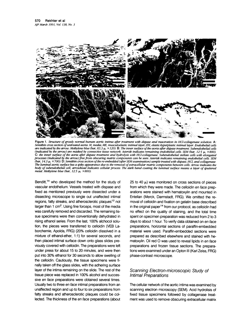
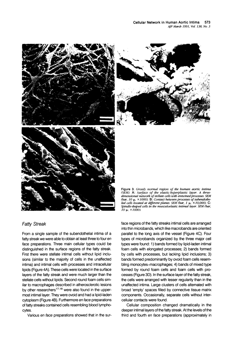
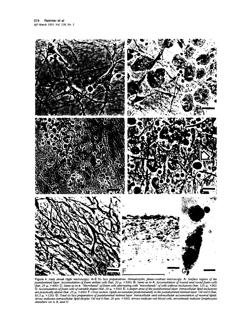
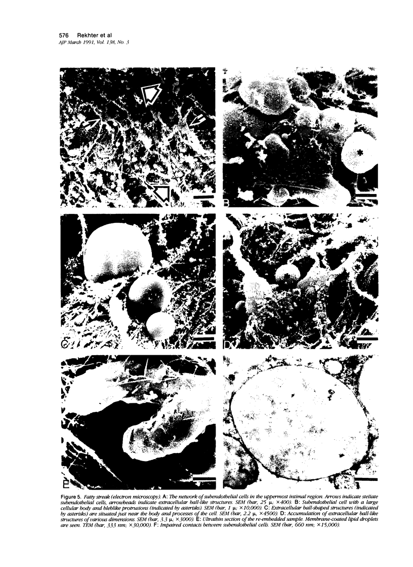
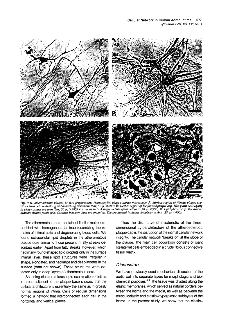
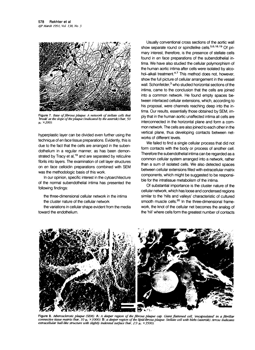
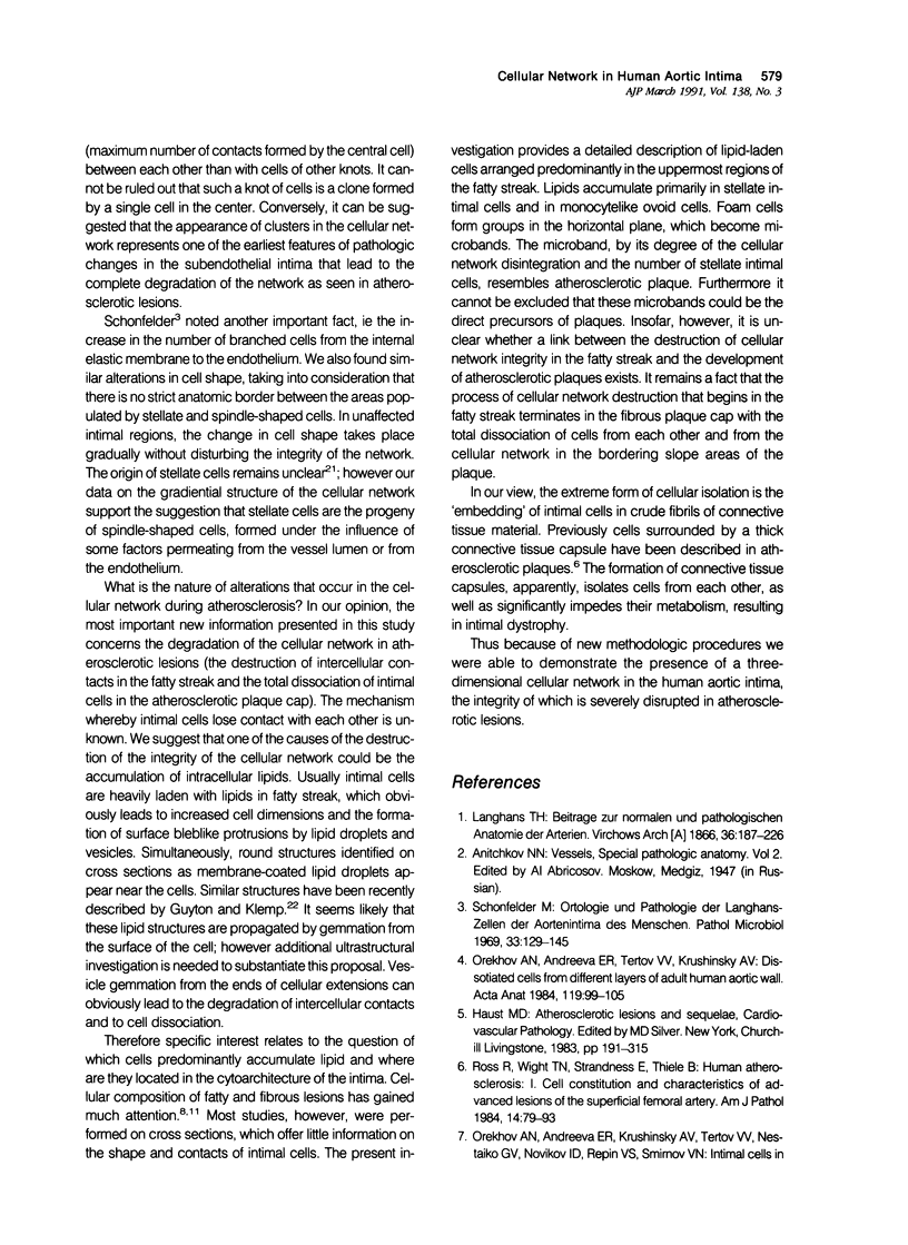
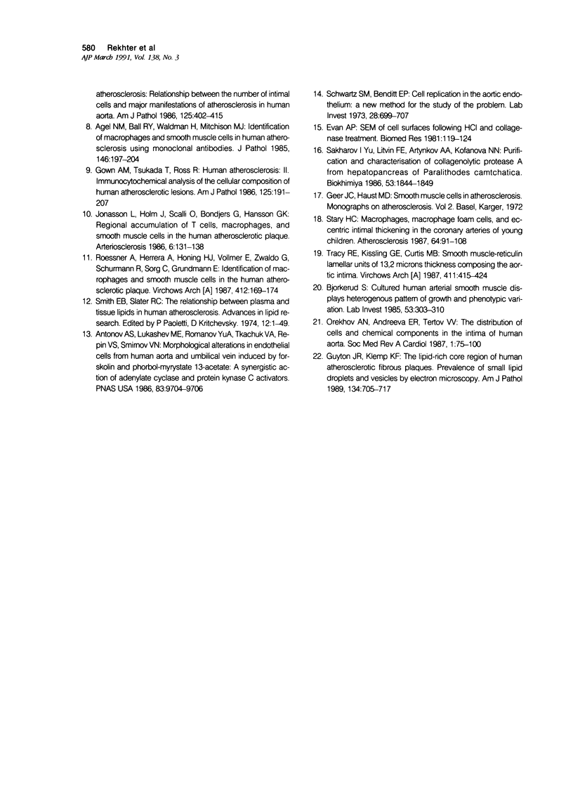
The three-dimensional cytoarchitecture of normal and atherosclerotic intima of human aorta was studied by light microscopy of consecutive en face preparations (Hautchen preparations) and by scanning electron microscopy. In unaffected intima, a three-dimensional network consisting of cells of variable shape and probably origin was demonstrated. Cellular shape changed from predominantly stellate in the luminal regions of the elastic-hyperplastic layer to elongated spindlelike cells in the musculoelastic layer of the intima. In the surface layers of the fatty streak, cellular contacts were severed, and lipid droplets were often seen between cellular processes. Along with stellate and elongated cells, the fatty streak also had a number of round monocytelike cells. Lipid inclusions were usually detected in stellate and ovoid cells. The integrity of the cellular network was preserved at the marginal zone of the atherosclerotic plaque, while at the slopes and in the central part of the plaque, cells practically lost all contact with each other. Giant stellate cells embedded in crude fibrillar connective tissue matrix were often found there. Disintegration of the cellular network during atherosclerosis is suggested to play an important role in the development of various lesions.
Full text
PDF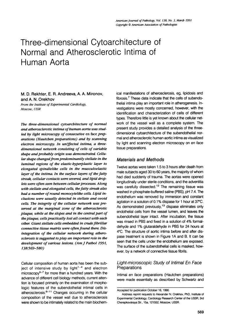
Images in this article
Selected References
These references are in PubMed. This may not be the complete list of references from this article.
- Antonov A. S., Lukashev M. E., Romanov Y. A., Tkachuk V. A., Repin V. S., Smirnov V. N. Morphological alterations in endothelial cells from human aorta and umbilical vein induced by forskolin and phorbol 12-myristate 13-acetate: a synergistic action of adenylate cyclase and protein kinase C activators. Proc Natl Acad Sci U S A. 1986 Dec;83(24):9704–9708. doi: 10.1073/pnas.83.24.9704. [DOI] [PMC free article] [PubMed] [Google Scholar]
- Aqel N. M., Ball R. Y., Waldmann H., Mitchinson M. J. Identification of macrophages and smooth muscle cells in human atherosclerosis using monoclonal antibodies. J Pathol. 1985 Jul;146(3):197–204. doi: 10.1002/path.1711460306. [DOI] [PubMed] [Google Scholar]
- Björkerud S. Cultivated human arterial smooth muscle displays heterogeneous pattern of growth and phenotypic variation. Lab Invest. 1985 Sep;53(3):303–310. [PubMed] [Google Scholar]
- Gown A. M., Tsukada T., Ross R. Human atherosclerosis. II. Immunocytochemical analysis of the cellular composition of human atherosclerotic lesions. Am J Pathol. 1986 Oct;125(1):191–207. [PMC free article] [PubMed] [Google Scholar]
- Guyton J. R., Klemp K. F. The lipid-rich core region of human atherosclerotic fibrous plaques. Prevalence of small lipid droplets and vesicles by electron microscopy. Am J Pathol. 1989 Mar;134(3):705–717. [PMC free article] [PubMed] [Google Scholar]
- Jonasson L., Holm J., Skalli O., Bondjers G., Hansson G. K. Regional accumulations of T cells, macrophages, and smooth muscle cells in the human atherosclerotic plaque. Arteriosclerosis. 1986 Mar-Apr;6(2):131–138. doi: 10.1161/01.atv.6.2.131. [DOI] [PubMed] [Google Scholar]
- Orekhov A. N., Andreeva E. R., Tertov V. V., Krushinsky A. V. Dissociated cells from different layers of adult human aortic wall. Acta Anat (Basel) 1984;119(2):99–105. doi: 10.1159/000145868. [DOI] [PubMed] [Google Scholar]
- Roessner A., Herrera A., Höning H. J., Vollmer E., Zwadlo G., Schürmann R., Sorg C., Grundmann E. Identification of macrophages and smooth muscle cells with monoclonal antibodies in the human atherosclerotic plaque. Virchows Arch A Pathol Anat Histopathol. 1987;412(2):169–174. doi: 10.1007/BF00716190. [DOI] [PubMed] [Google Scholar]
- Ross R., Wight T. N., Strandness E., Thiele B. Human atherosclerosis. I. Cell constitution and characteristics of advanced lesions of the superficial femoral artery. Am J Pathol. 1984 Jan;114(1):79–93. [PMC free article] [PubMed] [Google Scholar]
- Schwartz S. M., Benditt E. P. Cell replication in the aortic endothelium: a new method for study of the problem. Lab Invest. 1973 Jun;28(6):699–707. [PubMed] [Google Scholar]
- Schönfelder M. Orthologie und Pathologie der Langhans-Zellen der Aortenintima des Menschen. Pathol Microbiol (Basel) 1969;33(3):129–145. [PubMed] [Google Scholar]
- Smith E. B. The relationship between plasma and tissue lipids in human atherosclerosis. Adv Lipid Res. 1974;12(0):1–49. doi: 10.1016/b978-0-12-024912-1.50008-9. [DOI] [PubMed] [Google Scholar]
- Stary H. C. Macrophages, macrophage foam cells, and eccentric intimal thickening in the coronary arteries of young children. Atherosclerosis. 1987 Apr;64(2-3):91–108. doi: 10.1016/0021-9150(87)90234-6. [DOI] [PubMed] [Google Scholar]
- Tracy R. E., Kissling G. E., Curtis M. B. Smooth muscle cell-reticulin lamellar units of 13.2 microns thickness composing the aortic intima. Virchows Arch A Pathol Anat Histopathol. 1987;411(5):415–424. doi: 10.1007/BF00735222. [DOI] [PubMed] [Google Scholar]