

Abstract
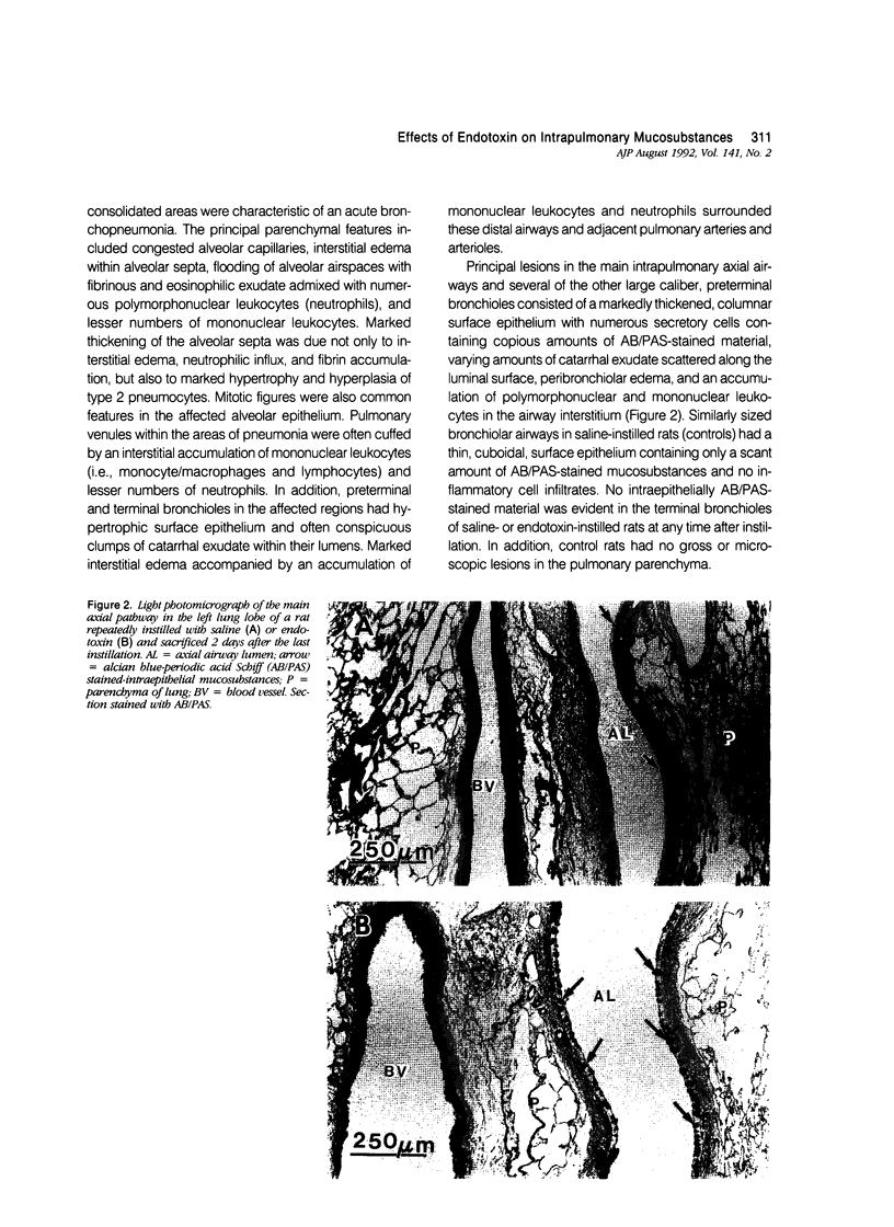
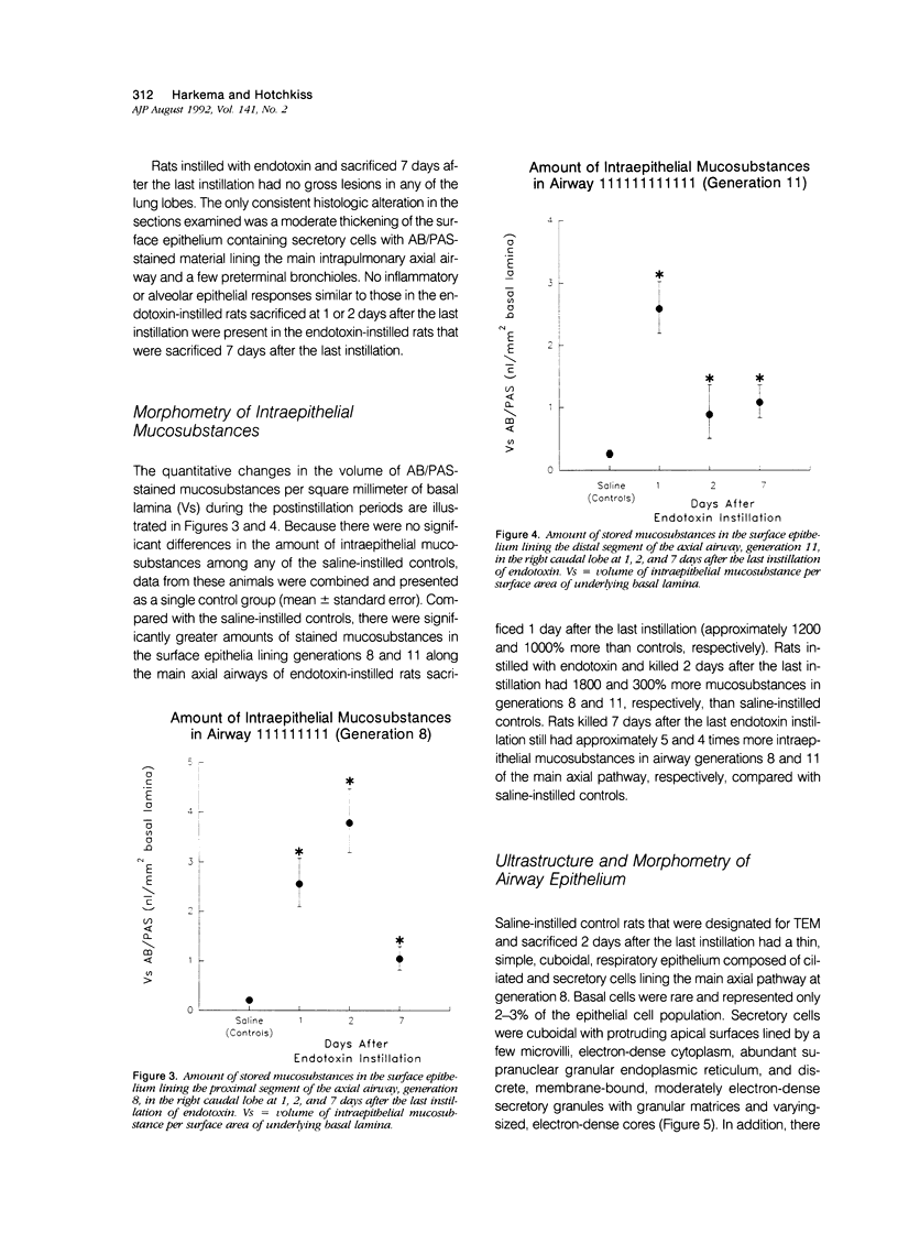
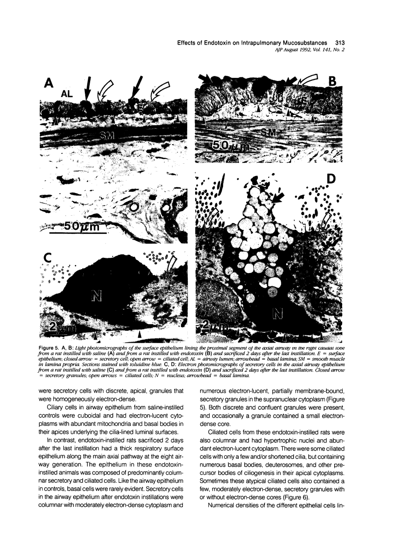
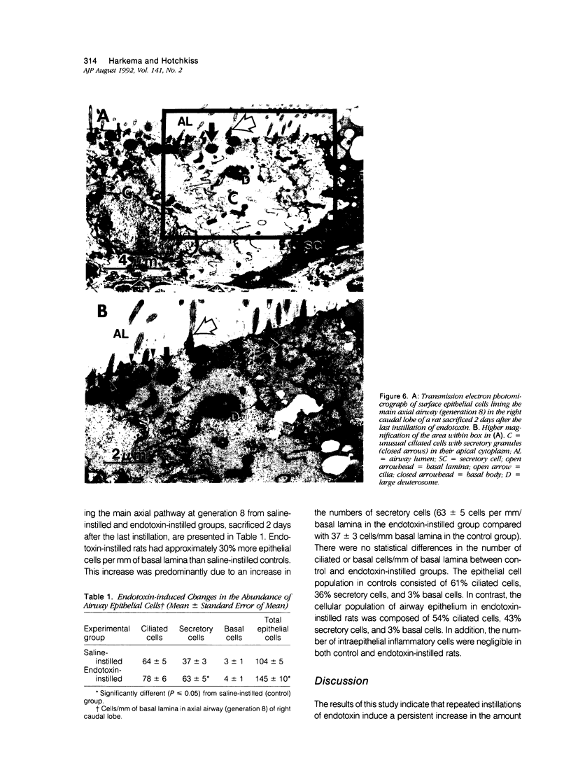
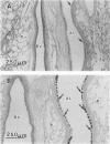
Bacteria-induced bronchopneumonias are often characterized by an influx of neutrophils and excess mucus in pulmonary airways. This study determined how endotoxin, a component of gram-negative bacteria and a potent inflammatory agent, affects the ultrastructure of the mucociliary apparatus and the amount of stored intraepithelial mucosubstances in the main axial airways within the lung. Rats were intranasally instilled, once a day for 3 days, with endotoxin or saline (controls). Animals were sacrificed 1, 2, or 7 days after the last instillation. Microdissected intrapulmonary axial airways (generations 8-11) from the right caudal lobes of infusion-fixed lungs were processed for light and electron microscopy. Morphometric techniques were used to determine the volume densities (Vs) of histochemically stained intraepithelial mucosubstances and numerical densities of airway epithelial cells. There were marked increases, compared with controls, in the amount of intraepithelial mucosubstances in the intrapulmonary axial airways at generations 8 and 11 in the right caudal lobes from endotoxin-instilled rats sacrificed 1, 2, and 7 days after the last instillation. There were significantly greater numbers of surface epithelial cells per length of basal lamina (i.e., hyperplasia) in endotoxin-exposed airways compared with airways from controls. This endotoxin-induced hyperplasia was due primarily to an increase in the number of mucus-secretory cells, which in endotoxin-exposed epithelium were columnar and contained numerous, large confluent, electronlucent, secretory granules composed of acidic and neutral glycoproteins. In contrast, secretory cells in airway epithelium from controls were cuboidal and contained small discrete, electron-dense, granules composed of only neutral glycoproteins. The numbers of ciliated cells and basal cells were similar in both control and endotoxin-exposed epithelium. Only endotoxin-exposed epithelium, however, contained atypical epithelial cells with numerous basal bodies, few cilia, and few apical secretory granules. These results indicate that repeated airway instillations of endotoxin induce an increase in the amount of intraepithelial mucosubstances, secretory cell hyperplasia, and excess luminal mucus in pulmonary airways. Therefore, endotoxin released from gram-negative bacteria may be partially responsible for the structural alterations, in the airway surface epithelium, which result in the excess luminal mucus observed in bacteria-induced bronchopneumonias.
Full text
PDF
Images in this article
Selected References
These references are in PubMed. This may not be the complete list of references from this article.
- Colditz I. G., Movat H. Z. Kinetics of neutrophil accumulation in acute inflammatory lesions induced by chemotaxins and chemotaxinigens. J Immunol. 1984 Oct;133(4):2169–2173. [PubMed] [Google Scholar]
- De Haller R., Reid L. Adult chronic bronchitis. Morphology, histochemistry and vascularisation of the bronchial mucous glands. Med Thorac. 1965;22(6):549–567. [PubMed] [Google Scholar]
- Flaherty D. K., Deck F. H., Cooper J., Bishop K., Winzenburger P. A., Smith L. R., Bynum L., Witmer W. B. Bacterial endotoxin isolated from a water spray air humidification system as a putative agent of occupation-related lung disease. Infect Immun. 1984 Jan;43(1):206–212. doi: 10.1128/iai.43.1.206-212.1984. [DOI] [PMC free article] [PubMed] [Google Scholar]
- GOCO R. V., KRESS M. B., BRANTIGAN O. C. Comparison of mucus glands in the tracheobronchial tree of man and animals. Ann N Y Acad Sci. 1963 Mar 30;106:555–571. doi: 10.1111/j.1749-6632.1963.tb16665.x. [DOI] [PubMed] [Google Scholar]
- Harkema J. R., Hotchkiss J. A., Harmsen A. G., Henderson R. F. In vivo effects of transient neutrophil influx on nasal respiratory epithelial mucosubstances. Quantitative histochemistry. Am J Pathol. 1988 Mar;130(3):605–615. [PMC free article] [PubMed] [Google Scholar]
- Harkema J. R., Hotchkiss J. A., Henderson R. F. Effects of 0.12 and 0.80 ppm ozone on rat nasal and nasopharyngeal epithelial mucosubstances: quantitative histochemistry. Toxicol Pathol. 1989;17(3):525–535. doi: 10.1177/019262338901700307. [DOI] [PubMed] [Google Scholar]
- Harkema J. R., Plopper C. G., Hyde D. M., St George J. A., Dungworth D. L. Effects of an ambient level of ozone on primate nasal epithelial mucosubstances. Quantitative histochemistry. Am J Pathol. 1987 Apr;127(1):90–96. [PMC free article] [PubMed] [Google Scholar]
- Harkema J. R., Plopper C. G., Hyde D. M., St George J. A. Regional differences in quantities of histochemically detectable mucosubstances in nasal, paranasal, and nasopharyngeal epithelium of the bonnet monkey. J Histochem Cytochem. 1987 Mar;35(3):279–286. doi: 10.1177/35.3.2434556. [DOI] [PubMed] [Google Scholar]
- Hotchkiss J. A., Harkema J. R., Henderson R. F. Effect of cumulative ozone exposure on ozone-induced nasal epithelial hyperplasia and secretory metaplasia in rats. Exp Lung Res. 1991 May-Jun;17(3):589–600. doi: 10.3109/01902149109062867. [DOI] [PubMed] [Google Scholar]
- Hudson A. R., Kilburn K. H., Halprin G. M., McKenzie W. N. Granulocyte recruitment to airways exposed to endotoxin aerosols. Am Rev Respir Dis. 1977 Jan;115(1):89–95. doi: 10.1164/arrd.1977.115.1.89. [DOI] [PubMed] [Google Scholar]
- Issekutz A. C., Bhimji S. Role for endotoxin in the leukocyte infiltration accompanying Escherichia coli inflammation. Infect Immun. 1982 May;36(2):558–566. doi: 10.1128/iai.36.2.558-566.1982. [DOI] [PMC free article] [PubMed] [Google Scholar]
- Jeffery P. K., Ayers M., Rogers D. The mechanisms and control of bronchial mucous cell hyperplasia. Adv Exp Med Biol. 1982;144:399–409. doi: 10.1007/978-1-4615-9254-9_62. [DOI] [PubMed] [Google Scholar]
- Jeffery P. K. Morphologic features of airway surface epithelial cells and glands. Am Rev Respir Dis. 1983 Aug;128(2 Pt 2):S14–S20. doi: 10.1164/arrd.1983.128.2P2.S14. [DOI] [PubMed] [Google Scholar]
- Jones R., Reid L. Beta-agonists and secretory cell number and intracellular glycoproteins in airway epithelium. The effect of isoproterenol and salbutamol. Am J Pathol. 1979 May;95(2):407–421. [PMC free article] [PubMed] [Google Scholar]
- Jones R., Reid L. Secretory cell hyperplasia and modification of intracellular glycoprotein in rat airways induced by short periods of exposure to tobacco smoke, and the effect of the antiinflammatory agent phenylmethyloxadiazole. Lab Invest. 1978 Jul;39(1):41–49. [PubMed] [Google Scholar]
- Keenan K. P., Combs J. W., McDowell E. M. Regeneration of hamster tracheal epithelium after mechanical injury. I. Focal lesions: quantitative morphologic study of cell proliferation. Virchows Arch B Cell Pathol Incl Mol Pathol. 1982;41(3):193–214. doi: 10.1007/BF02890281. [DOI] [PubMed] [Google Scholar]
- Keenan K. P., Combs J. W., McDowell E. M. Regeneration of hamster tracheal epithelium after mechanical injury. II. Multifocal lesions: stathmokinetic and autoradiographic studies of cell proliferation. Virchows Arch B Cell Pathol Incl Mol Pathol. 1982;41(3):215–229. doi: 10.1007/BF02890282. [DOI] [PubMed] [Google Scholar]
- Keenan K. P., Combs J. W., McDowell E. M. Regeneration of hamster tracheal epithelium after mechanical injury. III. Large and small lesions: comparative stathmokinetic and single pulse and continuous thymidine labeling autoradiographic studies. Virchows Arch B Cell Pathol Incl Mol Pathol. 1982;41(3):231–252. [PubMed] [Google Scholar]
- LEV R., SPICER S. S. A HISTOCHEMICAL COMPARISON OF HUMAN EPITHELIAL MUCINS IN NORMAL AND IN HYPERSECRETORY STATES INCLUDING PANCREATIC CYSTIC FIBROSIS. Am J Pathol. 1965 Jan;46:23–47. [PMC free article] [PubMed] [Google Scholar]
- Lamb D., Reid L. Mitotic rates, goblet cell increase and histochemical changes in mucus in rat bronchial epithelium during exposure to sulphur dioxide. J Pathol Bacteriol. 1968 Jul;96(1):97–111. doi: 10.1002/path.1700960111. [DOI] [PubMed] [Google Scholar]
- Lamb D., Reid L. The tracheobronchial submucosal glands in cystic fibrosis: a qualitative and quantitative histochemical study. Br J Dis Chest. 1972 Oct;66(4):239–247. doi: 10.1016/0007-0971(72)90042-3. [DOI] [PubMed] [Google Scholar]
- McDowell E. M., Barrett L. A., Glavin F., Harris C. C., Trump B. F. The respiratory epithelium. I. Human bronchus. J Natl Cancer Inst. 1978 Aug;61(2):539–549. [PubMed] [Google Scholar]
- McDowell E. M., Becci P. J., Schürch W., Trump B. F. The respiratory epithelium. VII. Epidermoid metaplasia of hamster tracheal epithelium during regeneration following mechanical injury. J Natl Cancer Inst. 1979 Apr;62(4):995–1008. [PubMed] [Google Scholar]
- Michel O., Duchateau J., Sergysels R. Effect of inhaled endotoxin on bronchial reactivity in asthmatic and normal subjects. J Appl Physiol (1985) 1989 Mar;66(3):1059–1064. doi: 10.1152/jappl.1989.66.3.1059. [DOI] [PubMed] [Google Scholar]
- Muittari A., Rylander R., Salkinoja-Salonen M. Endotoxin and bath-water fever. Lancet. 1980 Jul 12;2(8185):89–89. doi: 10.1016/s0140-6736(80)92965-7. [DOI] [PubMed] [Google Scholar]
- PERNIS B., VIGLIANI E. C., CAVAGNA C., FINULLI M. The role of bacterial endotoxins in occupational diseases caused by inhaling vegetable dusts. Br J Ind Med. 1961 Apr;18:120–129. doi: 10.1136/oem.18.2.120. [DOI] [PMC free article] [PubMed] [Google Scholar]
- PETERSON R. D., WICKLUNDS P. E., GOOD R. A. ENDOTOXIN ACTIVITY OF A HOUSE DUST EXTRACT. J Allergy. 1964 Mar-Apr;35:134–142. doi: 10.1016/0021-8707(64)90027-9. [DOI] [PubMed] [Google Scholar]
- Phalen R. F., Yeh H. C., Schum G. M., Raabe O. G. Application of an idealized model to morphometry of the mammalian tracheobronchial tree. Anat Rec. 1978 Feb;190(2):167–176. doi: 10.1002/ar.1091900202. [DOI] [PubMed] [Google Scholar]
- Pier G. B. Pulmonary disease associated with Pseudomonas aeruginosa in cystic fibrosis: current status of the host-bacterium interaction. J Infect Dis. 1985 Apr;151(4):575–580. doi: 10.1093/infdis/151.4.575. [DOI] [PubMed] [Google Scholar]
- Plopper C. G., Mariassy A. T., Lollini L. O. Structure as revealed by airway dissection. A comparison of mammalian lungs. Am Rev Respir Dis. 1983 Aug;128(2 Pt 2):S4–S7. doi: 10.1164/arrd.1983.128.2P2.S4. [DOI] [PubMed] [Google Scholar]
- Reyes M. P. The aerobic gram-negative bacillary pneumonias. Med Clin North Am. 1980 May;64(3):363–383. doi: 10.1016/s0025-7125(16)31598-x. [DOI] [PubMed] [Google Scholar]
- Reynolds H. Y., Merrill W. W. Airway changes in young smokers that may antedate chronic obstructive lung disease. Med Clin North Am. 1981 May;65(3):667–689. doi: 10.1016/s0025-7125(16)31518-8. [DOI] [PubMed] [Google Scholar]
- Rylander R., Haglind P. Airborne endotoxins and humidifier disease. Clin Allergy. 1984 Jan;14(1):109–112. doi: 10.1111/j.1365-2222.1984.tb02197.x. [DOI] [PubMed] [Google Scholar]
- Rylander R., Nordstrand A. Pulmonary cell reactions after exposure to cotton dust extract. Br J Ind Med. 1974 Jul;31(3):220–223. doi: 10.1136/oem.31.3.220. [DOI] [PMC free article] [PubMed] [Google Scholar]
- Tillotson J. R., Lerner A. M. Pneumonias caused by gram negative bacilli. Medicine (Baltimore) 1966 Jan;45(1):65–76. doi: 10.1097/00005792-196601000-00003. [DOI] [PubMed] [Google Scholar]
- Wilson D. W., Plopper C. G., Dungworth D. L. The response of the macaque tracheobronchial epithelium to acute ozone injury. A quantitative ultrastructural and autoradiographic study. Am J Pathol. 1984 Aug;116(2):193–206. [PMC free article] [PubMed] [Google Scholar]