Abstract
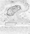
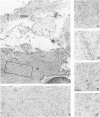
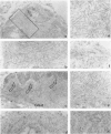
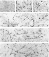
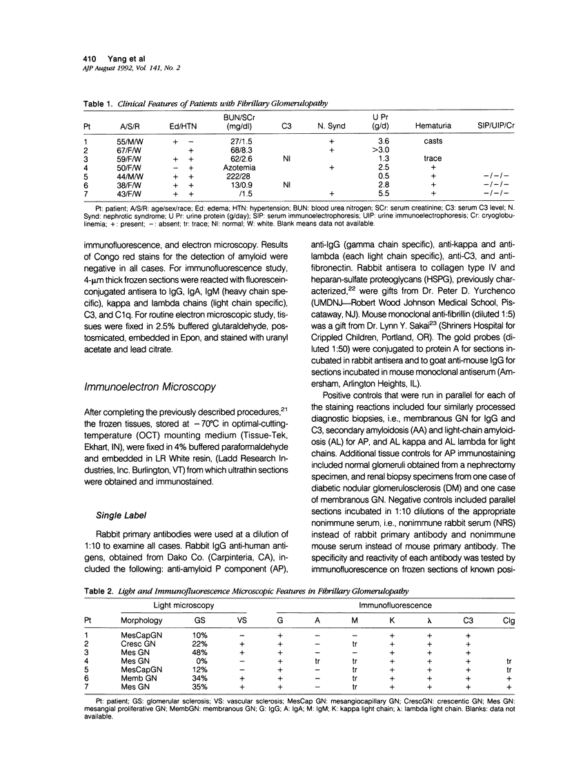
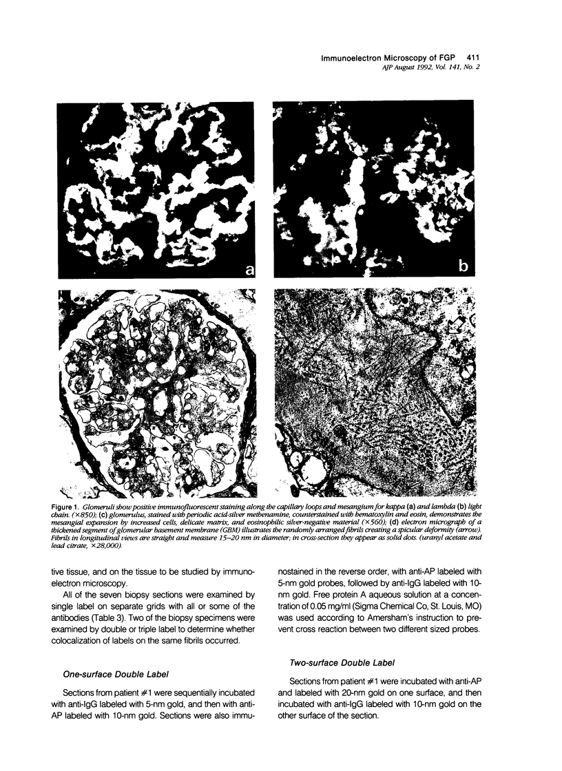
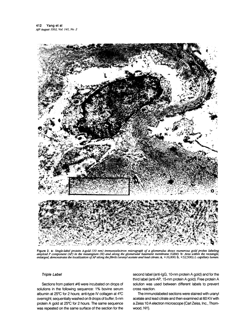
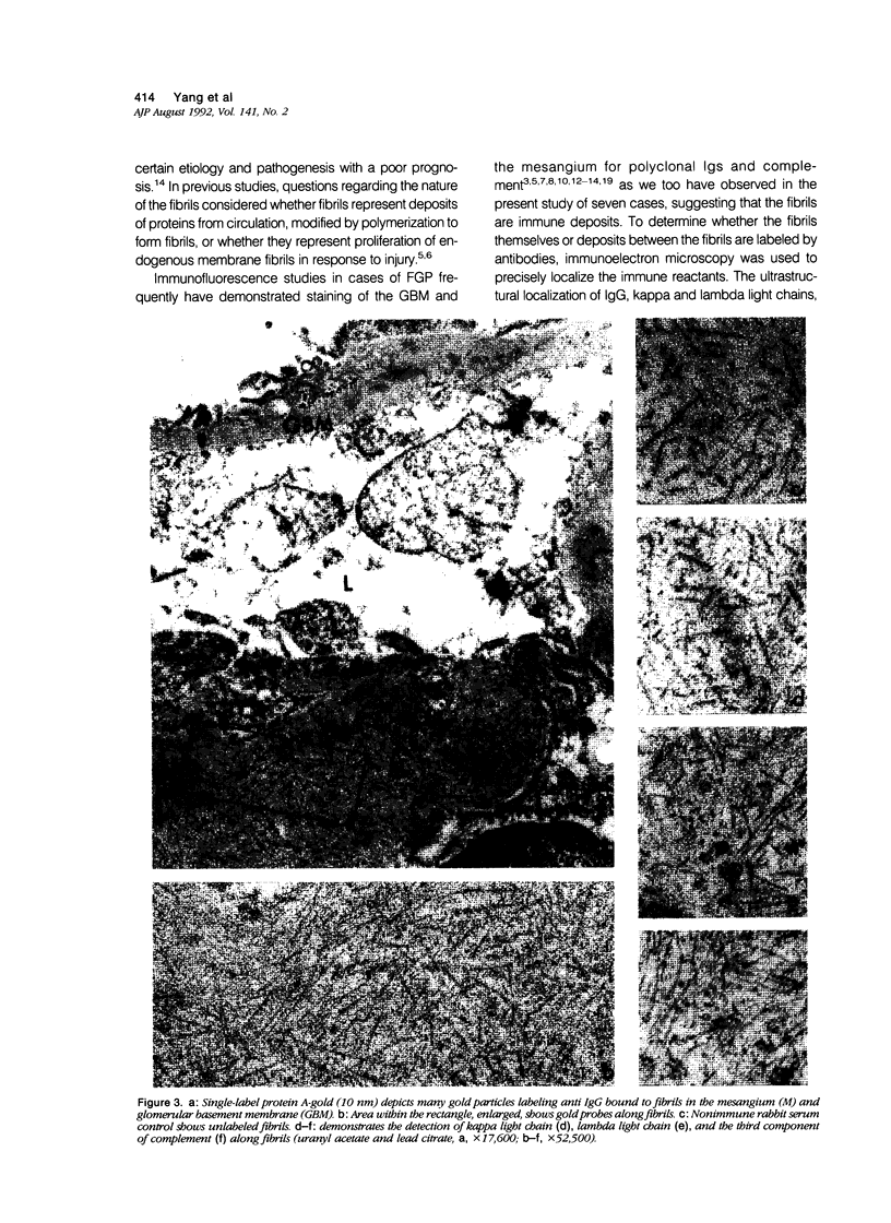
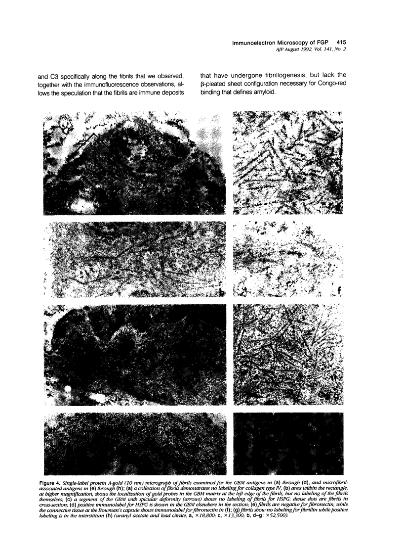
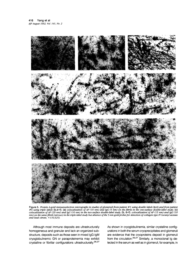
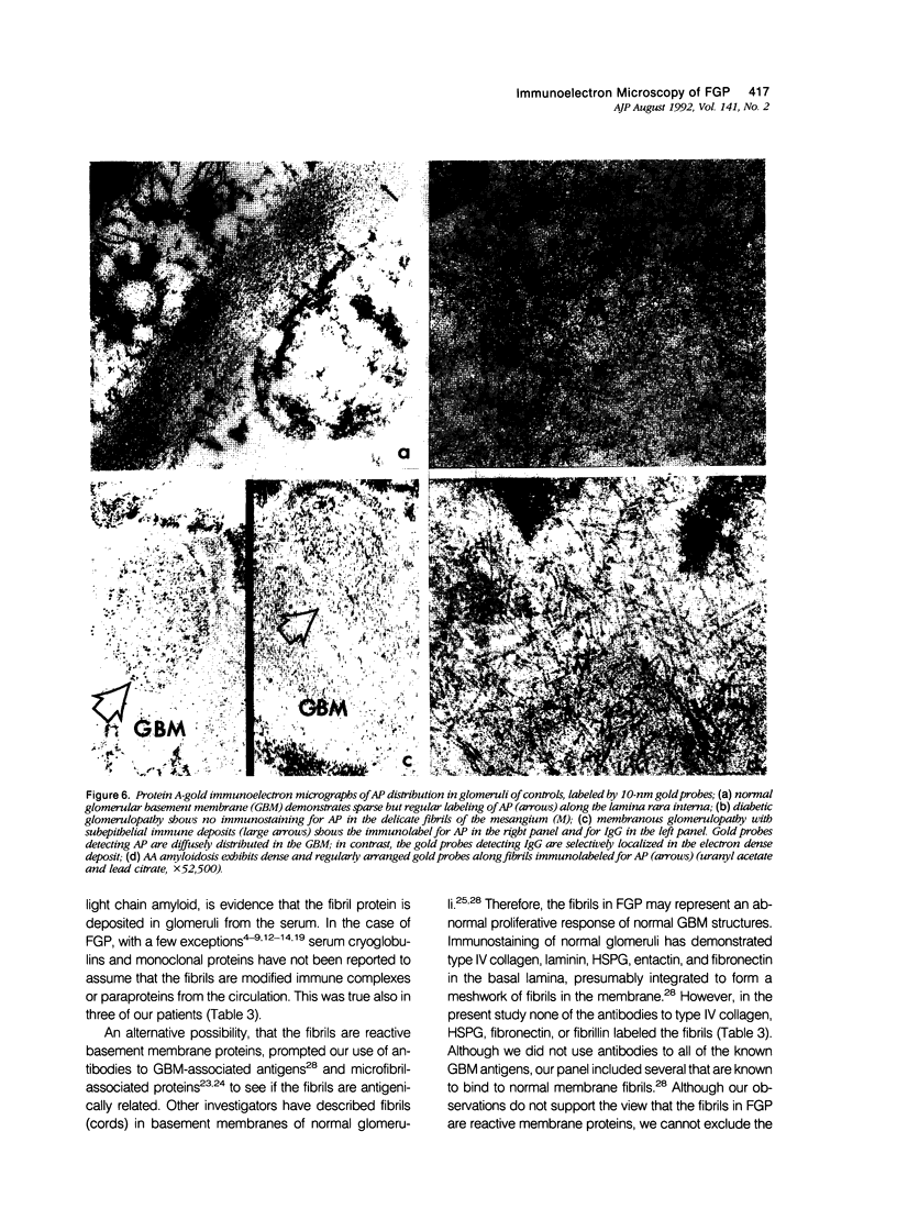
Renal biopsies from seven patients with Congo red-negative amyloid-like fibrillary glomerulopathy (FGP) were examined by protein A gold immuno-electron microscopy. Ultrastructurally, the fibrils in all cases exhibited positive immunostaining for IgG, both Ig light chains, C3, and amyloid P component (AP), but did not show positive immunostaining for glomerular basement membrane (GBM)-associated proteins (collagen type IV and heparan-sulfate proteoglycans) or microfibril-associated proteins (fibronectin and fibrillin). In a triple-label study, AP and IgG were colocalized along the same fibril, whereas the gold probes for the detection of collagen type IV were absent. The results suggest that the fibrils are comprised of polyclonal IgG and C3 that bind AP. AP was immunolocalized sparsely but regularly along the lamina rara interna of normal GBM. AP was absent in the fibrils in a case of diabetic glomerulopathy, was scattered randomly without specificity for the electron-dense deposits in the GBM of membranous glomerulopathy, and lined up regularly along the fibrils in amyloid deposits. FGP is an entity in which the fibrils bind AP but lack the beta-pleated sheet structure necessary for Congo red staining that is typical of amyloid.
Full text
PDF
Images in this article
Selected References
These references are in PubMed. This may not be the complete list of references from this article.
- Alpers C. E., Rennke H. G., Hopper J., Jr, Biava C. G. Fibrillary glomerulonephritis: an entity with unusual immunofluorescence features. Kidney Int. 1987 Mar;31(3):781–789. doi: 10.1038/ki.1987.66. [DOI] [PubMed] [Google Scholar]
- Bakir A. A., Ainis H., Rhee H. L. Nonamyloidotic fibrillar glomerulopathy and recurrent gestational anasarca. Am J Nephrol. 1990;10(4):333–338. doi: 10.1159/000168129. [DOI] [PubMed] [Google Scholar]
- Baltz M. L., De Beer F. C., Feinstein A., Pepys M. B. Calcium-dependent aggregation of human serum amyloid P component. Biochim Biophys Acta. 1982 Feb 18;701(2):229–236. doi: 10.1016/0167-4838(82)90118-2. [DOI] [PubMed] [Google Scholar]
- Breathnach S. M., Melrose S. M., Bhogal B., de Beer F. C., Dyck R. F., Tennent G., Black M. M., Pepys M. B. Amyloid P component is located on elastic fibre microfibrils in normal human tissue. Nature. 1981 Oct 22;293(5834):652–654. doi: 10.1038/293652a0. [DOI] [PubMed] [Google Scholar]
- Bürgin M., Hofmann E., Reutter F. W., Gürtler B. A., Matter L., Briner J., Gloor F. Familial glomerulopathy with giant fibrillar deposits. Virchows Arch A Pathol Anat Histol. 1980;388(3):313–326. doi: 10.1007/BF00430861. [DOI] [PubMed] [Google Scholar]
- Cordonnier D., Martin H., Groslambert P., Micouin C., Chenais F., Stoebner P. Mixed IgG-IgM cryoglobulinemia with glomerulonephritis. Immunochemical, fluorescent and ultrastructural study of kidney and in vitro cryoprecipitate. Am J Med. 1975 Dec;59(6):867–872. doi: 10.1016/0002-9343(75)90480-5. [DOI] [PubMed] [Google Scholar]
- Coria F., Castaño E., Prelli F., Larrondo-Lillo M., van Duinen S., Shelanski M. L., Frangione B. Isolation and characterization of amyloid P component from Alzheimer's disease and other types of cerebral amyloidosis. Lab Invest. 1988 Apr;58(4):454–458. [PubMed] [Google Scholar]
- Devaney K., Sabnis S. G., Antonovych T. T. Nonamyloidotic fibrillary glomerulopathy, immunotactoid glomerulopathy, and the differential diagnosis of filamentous glomerulopathies. Mod Pathol. 1991 Jan;4(1):36–45. [PubMed] [Google Scholar]
- Duffy J. L., Khurana E., Susin M., Gomez-Leon G., Churg J. Fibrillary renal deposits and nephritis. Am J Pathol. 1983 Dec;113(3):279–290. [PMC free article] [PubMed] [Google Scholar]
- Dyck R. F., Lockwood C. M., Kershaw M., McHugh N., Duance V. C., Baltz M. L., Pepys M. B. Amyloid P-component is a constituent of normal human glomerular basement membrane. J Exp Med. 1980 Nov 1;152(5):1162–1174. doi: 10.1084/jem.152.5.1162. [DOI] [PMC free article] [PubMed] [Google Scholar]
- Esparza A. R., Chazan J. A., Nayak R. N., Cavallo T. Fibrillary (immunotactoid) glomerulopathy. A possible role for kappa light chain in its etiology and/or pathogenesis. Am J Surg Pathol. 1991 Jul;15(7):632–643. doi: 10.1097/00000478-199107000-00004. [DOI] [PubMed] [Google Scholar]
- FARQUHAR M. G., WISSIG S. L., PALADE G. E. Glomerular permeability. I. Ferritin transfer across the normal glomerular capillary wall. J Exp Med. 1961 Jan 1;113:47–66. doi: 10.1084/jem.113.1.47. [DOI] [PMC free article] [PubMed] [Google Scholar]
- Feiner H., Gallo G. Ultrastructure in glomerulonephritis associated with cryoglobulinemia. A report of six cases and review of the literature. Am J Pathol. 1977 Jul;88(1):145–162. [PMC free article] [PubMed] [Google Scholar]
- Griffel B., Bernheim J. Glomerular deposits in idiopathic membranous glomerulopathy. Arch Pathol Lab Med. 1980 Jan;104(1):56–57. [PubMed] [Google Scholar]
- Hsu H. C., Churg J. Glomerular microfibrils in renal disease: a comparative electron microscopic study. Kidney Int. 1979 Oct;16(4):497–504. doi: 10.1038/ki.1979.155. [DOI] [PubMed] [Google Scholar]
- Inoue S., Leblond C. P., Rico P., Grant D. Association of fibronectin with the microfibrils of connective tissue. Am J Anat. 1989 Sep;186(1):43–54. doi: 10.1002/aja.1001860104. [DOI] [PubMed] [Google Scholar]
- Inoue S., Leblond C. P. The basement-membrane-like matrix of the mouse EHS tumor: III. Immunodetection of the amyloid P component in basotubules. Am J Anat. 1985 Dec;174(4):399–407. doi: 10.1002/aja.1001740404. [DOI] [PubMed] [Google Scholar]
- Inoué S., Leblond C. P., Grant D. S., Rico P. The microfibrils of connective tissue: II. Immunohistochemical detection of the amyloid P component. Am J Anat. 1986 Jun;176(2):139–152. doi: 10.1002/aja.1001760204. [DOI] [PubMed] [Google Scholar]
- Kobayashi Y., Fujii K., Kurokawa A., Kamiyama M., Miyazawa H. Glomerulonephropathy with amyloid-stain-negative microfibrillar glomerular deposits. Nephron. 1988;48(1):33–39. doi: 10.1159/000184865. [DOI] [PubMed] [Google Scholar]
- Korbet S. M., Schwartz M. M., Lewis E. J. Immunotactoid glomerulopathy. Am J Kidney Dis. 1991 Mar;17(3):247–257. doi: 10.1016/s0272-6386(12)80469-0. [DOI] [PubMed] [Google Scholar]
- Korbet S. M., Schwartz M. M., Rosenberg B. F., Sibley R. K., Lewis E. J. Immunotactoid glomerulopathy. Medicine (Baltimore) 1985 Jul;64(4):228–243. doi: 10.1097/00005792-198507000-00004. [DOI] [PubMed] [Google Scholar]
- Laurie G. W., Leblond C. P., Inoue S., Martin G. R., Chung A. Fine structure of the glomerular basement membrane and immunolocalization of five basement membrane components to the lamina densa (basal lamina) and its extensions in both glomeruli and tubules of the rat kidney. Am J Anat. 1984 Apr;169(4):463–481. doi: 10.1002/aja.1001690408. [DOI] [PubMed] [Google Scholar]
- Mazzucco G., Casanova S., Donini U., Rollino C., Zucchelli P., Piccoli G., Monga G. Glomerulonephritis with organized deposits: a new clinicopathological entity? Light-, electron-microscopic and immunofluorescence study of 12 cases. Am J Nephrol. 1990;10(1):21–30. doi: 10.1159/000168049. [DOI] [PubMed] [Google Scholar]
- Neill J., Rubin J. Nonamyloidotic fibrillary glomerulopathy. Arch Pathol Lab Med. 1989 May;113(5):553–555. [PubMed] [Google Scholar]
- Olesnicky L., Doty S. B., Bertani T., Pirani C. L. Tubular microfibrils in the glomeruli of membranous nephropathy. Arch Pathol Lab Med. 1984 Nov;108(11):902–905. [PubMed] [Google Scholar]
- Olsen S., Bohman S. O., Petersen V. P. Ultrastructure of the glomerular basement membrane in long term renal allografts with transplant glomerular disease. Lab Invest. 1974 Feb;30(2):176–189. [PubMed] [Google Scholar]
- Panner B. J. Rapidly progressive glomerulonephritis and possible amyloidosis. Arch Pathol Lab Med. 1980 Nov;104(11):603–609. [PubMed] [Google Scholar]
- Pinteric L., Painter R. H. Electron microscopy of serum amyloid protein in the presence of calcium; alternative forms of assembly of pentagonal molecules in two-dimensional lattices. Can J Biochem. 1979 Jun;57(6):727–736. doi: 10.1139/o79-091. [DOI] [PubMed] [Google Scholar]
- Rosenmann E., Brisson M. L., Bercovitch D. D., Rosenberg A. Atypical membranous glomerulonephritis with fibrillar subepithelial deposits in a patient with malignant lymphoma. Nephron. 1988;48(3):226–230. doi: 10.1159/000184917. [DOI] [PubMed] [Google Scholar]
- Rosenmann E., Eliakim M. Nephrotic syndrome associated with amyloid-like glomerular deposits. Nephron. 1977;18(5):301–308. doi: 10.1159/000180846. [DOI] [PubMed] [Google Scholar]
- Sadjadi S. A., Sobel H. J. Congo red-negative amyloidosis-like glomerulopathy: report of a case. Am J Kidney Dis. 1987 Mar;9(3):231–234. doi: 10.1016/s0272-6386(87)80060-4. [DOI] [PubMed] [Google Scholar]
- Sakai L. Y., Keene D. R., Engvall E. Fibrillin, a new 350-kD glycoprotein, is a component of extracellular microfibrils. J Cell Biol. 1986 Dec;103(6 Pt 1):2499–2509. doi: 10.1083/jcb.103.6.2499. [DOI] [PMC free article] [PubMed] [Google Scholar]
- Sanders P. W., Herrera G. A., Kirk K. A., Old C. W., Galla J. H. Spectrum of glomerular and tubulointerstitial renal lesions associated with monotypical immunoglobulin light chain deposition. Lab Invest. 1991 Apr;64(4):527–537. [PubMed] [Google Scholar]
- Shirahama T., Skinner M., Sipe J. D., Cohen A. S. Widespread occurrence of AP in amyloidotic tissues. An immunohistochemical observation. Virchows Arch B Cell Pathol Incl Mol Pathol. 1985;48(3):197–206. doi: 10.1007/BF02890128. [DOI] [PubMed] [Google Scholar]
- Skinner M., Sipe J. D., Yood R. A., Shirahama T., Cohen A. S. Characterization of P-component (AP) isolated from amyloidotic tissue: half-life studies of human and murine AP. Ann N Y Acad Sci. 1982;389:190–198. doi: 10.1111/j.1749-6632.1982.tb22137.x. [DOI] [PubMed] [Google Scholar]
- Snow A. D., Willmer J., Kisilevsky R. A close ultrastructural relationship between sulfated proteoglycans and AA amyloid fibrils. Lab Invest. 1987 Dec;57(6):687–698. [PubMed] [Google Scholar]
- Sohar E., Ravid M., Ben-Shaul Y., Reshef T., Gafni J. Diabetic fibrillosis. A report of three cases. Am J Med. 1970 Jul;49(1):64–69. doi: 10.1016/s0002-9343(70)80114-0. [DOI] [PubMed] [Google Scholar]
- Sturgill B. C., Bolton W. K., Griffith K. M. Congo red-negative amyloidosis-like glomerulopathy. Hum Pathol. 1985 Mar;16(3):220–224. doi: 10.1016/s0046-8177(85)80005-8. [DOI] [PubMed] [Google Scholar]
- Yang G. C., Gallo G. R. Protein A-gold immunoelectron microscopic study of amyloid fibrils, granular deposits, and fibrillar luminal aggregates in renal amyloidosis. Am J Pathol. 1990 Nov;137(5):1223–1231. [PMC free article] [PubMed] [Google Scholar]
- Yurchenco P. D., Ruben G. C. Basement membrane structure in situ: evidence for lateral associations in the type IV collagen network. J Cell Biol. 1987 Dec;105(6 Pt 1):2559–2568. doi: 10.1083/jcb.105.6.2559. [DOI] [PMC free article] [PubMed] [Google Scholar]


