

Abstract
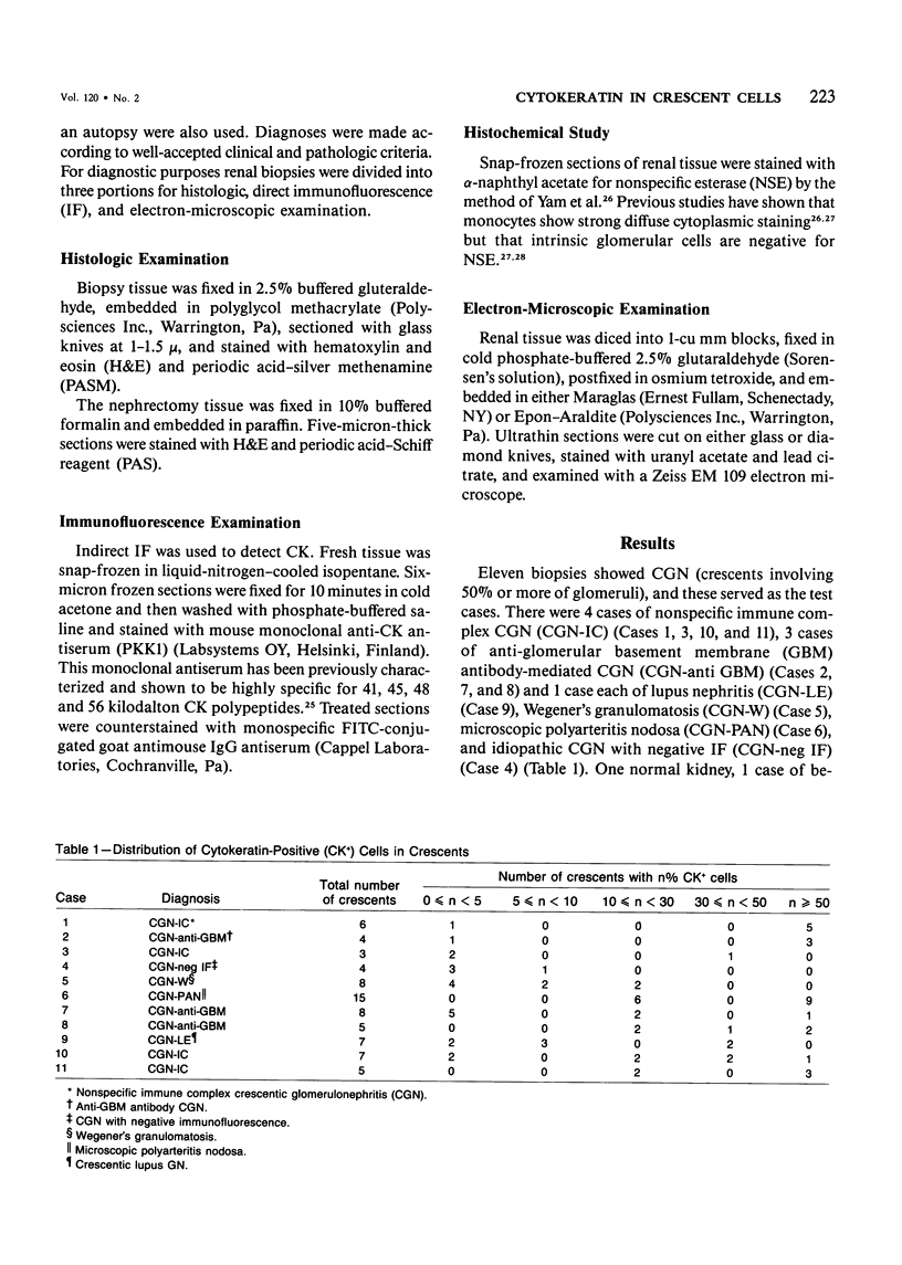
Recent studies have suggested that crescents are primarily of monocytic origin and that epithelial cells are a minor factor in their composition. Frozen sections of renal biopsies from 11 cases of crescentic glomerulonephritis (CGN) and 5 controls (2 acute interstitial nephritis, 1 focal glomerulosclerosis, 1 benign recurrent hematuria, 1 normal kidney) were stained for intracellular cytokeratin (CK) with a mouse monoclonal anti-CK antiserum (PKK1) and nonspecific esterase (NSE) activity. Indirect immunofluorescence with PKK1 antiserum showed that in all biopsies there was positive staining of collecting duct and proximal and distal tubular epithelium but no reactions in blood vessels or interstitium. In control case glomeruli there was no staining of the tuft, including the visceral epithelium. In all cases some parietal epithelium was CK-positive. In 4 CGN biopsies the majority of the crescents showed cytoplasmic staining for CK in more than 50% of the crescent cells. In 2 cases most crescents contained between 10-50% CK-positive cells, whereas in 5 biopsies little or no CK was present in the majority of crescents. In all but one CGN case the majority of crescents contained fewer than 30% NSE-positive cells (monocytes). Electron microscopy demonstrated intermediate filaments in many crescent cells and scattered desmosomes within crescents. The results indicate that epithelial cells, probably of parietal epithelial origin, contribute significantly to crescent formation.
Full text
PDF


Images in this article


Selected References
These references are in PubMed. This may not be the complete list of references from this article.
- Altmannsberger M., Osborn M., Schauer A., Weber K. Antibodies to different intermediate filament proteins. Cell type-specific markers on paraffin-embedded human tissues. Lab Invest. 1981 Nov;45(5):427–434. [PubMed] [Google Scholar]
- Atkins R. C., Holdsworth S. R., Glasgow E. F., Matthews F. E. The macrophagen in human rapidly progressive glomerulonephritis. Lancet. 1976 Apr 17;1(7964):830–832. doi: 10.1016/s0140-6736(76)90480-3. [DOI] [PubMed] [Google Scholar]
- Bacani R. A., Velasquez F., Kanter A., Pirani C. L., Pollak V. E. Rapidly progressive (nonstreptococcal) glomerulonephritis. Ann Intern Med. 1968 Sep;69(3):463–485. doi: 10.7326/0003-4819-69-3-463. [DOI] [PubMed] [Google Scholar]
- Bachmann S., Kriz W., Kuhn C., Franke W. W. Differentiation of cell types in the mammalian kidney by immunofluorescence microscopy using antibodies to intermediate filament proteins and desmoplakins. Histochemistry. 1983;77(3):365–394. doi: 10.1007/BF00490899. [DOI] [PubMed] [Google Scholar]
- Beirne G. J., Wagnild J. P., Zimmerman S. W., Macken P. D., Burkholder P. M. Idiopathic crescentic glomerulonephritis. Medicine (Baltimore) 1977 Sep;56(5):349–381. doi: 10.1097/00005792-197709000-00001. [DOI] [PubMed] [Google Scholar]
- Bohman S. O., Olsen S., Petersen V. P. Glomerular ultrastructure in extracapillary glomerulonephritis. Acta Pathol Microbiol Scand Suppl. 1974;Suppl 249:29–54. [PubMed] [Google Scholar]
- Cattell V., Arlidge S. The origin of proliferating cells in the glomerulus and Bowman's capsule in nephrotoxic serum nephritis: effects of unilateral renal irradiation. Br J Exp Pathol. 1981 Dec;62(6):669–675. [PMC free article] [PubMed] [Google Scholar]
- Franke W. W., Schiller D. L., Moll R., Winter S., Schmid E., Engelbrecht I., Denk H., Krepler R., Platzer B. Diversity of cytokeratins. Differentiation specific expression of cytokeratin polypeptides in epithelial cells and tissues. J Mol Biol. 1981 Dec 25;153(4):933–959. doi: 10.1016/0022-2836(81)90460-5. [DOI] [PubMed] [Google Scholar]
- Franke W. W., Weber K., Osborn M., Schmid E., Freudenstein C. Antibody to prekeratin. Decoration of tonofilament like arrays in various cells of epithelial character. Exp Cell Res. 1978 Oct 15;116(2):429–445. doi: 10.1016/0014-4827(78)90466-4. [DOI] [PubMed] [Google Scholar]
- Glenn K. C., Ross R. Human monocyte-derived growth factor(s) for mesenchymal cells: activation of secretion by endotoxin and concanavalin A. Cell. 1981 Sep;25(3):603–615. doi: 10.1016/0092-8674(81)90168-9. [DOI] [PubMed] [Google Scholar]
- Gown A. M., Vogel A. M. Monoclonal antibodies to human intermediate filament proteins. II. Distribution of filament proteins in normal human tissues. Am J Pathol. 1984 Feb;114(2):309–321. [PMC free article] [PubMed] [Google Scholar]
- Hancock W. W., Atkins R. C. Cellular composition of crescents in human rapidly progressive glomerulonephritis identified using monoclonal antibodies. Am J Nephrol. 1984;4(3):177–181. doi: 10.1159/000166800. [DOI] [PubMed] [Google Scholar]
- Hancock W. W., Atkins R. C. Monoclonal antibodies to human glomerular cells: a marker for glomerular epithelial cells. Nephron. 1983;33(2):83–90. doi: 10.1159/000182918. [DOI] [PubMed] [Google Scholar]
- Holdsworth S. R., Allen D. E., Thomson N. M., Glasgow E. F., Atkins R. C. Histochemistry of glomerular cells in animal models of crescentic glomerulonephritis. Pathology. 1980 Jul;12(3):339–346. doi: 10.3109/00313028009077095. [DOI] [PubMed] [Google Scholar]
- Holdsworth S. R., Thomson N. M., Glasgow E. F., Atkins R. C. The effect of defibrination on macrophage participation in rabbit nephrotoxic nephritis: studies using glomerular culture and electronmicroscopy. Clin Exp Immunol. 1979 Jul;37(1):38–43. [PMC free article] [PubMed] [Google Scholar]
- Holdsworth S. R., Thomson N. M., Glasgow E. F., Dowling J. P., Atkins R. C. Tissue culture of isolated glomeruli in experimental crescentic glomerulonephritis. J Exp Med. 1978 Jan 1;147(1):98–109. doi: 10.1084/jem.147.1.98. [DOI] [PMC free article] [PubMed] [Google Scholar]
- Holthöfer H., Miettinen A., Lehto V. P., Lehtonen E., Virtanen I. Expression of vimentin and cytokeratin types of intermediate filament proteins in developing and adult human kidneys. Lab Invest. 1984 May;50(5):552–559. [PubMed] [Google Scholar]
- Holthöfer H., Miettinen A., Paasivuo R., Lehto V. P., Linder E., Alfthan O., Virtanen I. Cellular origin and differentiation of renal carcinomas. A fluorescence microscopic study with kidney-specific antibodies, antiintermediate filament antibodies, and lectins. Lab Invest. 1983 Sep;49(3):317–326. [PubMed] [Google Scholar]
- Leibovich S. J., Ross R. A macrophage-dependent factor that stimulates the proliferation of fibroblasts in vitro. Am J Pathol. 1976 Sep;84(3):501–514. [PMC free article] [PubMed] [Google Scholar]
- Magil A. B. Drug-induced acute interstitial nephritis with granulomas. Hum Pathol. 1983 Jan;14(1):36–41. doi: 10.1016/s0046-8177(83)80044-6. [DOI] [PubMed] [Google Scholar]
- Magil A. B., Wadsworth L. D., Loewen M. Monocytes and human renal glomerular disease: a quantitative evaluation. Lab Invest. 1981 Jan;44(1):27–33. [PubMed] [Google Scholar]
- Magil A. B., Wadsworth L. D. Monocyte involvement in glomerular crescents: a histochemical and ultrastructural study. Lab Invest. 1982 Aug;47(2):160–166. [PubMed] [Google Scholar]
- Martinez-Hernandez A., Amenta P. S. The basement membrane in pathology. Lab Invest. 1983 Jun;48(6):656–677. [PubMed] [Google Scholar]
- Min K. W., Györkey F., Györkey P., Yium J. J., Eknoyan G. The morphogenesis of glomerular crescents in rapidly progressive glomerulonephritis. Kidney Int. 1974 Jan;5(1):47–56. doi: 10.1038/ki.1974.6. [DOI] [PubMed] [Google Scholar]
- Monga G., Mazzucco G., di Belgiojoso G. B., Busnach G. The presence and possible role of monocyte infiltration in human chronic proliferative glomerulonephritides. Light microscopic, immunofluorescence, and histochemical correlations. Am J Pathol. 1979 Feb;94(2):271–284. [PMC free article] [PubMed] [Google Scholar]
- Morel-Maroger Striker L., Killen P. D., Chi E., Striker G. E. The composition of glomerulosclerosis. I. Studies in focal sclerosis, crescentic glomerulonephritis, and membranoproliferative glomerulonephritis. Lab Invest. 1984 Aug;51(2):181–192. [PubMed] [Google Scholar]
- Morita T., Suzuki Y., Churg J. Structure and development of the glomerular crescent. Am J Pathol. 1973 Sep;72(3):349–368. [PMC free article] [PubMed] [Google Scholar]
- Naish P. F., Thomson N. M., Simpson I. J., Peters D. K. The role of polymorphonuclear leucocytes in the autologous phase of nephrotoxic nephritis. Clin Exp Immunol. 1975 Oct;22(1):102–111. [PMC free article] [PubMed] [Google Scholar]
- Sarno E. N., Alvarenga F. B., Ruzany F., Gattass C. R. Distribution of mononuclear phagocytes in glomerulonephritis with crescents. Nephron. 1982;32(3):265–265. doi: 10.1159/000182859. [DOI] [PubMed] [Google Scholar]
- Scheinman J. I., Foidart J. M., Michael A. F. The immunohistology of glomerular antigens. V. The collagenous antigens of the glomerulus. Lab Invest. 1980 Oct;43(4):373–381. [PubMed] [Google Scholar]
- Schiffer M. S., Michael A. F. Renal cell turnover studied by Y chromosome (Y body) staining of the transplanted human kidney. J Lab Clin Med. 1978 Dec;92(6):841–848. [PubMed] [Google Scholar]
- Schlegel R., Banks-Schlegel S., Pinkus G. S. Immunohistochemical localization of keratin in normal human tissues. Lab Invest. 1980 Jan;42(1):91–96. [PubMed] [Google Scholar]
- Tanaka K., Shibata N., Tatsumi N. Electron microscopic studies on the myofibrils in the epithelial cells of the Bowman's capsule and of proximal tubules in rat renal cortex (first report). Jpn Circ J. 1977 Dec;41(12):1329–1336. doi: 10.1253/jcj.41.1329. [DOI] [PubMed] [Google Scholar]
- Thomson N. M., Holdsworth S. R., Glasgow E. F., Atkins R. C. The macrophage in the development of experimental crescentic glomerulonephritis. Studies using tissue culture and electron microscopy. Am J Pathol. 1979 Feb;94(2):223–240. [PMC free article] [PubMed] [Google Scholar]
- Thomson N. M., Moran J., Simpson I. J., Peters D. K. Defibrination with ancrod in nephrotoxic nephritis in rabbits. Kidney Int. 1976 Nov;10(5):343–347. doi: 10.1038/ki.1976.120. [DOI] [PubMed] [Google Scholar]
- Thomson N. M., Simpson I. J., Peters D. K. A quantitative evaluation of anticoagulants in experimental nephrotoxic nephritis. Clin Exp Immunol. 1975 Feb;19(2):301–308. [PMC free article] [PubMed] [Google Scholar]
- VASSALLI P., MCCLUSKEY R. T. THE PATHOGENIC ROLE OF THE COAGULATION PROCESS IN RABBIT MASUGI NEPHRITIS. Am J Pathol. 1964 Oct;45:653–677. [PMC free article] [PubMed] [Google Scholar]
- Webber W. A., Blackbourn J. The permeability of the parietal layer of Bowman's capsule. Lab Invest. 1971 Nov;25(5):367–373. [PubMed] [Google Scholar]
- Yam L. T., Li C. Y., Crosby W. H. Cytochemical identification of monocytes and granulocytes. Am J Clin Pathol. 1971 Mar;55(3):283–290. doi: 10.1093/ajcp/55.3.283. [DOI] [PubMed] [Google Scholar]