

Abstract
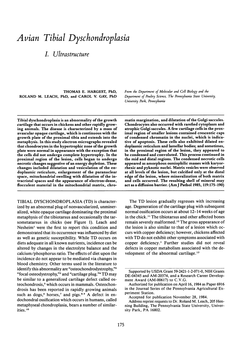
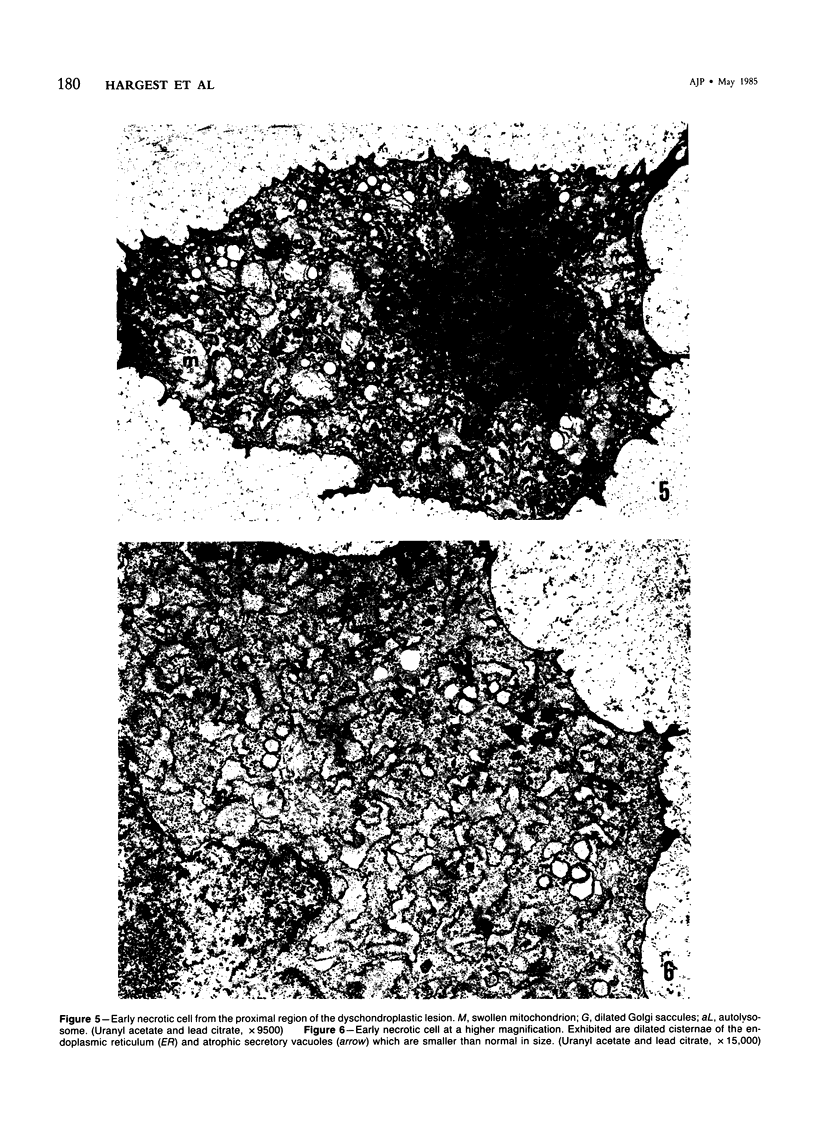
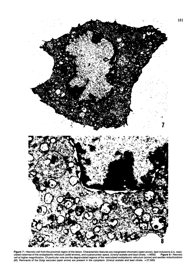
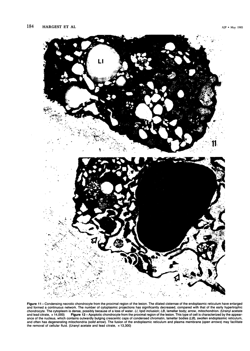
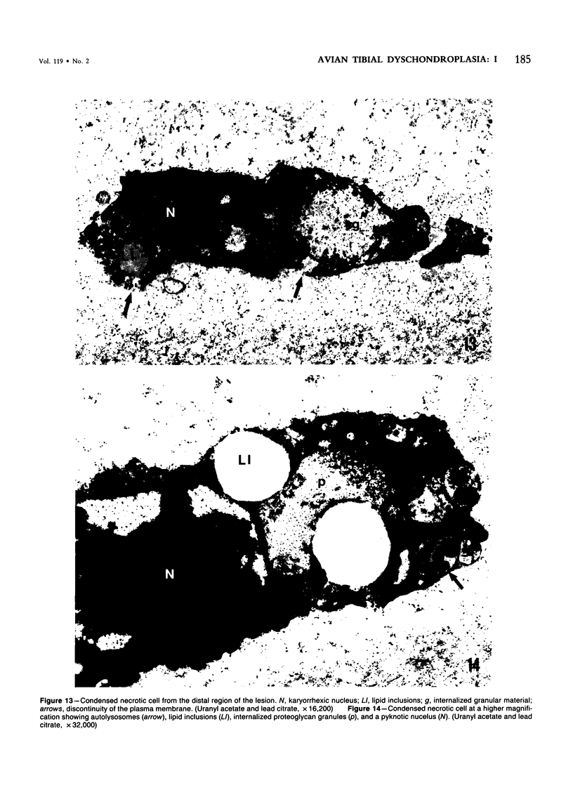
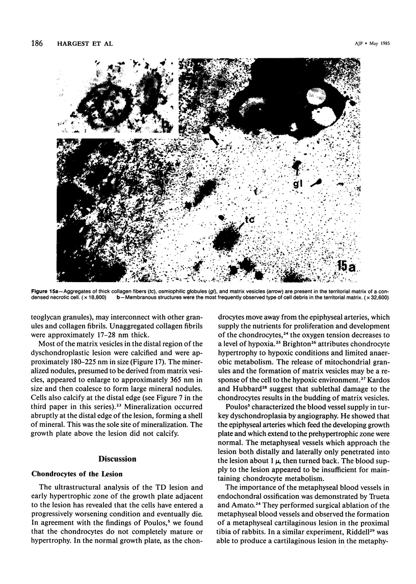
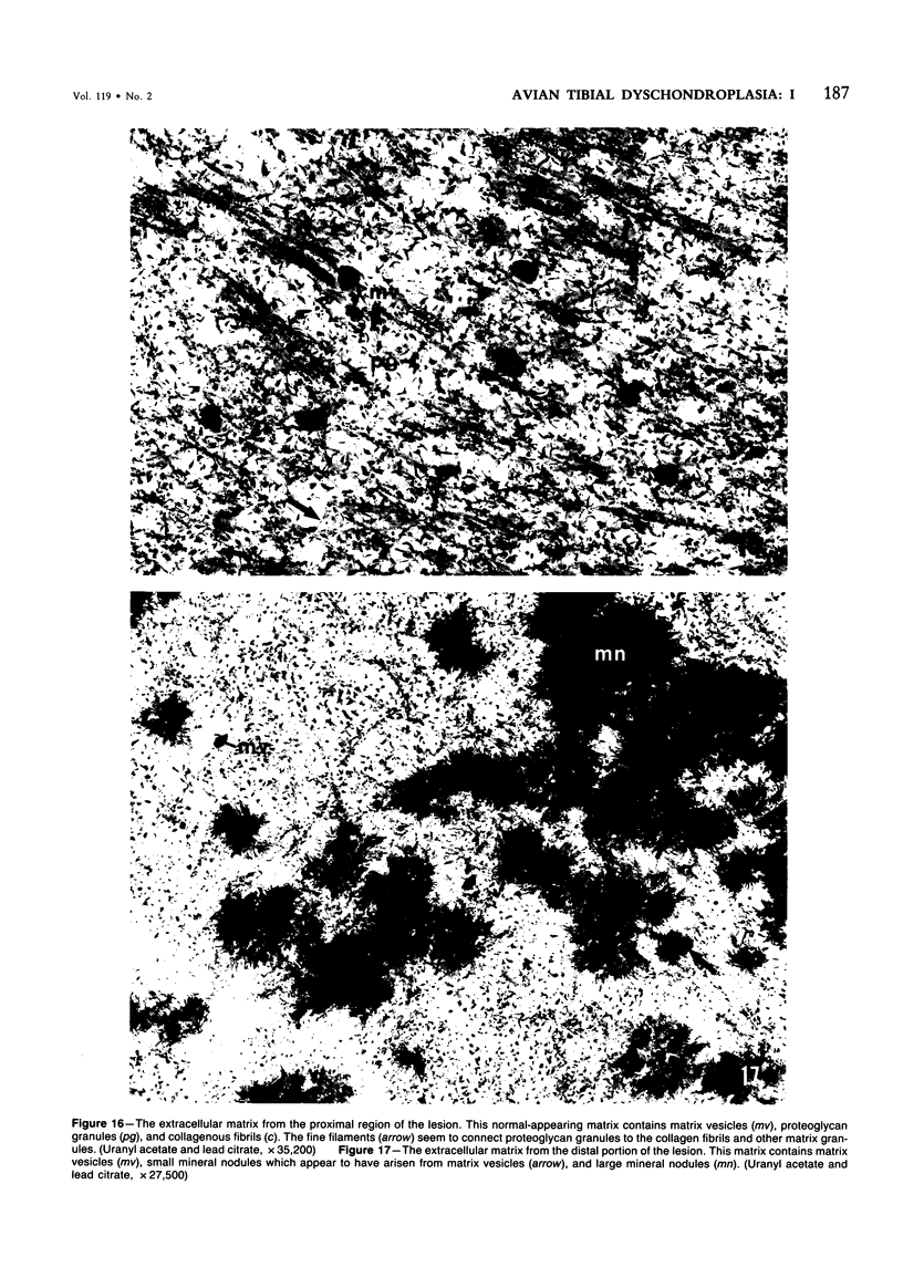
Tibial dyschondroplasia is an abnormality of the growth cartilage that occurs in chickens and other rapidly growing animals. The disease is characterized by a mass of avascular opaque cartilage, which is continuous with the growth plate of the proximal tibia and extends into the metaphysis. In this study electron micrographs revealed that chondrocytes in the hypertrophic zone of the growth plate were normal in appearance with the exception that the cells did not undergo complete hypertrophy. In the proximal region of the lesion, cells began to undergo necrotic changes suggestive of an energy depletion. These changes included dilatation and vesiculation of the endoplasmic reticulum, enlargement of the paranuclear space, mitochondrial swelling with dilatation of the intracristal spaces and the appearance of electron-dense, flocculent material in the mitochondrial matrix, chromatin margination, and dilatation of the Golgi saccules. Chondrocytes also occurred with rarefied cytoplasm and atrophic Golgi saccules. A few cartilage cells in the proximal region of smaller lesions contained crescentic caps of condensed chromatin in the nuclei, which is indicative of apoptosis. These cells also exhibited dilated endoplasmic reticulum and lamellar bodies; and sometimes, in the proximal region of the lesion, they appeared to be condensed and convoluted. This process continued in the mid and distal regions. The condensed necrotic cells appeared as amorphous osmiophilic masses with karyorrhexic and pyknotic nuclei. Matrix vesicles were observed at all levels of the lesion, but calcified only at the distal edge of the lesion, where mineralization of both matrix and cells occurred. The resulting shell of mineral may act as a diffusion barrier.
Full text
PDF


Images in this article


Selected References
These references are in PubMed. This may not be the complete list of references from this article.
- Brighton C. T., Heppenstall R. B. Oxygen tension in zones of the epiphyseal plate, the metaphysis and diaphysis. An in vitro and in vivo study in rats and rabbits. J Bone Joint Surg Am. 1971 Jun;53(4):719–728. [PubMed] [Google Scholar]
- Brighton C. T. Structure and function of the growth plate. Clin Orthop Relat Res. 1978 Oct;(136):22–32. [PubMed] [Google Scholar]
- Cooper R. R., Ponseti I. V. Metaphyseal dysotosis: description of an ultrastructural defect in the epiphyseal plate chondrocytes. J Bone Joint Surg Am. 1973 Apr;55(3):485–495. [PubMed] [Google Scholar]
- DAVID H., KETTLER L. H. [Degeneration of liver mitochondria after ammonia poisoning]. Z Zellforsch Mikrosk Anat. 1961;53:857–866. [PubMed] [Google Scholar]
- Del Conte E. Ultrastructural aspects of degradation and necrosis of Leydig cells in lizards by effect of metyrapone. Gen Comp Endocrinol. 1979 Jan;37(1):101–110. doi: 10.1016/0016-6480(79)90051-0. [DOI] [PubMed] [Google Scholar]
- Freedman B. D., Gay C. V., Leach R. M. Avian tibial dyschondroplasia. II. Biochemical changes. Am J Pathol. 1985 May;119(2):191–198. [PMC free article] [PubMed] [Google Scholar]
- Goracci G., Porcellati G., Woelk H. Subcellular localization and distribution of phospholipases A in liver and brain tissue. Adv Prostaglandin Thromboxane Res. 1978;3:55–67. [PubMed] [Google Scholar]
- Hargest T. E., Gay C. V., Leach R. M. Avian tibial dyschondroplasia. III. Electron probe analysis. Am J Pathol. 1985 May;119(2):199–209. [PMC free article] [PubMed] [Google Scholar]
- Hay E. D. Extracellular matrix. J Cell Biol. 1981 Dec;91(3 Pt 2):205s–223s. doi: 10.1083/jcb.91.3.205s. [DOI] [PMC free article] [PubMed] [Google Scholar]
- Hilley H. D. Skeletal abnormalities in the pig. Vet Clin North Am Large Anim Pract. 1982 Nov;4(2):225–258. doi: 10.1016/s0196-9846(17)30104-0. [DOI] [PubMed] [Google Scholar]
- Hirsch J. G., Fedorko M. E. Ultrastructure of human leukocytes after simultaneous fixation with glutaraldehyde and osmium tetroxide and "postfixation" in uranyl acetate. J Cell Biol. 1968 Sep;38(3):615–627. doi: 10.1083/jcb.38.3.615. [DOI] [PMC free article] [PubMed] [Google Scholar]
- Holtrop M. E. The ultrastructure of the epiphyseal plate. I. The flattened chondrocyte. Calcif Tissue Res. 1972;9(2):131–139. doi: 10.1007/BF02061951. [DOI] [PubMed] [Google Scholar]
- Howlett C. R. The fine structure of the proximal growth plate of the avian tibia. J Anat. 1979 Mar;128(Pt 2):377–399. [PMC free article] [PubMed] [Google Scholar]
- Jennings R. B., Shen A. C., Hill M. L., Ganote C. E., Herdson P. B. Mitochondrial matrix densities in myocardial ischemia and autolysis. Exp Mol Pathol. 1978 Aug;29(1):55–65. doi: 10.1016/0014-4800(78)90026-6. [DOI] [PubMed] [Google Scholar]
- LEACH R. M., Jr, NESHEIM M. C. NUTRITIONAL GENETIC AND MORPHOLOGICAL STUDIES OF AN ABNORMAL CARTILAGE FORMATION IN YOUNG CHICKS. J Nutr. 1965 Jul;86:236–244. doi: 10.1093/jn/86.3.236. [DOI] [PubMed] [Google Scholar]
- Lilburn M. S., Leach R. M., Jr Metabolism of abnormal cartilage cells associated with tibial dyschondroplasia. Poult Sci. 1980 Aug;59(8):1892–1896. doi: 10.3382/ps.0591892. [DOI] [PubMed] [Google Scholar]
- Lowther D. A., Robinson H. C., Dolman J. W., Thomas K. W. Cartilage matrix components in chickens with tibial dyschondroplasia. J Nutr. 1974 Jul;104(7):922–929. doi: 10.1093/jn/104.7.922. [DOI] [PubMed] [Google Scholar]
- Mergner W. J., Smith M. W., Sahaphong S., Trump B. F. Studies on the pathogenesis of ischemic cell injury. VI. Accumulation of calcium by isolated mitochondria of ischemic rat kidney cortex. Virchows Arch B Cell Pathol. 1977 Nov 30;26(1):1–16. [PubMed] [Google Scholar]
- Poulos P. W., Jr Tibial dyschondroplasia (osteochondrosis) in the turkey. A morphologic investigation. Acta Radiol Suppl. 1978;358:197–227. [PubMed] [Google Scholar]
- Reiland S. Pathology of so-called leg weakness in the pig. Acta Radiol Suppl. 1978;358:23–44. [PubMed] [Google Scholar]
- Rejnö S., Strömberg B. Osteochondrosis in the horse. II. Pathology. Acta Radiol Suppl. 1978;358:153–178. [PubMed] [Google Scholar]
- Riddell C. Studies on the pathogenesis of tibial dyschondroplasia in chickens. I. Production of a similar defect by surgical interference. Avian Dis. 1975 Jul-Sep;19(3):483–489. [PubMed] [Google Scholar]
- Riddell C. The development of tibial dyschondroplasia in broiler chickens. Avian Dis. 1975 Jul-Sep;19(3):443–462. [PubMed] [Google Scholar]
- Rimoin D. L., Hollister D. W., Lachman R. S., Kaufman R. L., McAlister W. H., Rosenthal R. E., Hughes G. N. Histologic studies in the chondrodystrophies. Birth Defects Orig Artic Ser. 1974;10(12):274–295. [PubMed] [Google Scholar]
- Rimoin D. L., Silberberg R., Hollister D. W. Chondro-osseous pathology in the chondrodystrophies. Clin Orthop Relat Res. 1976 Jan-Feb;(114):137–152. [PubMed] [Google Scholar]
- Silberberg R. Ultrastructure of cartilage in chondrodystrophies. Birth Defects Orig Artic Ser. 1974;10(12):306–313. [PubMed] [Google Scholar]
- Siller W. G. Tibial dyschondroplasia in the fowl. J Pathol. 1970 May;101(1):39–46. doi: 10.1002/path.1711010105. [DOI] [PubMed] [Google Scholar]
- Trueta J., Amato V. P. The vascular contribution to osteogenesis. III. Changes in the growth cartilage caused by experimentally induced ischaemia. J Bone Joint Surg Br. 1960 Aug;42-B:571–587. doi: 10.1302/0301-620X.42B3.571. [DOI] [PubMed] [Google Scholar]
- Wuthier R. E. A review of the primary mechanism of endochondral calcification with special emphasis on the role of cells, mitochondria and matrix vesicles. Clin Orthop Relat Res. 1982 Sep;(169):219–242. [PubMed] [Google Scholar]