Abstract
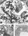
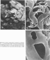
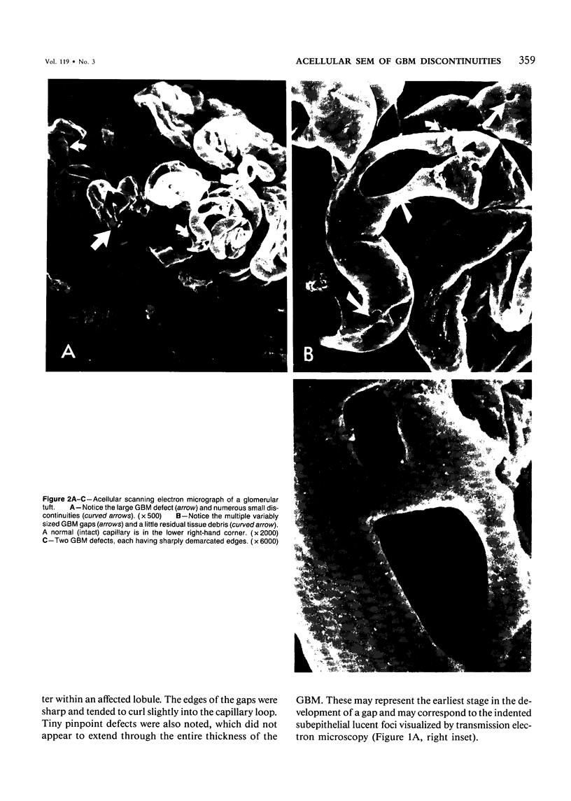
Glomerulonephritides which develop necrotizing and crescentic lesions usually have glomerular basement membrane (GBM) disruptions when carefully examined by light microscopy or transmission electron microscopy. Despite numerous excellent and detailed ultrastructural investigations of GBM discontinuities, a complete appreciation of their actual number, appearance, and distribution within a glomerulus has been difficult to achieve by reconstruction of two-dimensional light or transmission electron microscopic images. Selective removal of podocytes by a sequence of lytic and solubilization procedures has been developed which exposes any structural alteration of the GBM to direct examination by scanning electron microscopy. A case of idiopathic, immune-complex-negative, focal-segmental necrotizing glomerulonephritis has been studied by this technique, permitting three-dimensional visualization of the GBM defects which result in free communication between the vascular and urinary spaces. These disruptions were distinctive by their frequency within an affected lobule, variable size, and sharply demarcated edges. Application of this technique to human renal biopsies is capable of enhancing our understanding of the morphologic alterations occurring in human glomerulonephritis.
Full text
PDF
Images in this article
Selected References
These references are in PubMed. This may not be the complete list of references from this article.
- Bohman S. O., Olsen S., Petersen V. P. Glomerular ultrastructure in extracapillary glomerulonephritis. Acta Pathol Microbiol Scand Suppl. 1974;Suppl 249:29–54. [PubMed] [Google Scholar]
- Bonsib S. M. Scanning electron microscopy of acellular glomeruli in human glomerulonephritis: application of a technique. Ultrastruct Pathol. 1984;7(2-3):215–217. doi: 10.3109/01913128409141479. [DOI] [PubMed] [Google Scholar]
- Burkholder P. M. Ultrastructural demonstration of injury and perforation of glomerular capillary basement membrane in acute proliferative glomerulonephritis. Am J Pathol. 1969 Aug;56(2):251–265. [PMC free article] [PubMed] [Google Scholar]
- Couser W. G. Idiopathic rapidly progressive glomerulonephritis. Am J Nephrol. 1982;2(2):57–69. doi: 10.1159/000166586. [DOI] [PubMed] [Google Scholar]
- Min K. W., Györkey F., Györkey P., Yium J. J., Eknoyan G. The morphogenesis of glomerular crescents in rapidly progressive glomerulonephritis. Kidney Int. 1974 Jan;5(1):47–56. doi: 10.1038/ki.1974.6. [DOI] [PubMed] [Google Scholar]
- Morita T., Suzuki Y., Churg J. Structure and development of the glomerular crescent. Am J Pathol. 1973 Sep;72(3):349–368. [PMC free article] [PubMed] [Google Scholar]
- SPIRO D. The structural basis of proteinuria in man; electron microscopic studies of renal biopsy specimens from patients with lipid nephrosis, amyloidosis, and subacute and chronic glomerulonephritis. Am J Pathol. 1959 Jan-Feb;35(1):47–73. [PMC free article] [PubMed] [Google Scholar]
- Stejskal J., Pirani C. L., Okada M., Mandelanakis N., Pollak V. E. Discontinuities (gaps) of the glomerular capillary wall and basement membrane in renal diseases. Lab Invest. 1973 Feb;28(2):149–169. [PubMed] [Google Scholar]


