

Abstract
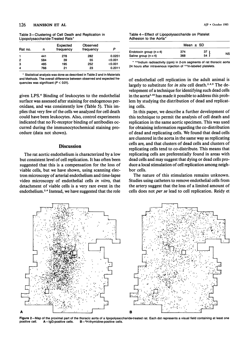
Endothelial cell death and replication was analyzed in the rat aorta with the use of combined IgG immunocytochemistry and 3H-thymidine autoradiography. Dead and replicating cells were clustered in the aortic endothelium of normal rats with a low level of spontaneous cell death and replication. Death and replication were also clustered in rats treated with endotoxin (Escherichia coli lipopolysaccharide [LPS]), a substance which is cytotoxic to endothelial cells in culture. LPS caused a dramatic increase both in endothelial cell death and replication, while no endothelial denudation could be discerned with the use of the 111In-labeled platelet technique. We conclude that endothelial cell death and replication are topographically clustered phenomena, which may imply that cell death leads to a local stimulation of cell replication. Furthermore, LPS induces a massive cell death, which is paralleled by cell replication, yet does not cause any significant increase in denudation. This suggests that the primary effect of LPS on the vessel wall is its cytotoxicity for endothelial cells. Endothelial cell death appears to provide the stimulus for cell replication both in normal and in LPS-treated rats.
Full text
PDF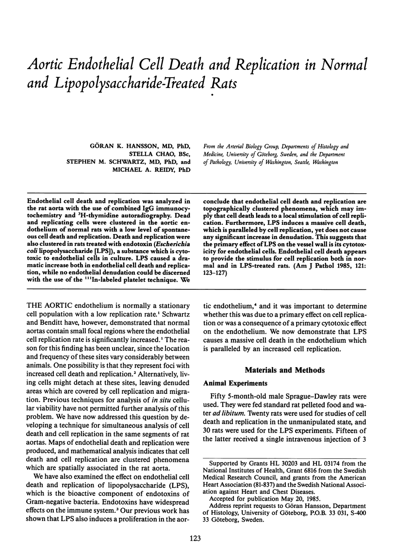
Selected References
These references are in PubMed. This may not be the complete list of references from this article.
- Bradley S. G. Cellular and molecular mechanisms of action of bacterial endotoxins. Annu Rev Microbiol. 1979;33:67–94. doi: 10.1146/annurev.mi.33.100179.000435. [DOI] [PubMed] [Google Scholar]
- Evensen S. A., Shepro D. DNA synthesis in rat aortic endothelium; effect of bacterial endotoxin and trauma. Microvasc Res. 1974 Jul;8(1):90–96. doi: 10.1016/0026-2862(74)90067-3. [DOI] [PubMed] [Google Scholar]
- Gaynor E. Increased mitotic activity in rabbit endothelium after endotoxin. An autoradiographic study. Lab Invest. 1971 Apr;24(4):318–320. [PubMed] [Google Scholar]
- Gerrity R. G., Caplan B. A., Richardson M., Cade J. F., Hirsh J., Schwartz C. J. Endotoxin-induced endothelial injury and repair. I. Endothelial cell turnover in the aorta of the rabbit. Exp Mol Pathol. 1975 Dec;23(3):379–385. doi: 10.1016/0014-4800(75)90031-3. [DOI] [PubMed] [Google Scholar]
- Hansson G. K., Schwartz S. M. Evidence for cell death in the vascular endothelium in vivo and in vitro. Am J Pathol. 1983 Sep;112(3):278–286. [PMC free article] [PubMed] [Google Scholar]
- Hansson G. K., Starkebaum G. A., Benditt E. P., Schwartz S. M. Fc-mediated binding of IgG to vimentin-type intermediate filaments in vascular endothelial cells. Proc Natl Acad Sci U S A. 1984 May;81(10):3103–3107. doi: 10.1073/pnas.81.10.3103. [DOI] [PMC free article] [PubMed] [Google Scholar]
- Harlan J. M., Harker L. A., Reidy M. A., Gajdusek C. M., Schwartz S. M., Striker G. E. Lipopolysaccharide-mediated bovine endothelial cell injury in vitro. Lab Invest. 1983 Mar;48(3):269–274. [PubMed] [Google Scholar]
- Reidy M. A., Bowyer D. E. Scanning electron microscopy: morphology of aortic endothelium following injury by endotoxin and during subsequent repair. Atherosclerosis. 1977 Mar;26(3):319–328. doi: 10.1016/0021-9150(77)90084-3. [DOI] [PubMed] [Google Scholar]
- Reidy M. A., Schwartz S. M. Endothelial injury and regeneration. IV. Endotoxin: a nondenuding injury to aortic endothelium. Lab Invest. 1983 Jan;48(1):25–34. [PubMed] [Google Scholar]
- Reidy M. A., Schwartz S. M. Endothelial regeneration. III. Time course of intimal changes after small defined injury to rat aortic endothelium. Lab Invest. 1981 Apr;44(4):301–308. [PubMed] [Google Scholar]
- Schwartz S. M., Benditt E. P. Cell replication in the aortic endothelium: a new method for study of the problem. Lab Invest. 1973 Jun;28(6):699–707. [PubMed] [Google Scholar]
- Schwartz S. M., Benditt E. P. Clustering of replicating cells in aortic endothelium. Proc Natl Acad Sci U S A. 1976 Feb;73(2):651–653. doi: 10.1073/pnas.73.2.651. [DOI] [PMC free article] [PubMed] [Google Scholar]
- Wright H. P. Mitosis patterns in aortic endothelium. Atherosclerosis. 1972 Jan-Feb;15(1):93–100. doi: 10.1016/0021-9150(72)90042-1. [DOI] [PubMed] [Google Scholar]