Abstract
In this report, two patients sustained a recurrent supracondylar humerus fracture following malunion of a previous supracondylar humerus fracture. The patients were treated for their first fracture at 5 and 6 years of age, respectively. One underwent open reduction with percutaneous pinning, and the other was treated with closed reduction with casting. Both patients healed in a moderate degree of extension after the first fracture.Two years later, both sustained a second fracture of the supracondylar humerus. Both had closed reduction with percutaneous pinning and went on to heal uneventfully. We speculate that ensuing post-traumatic extension deformity may accentuate a child's tendency for elbow hyperextension. Extension malunion may place the child at increased risk for a second fracture via similar mechanisms of injury.
INTRODUCTION
Supracondylar fractures of the humerus account for 3% of all fractures in children.6 However, they are the most common fractures occurring in the pediatric elbow, with a reported incidence of 75%.3 These fractures can be associated with serious complications such as nerve injuries, compartment syndrome, and angular deformities such as cubitus varus.1, 2, 5, 7, 9 It is well known that most malunions result in cosmetic adnormalities with minimal functional impairment. The current literature contains a few reports that describe lateral condyle fractures of the distal humerus as late complications of post-traumatic varus malunion following supracondylar humerus fracture.2, 4, 10, 11 In this report, we describe the cases of two patients treated initially for a supracondylar humerus fracture, both of whom went on to develop malunion with extension and then sustained a second supracondylar humerus fracture.
CASE EXAMPLES
Case 1
A 5-year-old female fell from the monkey bars and sustained a closed supracondylar fracture of the left humerus. In the emergency room, radiographs demonstrated a severely displaced Type III fracture of the left humerus (Figure 1). She was then taken to the operating room and underwent unsuccessful closed reduction under anesthesia. Therefore, the fracture site was opened, reduced and the fracture was pinned with crossed K-wires. Postoperatively, the patient was noted to have a clear decrease in ulnar nerve function and the dressing was split and the medial pin was removed. Unfortunately, subsequent radiographs revealed loss in reduction. She was taken back to the operating room where the fracture site was re-opened, reduced and K-wires were replaced to stabilize the fracture. Postoperatively ulnar nerve function returned and the patient was maintained in a splint followed by a cast for a total of five weeks. Radiographs showed her fracture site to be well-healed in moderate extension (Figure 2). Four months later, the patient had full range of motion of her elbow and her surgical scar was healing well. She did report some occasional mild throbbing in her small finger, but her neurosensory exam was intact.
Figure 1.
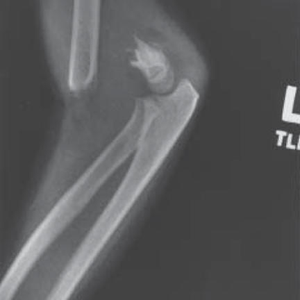
Lateral radiograph of a severely displaced Type III fracture of the left distal humerus in a 5-year-old female after falling off the monkey bars.
Figure 2.
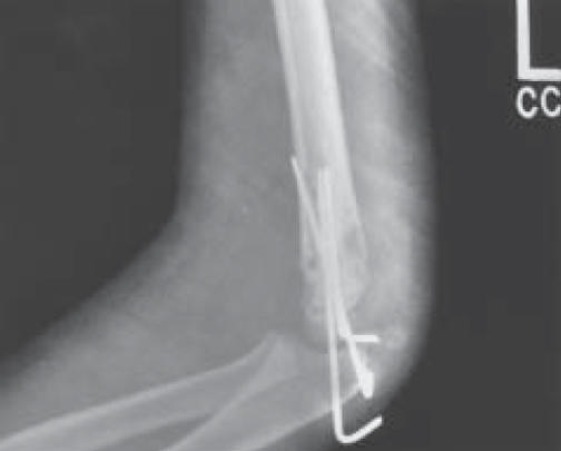
5 weeks after injury, a lateral radiograph shows the fracture site is well-healed in extension.
Two years later, the patient again fell down on a school playground and was taken to the emergency room complaining of bilateral elbow pain. Radiographs revealed a recurrent left supracondylar fracture of the humerus (Figure 3). Radiographs of the right elbow demonstrated a positive fat pad sign but were negative for fracture. She was taken to the operating room and underwent a closed reduction with percutaneous pinning of the left elbow. An arthrocentesis and arthrogram of the right elbow yielded a bloody aspirate but demonstrated no fracture and her right arm was placed in a long-arm splint. One month later, the pins in the left arm were removed and the arm was put in a long-arm cast for an additional two weeks and went on to heal uneventfully (Figure 4). The patient went on to regain motion of both joints.
Figure 3.
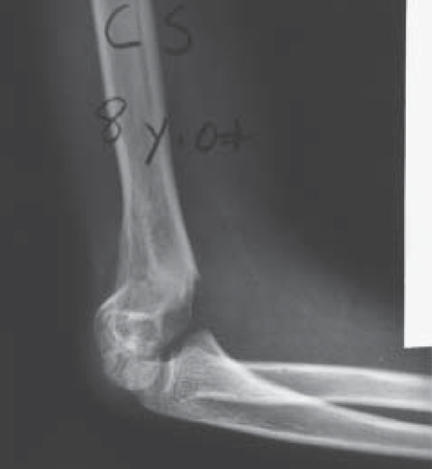
Two years later she suffered a recurrent supracondylar fracture of the left humerus after falling on the playground. Lateral radiographs demonstrate fracture through the previous fracture site.
Figure 4.
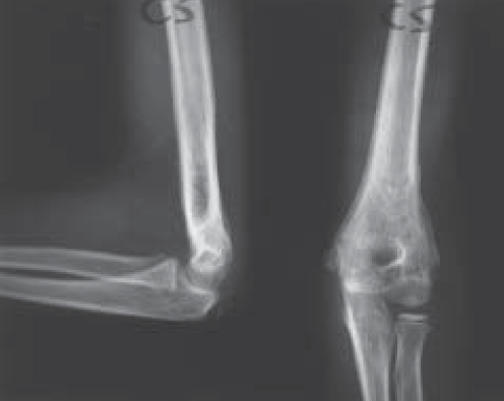
Anteroposterior and lateral radiographs 1 month after removal of percutaneous pins and long-arm cast demonstrate that the fracture is well-healed.
Case 2
A 6-year-old male fell from the swing set at school. He was taken to the emergency room complaining of severe left elbow pain. Radiographs revealed a Type II, supracondylar fracture of the left humerus (Figure 5). Although the fracture was moderately extended, no reduction was attempted and he was placed in a long-arm splint for three days, followed by a long-arm cast for a period of 6 weeks. At complete healing his distal humeral fragment was in an extended position with mild varus deformity (Figure 6).
Figure 5.
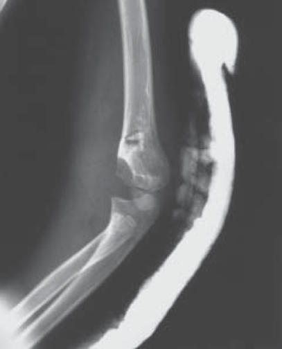
Oblique radiograph of a Type II supracondylar fracture of the left distal humerus in a 6-year-old boy after falling from swing set.
Figure 6.
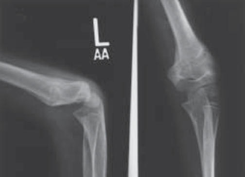
5 weeks later the fracture is well-healed but in a significantly extended position with mild varus noted on anteroposterior and lateral radiographs.
Two years later, the patient fell from a tree and sustained another supracondylar fracture of the left humerus (Figure 7), and a left distal radius fracture which was 100% displaced and 2 centimeters shortened. Review of the radiographs from his previous supracondylar fracture revealed this most recent fracture to have occurred through the same fracture site as the previous fracture. The patient was taken to the operating room for closed reduction and pinning of the supracondylar fracture and closed reduction and pinning of the distal radius. Similar to the first case, the refracture was reduced to the previous malunited position. No attempt was made to flex the refracture in order to reproduce the normal orientation of the distal humerus. He remained in his cast for 6 weeks. Serial radiographs obtained throughout the follow-up period demonstrated no change in the fracture alignment, with good healing of the supracondylar and radius fractures (Figure 8). At the most recent follow-up his fractures were fully healed and he had no functional concerns about elbow function in general.
Figure 7.
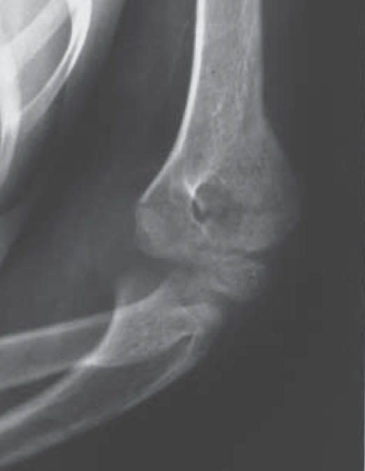
A recurrent supracondylar fracture of the left distal humerus is noted after falling from a tree 2 years after sustaining the previous fracture. The patient also sustained a left distal radius fracture.
Figure 8.
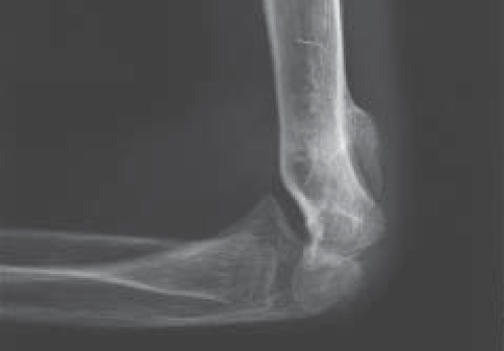
After treatment, radiographs reveal good healing of the supracondylar fracture. The fracture has been reduced and pinned in the original malunited position.
DISCUSSION
Second fractures of the distal humerus after varus malunion of a supracondylar humerus fracture have been described. In 1994, Davids et al, presented a review of lateral condyle fractures of the humerus due to malunion of supracondylar humerus fractures as well as varus as a result of overgrowth of a lateral condyle fracture. This complication results in decreased range of motion of the elbow, and can predispose to a fracture of the lateral condyle. In this report, biomechanical analysis suggested that torsional moment and shear forces are increased after varus malunion.2 Recently, Takahara et al, reported 9 children that sustained a second fracture of the distal humerus after an ipsilateral supracondylar fracture. They found that all of the patients had a varus malunion of the first fracture in the supracondylar region. They also hypothesized that varus malunion predisposed the patients to a second fracture. In this series all of the refractures were variations of lateral condyle fractures of which the more severe were trans-epiphyseal injuries of the distal humerus associated with a fracture involving the lateral metaphysis below the supracondylar fracture. Their findings suggest that the physis and epiphysis tend to be more prone to re-injury than the metaphysis of the distal humerus in children after a supracondylar fracture. They implied that the healed supracondylar humerus fractures result in a thickened metaphysis protecting the area from further injury. Conversely, the growth plate becomes more vulnerable, especially in cases of varus positioning.11
Varus malunion of the distal humerus usually results in cosmetic deformity with cubitus varus providing little functional deficit. As described above, this malunion may result in increased risk of lateral condyle fractures. Additional hyperextension deformity may accentuate the cosmetic appearance after a cubitus varus malunion. However, isolated extension malunion as a consequence of a true Type II fracture without medial or lateral column collapse usually results in little varus deformity and no functional limitation. In these cases, careful exam may demonstrate similar total range of motion in comparison to the contralateral side. In our experience, the patient experiences decreased flexion but increased extension corresponding to the degree of extension malunion.
Although in 1990 Wilkins suggested that residual hyperextension deformity may predispose for a refracture, we could find no reported cases of recurrent supracondylar humerus fractures following an extension malunion. Elbow hyperextension as a result of ligamentous laxity has long been considered a predisposing risk factor for supracondylar humerus fractures.12 Recently Nork et al studied the effect of hyperextension and ligamentous laxity on the incidence of fractures in the upper extremity. They concluded that a child who demonstrates ligamentous laxity is more likely to sustain an extension supracondylar humerus fracture as opposed to a displaced forearm fracture.8
In our review, both patients sustained an initial transverse fracture through the metaphysis of the distal humerus. They both went on to develop a malunion with a moderate component of extension. Despite good healing and a return to full activities, both patients sustained a refracture of the ipsilateral humerus at the same site as the first fracture two years later. Perhaps distal humeral extension deformity increases the ability to hyperextend the forearm in relationship to the humerus and thus increases the risk of a supracondylar humerus fracture. In these cases, a refracture may be more likely due to an accentuated ability to hyperextend allowing the olecranon into a bending fulcrum at the anatomically weak supracondylar area.12 Although the patients have no known bone fragility disorder, it is possible that the second fracture is closely associated to a point of weakness in the supracondylar area.
In summary, children who have sustained a supracondylar humerus fracture resulting in an extension malunion may be at increased risk of sustaining another supracondylar humerus fracture. Perhaps this is a result of the abnormal joint mechanics associated with a post-traumatic extension deformity, a child's normal state of ligamentous laxity, an anatomically weak supracondylar area, and a high level of physical activity.
References
- 1.Arnold JA, Nasca RJ, Nelson CL. Supracondylar Fractures of the Humerus: The Role of Dynamic Factors in Prevention of Deformity. Journal of Bone and Joint Surgery. 1977;59(A):589–595. [PubMed] [Google Scholar]
- 2.Davids JR, Maguire MF, Mubarak SJ, Wenger DR. Lateral Condyle Fracture of the Humerus Following Post-traumatic Cubitus Varus. Journal of Pediatric Orthopaedics. 1994;14:466–470. doi: 10.1097/01241398-199407000-00009. [DOI] [PubMed] [Google Scholar]
- 3.Devito DP. Management of Fractures and Their Complications. In: Morrissey RT, Weinstein SL, editors. Lovell and Winter's Pediatric Orthopaedics. 4th ed. Philadelphia: Lippincott-Raven Press; 1996. pp. 1241–1256. [Google Scholar]
- 4.Herring JA, Fitch RD. Lateral Condyle Fracture of the Elbow. Journal of Pediatric Orthopaedics. 1986;6:724–727. doi: 10.1097/01241398-198611000-00015. [DOI] [PubMed] [Google Scholar]
- 5.Labelle H, Bunnell WP, Duhaime M, Poitras B. Cubitus Varus Deformity Following Supracondylar Fractures of the Humerus in Children. Journal of Pediatric Orthopaedics. 1982;2:539–546. doi: 10.1097/01241398-198212000-00014. [DOI] [PubMed] [Google Scholar]
- 6.Minkowitz B, Busch MT. Supracondylar Humerus Fractures: Current Trends and Controversies. Orthopedic Clinics of North America. 1994;25(4):581–593. [PubMed] [Google Scholar]
- 7.Morrissey RT, Wilkins KE. Deformity Following Distal Humeral Fracture in Childhood. Journal of Bone and Joint Surgery. 1984;66-A( 4):557–562. [PubMed] [Google Scholar]
- 8.Nork SE, Hennrikus WL, Loncarich DP, Gillingham BL, Lapinsky AS. Relationship Between Ligamentous Laxity and the Site of the Upper Extremity Fractures in Children: Extension Supracondylar Fracture Versus Distal Forearm Fracture. Journal of Pediatric Orthopaedics. 1999;8(2) Part B:90–92. [PubMed] [Google Scholar]
- 9.Otsuka NY, Kasser JR. Supracondylar Fractures of the Humerus in Children. Journal of the American Academy of Orthopaedic Surgeons. 1997;5(1):19–26. doi: 10.5435/00124635-199701000-00003. [DOI] [PubMed] [Google Scholar]
- 10.Smith L. Deformity Following Supracondylar Fracture of the Humerus. Journal of Bone and Joint Surgery. 1960;42-A:235–252. [PubMed] [Google Scholar]
- 11.Takahara M, Sasaki I, Kimura T, Kato H, Minami A, Ogino T. Second Fracture of the Distal Humerus after Varus Malunion of a Supracondylar Fracture in Children. Journal of Bone and Joint Surgery. 1988;80-B(5):791–797. doi: 10.1302/0301-620x.80b5.8831. [DOI] [PubMed] [Google Scholar]
- 12.Wilkins KE, Beaty JH, Chambers HG, Toniolo RM. Fractures and Dislocations of the Elbow Region. In: Rockwood CA, Wilkins KE, Beaty JH, editors. Fractures in Children. 4th ed. Philadelphia: Lippincott-Raven; 1996. pp. 653–752. [Google Scholar]
- 13.Wilkins KE. Residuals of Elbow Trauma in Children. Orthopaedic Clinics of North America. 1990;21:291–314. [PubMed] [Google Scholar]


