

Abstract
Isolated PCL injuries have become more prevalent in recent years, possibly as a result of improved awareness and clinical recognition. However, the diagnosis can be difficult, and many of these injuries continue to go undiagnosed. Several clinical tests for PCL laxity have been described over the years, with varying degrees of sensitivity and clinical applicability. These include the posterior drawer,1 the Muller Quadriceps Active Test,2,3 Godfrey's Test,4 Trillat's reversal achman/total translation test,5 and the Dynamic Posterior Shift.6 All of these tests require significant posterior laxity associated with complete PCL disruption to be positive. Use of the KT-1000 arthrometer, and several radiographic tests have also been developed to help with diagnosis and quantification of laxity. It is the purpose of this paper to review the technique and application of the established diagnostic tests for PCL deficiency, and to introduce two new tests employed by the senior author for nearly three decades. It is the authors' experience that these new tests are sufficiently sensitive to allow the examiner to detect the presence of PCL insufficiency even in the most difficult diagnostic situations with subtle laxity.
TESTS CURRENTLY DESCRIBED IN THE LITERATURE SENSITIVE FOR GROSS LAXITY
The posterior drawer test is a classic exam that was described in the modern American literature by Hughston, et al in 1976,14 and again by Clancy, et al in 1983.1 Clancy's group performed the test on a supine patient with hip flexed 45° and the knee flexed 90°. The examiner sits on the patient's foot to fix this to the table. Each hand is then placed on the proximal anterior tibia, with a thumb on the respective medial and lateral joint lines. The proximal tibia is then pushed posteriorly, and an estimation of posterior plateau translation is determined. The test is first performed with the foot in neutral rotation. It is then repeated with the foot internally, and then externally rotated. The entire sequence is then compared to the contralateral side. Posterior translation is graded as a function of posterior motion of the proximal tibia relative to the opposite side, with a grade 1+ indicating 0 to 5mm greater translation, grade 2+ a 6 to 10mm greater translation, and a grade 3+ meaning 11mm or more.15 Another method to assess posterior laxity is to grade position of the anterior edge of the tibial plateau with respect to the ipsilateral femoral condyles during a forced posterior drawer. Posterior laxity of 1+ means the anteromedial edge of the plateau remains anterior to the medial condyle, 2+ is flush with the condyle and no step-off exists, and with 3+ the edge of the plateau is under, or posterior, to the condyle. Clancy, et al found that with isolated PCL injuries, the drawer decreased with the foot in internal rotation. This is opposite to Hughston's description and is presumably secondary to tightening of one or both of the menisco-femoral ligaments (i.e. Humphrey or Wrisberg), which were found to be intact at the time of surgery in these patients.
The Muller test is performed with the patient supine, and in the same position as the posterior drawer. The first part of the test is to examine the anterior silhouette of the proximal tibia from the side, and compare this to the uninjured, contralateral knee. The patient is then asked to raise his or her foot off the table. A positive test reveals posterior sag of the proximal tibia initially, and anterior translation of the proximal tibia prior to the foot leaving the table with attempted elevation of the foot. This anterior translation can be quantified and compared to the opposite knee. This test has also been termed the Quadriceps Active Test by Daniel, et al.2
Godfrey's test for posterior laxity is similar to the Muller test, but places the hip in 90° of flexion rather than 45° . A hand supports the leg under the lower cal for heel, suspending it in the air. The initial posterior sag may be more visible in this position, secondary to a greater gravitational pull, and may be useful in subtle cases. Again, the patient is asked to raise the foot, and anterior translation of the proximal tibia indicates a positive result.
There really is no true reverse Lachman test, but Trillat5 has described examining the knee in 20-30° flexion determining total anterior/posterior translation. A soft or absent endpoint determines direction of laxity, and this total translation is compared to the contralateral side.
Shelbourne, et al, described the Dynamic Posterior Shift test in 1989.6 The patient lies supine and the examiner stands on the side of the affected knee. The hip and knee are flexed to 90°, and the examiner's hand supports the anterolateral thigh, to maintain neutral rotation. The examiner's other hand supports the heel and the knee is then slowly extended by the examiner. The test is positive if a palpable and audible clunk, or jerk, occurs near extension, as the tibia reduces on the femoral condyles. This test requires the hamstrings to remain taught as the knee approaches full extension.
In a recent investigation, the posterior drawer test was found to be the most accurate clinical test in the diagnosis of PCL laxity.7 Rubinstein's study used a controlled blinded experiment design to assess the accuracy of the PCL clinical tests, for a group of sports medicine-trained orthopaedists. They evaluated the posterior drawer, posterior sagittal sign (first part of Mulleror Godfrey's test), reverse lachman, dynamic posterior shift, quadriceps active test (second part of Muller or Godfrey), reverse lachman endpoint and two tests for posterolateral rotatory instability, the reverse pivot shift and the external rotation recurvatum test. Their findings revealed a high specificity for each of the tests utilized, with a range of 89 to 100%. The sensitivity, however, varied greatly with a range of 37 to 90%. The posterior drawer was found in this study to be the most accurate test with a 90% sensitivity and 99% specificity. For Grade 1 laxity, however, this test was found to be only 70% sensitive. No other test had an overall sensitivity of 80% or higher. A combination of tests added in series would theoretically increase the ability of clinically diagnosing a PCL injury definitively.
Arthrometric evaluation with KT-1000 instrumentation has been shown to have a diagnostic sensitivity of 76-90% and accuracy of 89-96%7,8,9 in experienced hands, and is very user dependent. It is not as accurate with PCL laxity as it is for ACL laxity, and is not widely used in clinical applications.
Radiographic evaluation can be of some value in subtle cases, with posterior stress laterals at 70-90°, and has been shown in one study8 to be more accurate than KT-1000 and the posterior drawer test. This was especially true in the case of partial tears. MRI evaluation has become the gold standard for detection of PCL tears. A paper by Gross, et al10 reported accuracy approaching 100% in a study of over 200 patients with surgically confirmed diagnoses. The diagnosis, however, can usually be determined clinically with a careful history and thorough examination. Use of the MRI should be reserved for confirmation of the diagnosis and assessment of other suspected intra-articular injuries.
PREVIOUSLY UNPUBLISHED TESTS-SENSITIVE IN ABSENCE OF GROSS LAXITY
To follow is the description of two functional tests, as-of-yet undescribed in the literature, that can be used as adjuncts for the diagnosis of a PCL injury: the Posterior Functional Drawer test (PFD), and the Proximal Tibial Percussion test (PTP). These tests are very easily applied and interpreted, even by the most inexperienced health care providers. The tests are graded as either positive or negative, and indicate functional posterior instability.
Posterior Functional Drawer Test
The Posterior Functional Drawer test can be performed on a patient in either the prone or supine position. We have found it easier to examine both extremities simultaneously in the prone position, and will describe this technique first. With the patient prone, the knees are positioned at the lower edge of the examining table. Full manual resistance of knee flexion is performed at 90° of knee flexion and 0 of hip flexion (Figure 1). Amount of posterior pain and hamstring strength is compared to the contralateral side. The resistance is then repeated at 20 to 30° of knee flexion(Figure 2), and again compared to the other side. A positive test reveals posterior pain and significant weakness at 90°, versus the contralateral side, which is either not present, or is greatly reduced in the more extended position. In other words, resisted knee flexion hurts and there is weakness at 90°, while strength is maintained and pain decreased at 20-30°. The presumption is that at 90° the hamstrings pull the proximal tibia posteriorly into a subluxated position causing pain, with weakness probably from a pain inhibition mechanism. With the knee at 20°, the proximal tibia is reduced, and the component of the hamstring force vector acting posteriorly is insufficient to subluxate the plateau against secondary restraints in this position. Therefore the patient is able to generate pain-free hamstring power. The supine variation of this exam places the hip at 45° and the knee at 90°. One hand holds the heel and resists knee flexion, while the other palpates the anterior plateau to assess translation with resisted flexion (Figure 3). The test is then repeated with the knee at 20-30° (Figure 4). Further, an anterior drawer can be applied to the proximal tibia at 90° and the test repeated (Figure 5). A positive test is determined if an anterior drawer significantly reduces the pain and weakness found in the first part of the examination.
Figure 1. Performance of the Posterior Functional Drawer Test in the prone position at 90°.

Examiner compares strength of hamstrings and patient reports the presence of posterior pain.
Figure 2. Performance of the Posterior Functional Drawer Test in the prone position at 20°.

Examiner compares strength of hamstrings to each other, and to the 90° test, and patient reports the presence of posterior pain.
Figure 3. Performance of the Posterior Functional Drawer Test in the supine position at 90°.

Examiner compares strength of hamstrings and patient reports the presence of posterior pain.
Figure 4. Performance of the Posterior Functional Drawer Test in the prone position at 20°.

Examiner compares strength of hamstrings to each other, and to the 90° test, and patient reports the presence of posterior pain.
Figure 5. Posterior Functional Drawer test with an anterior drawer applied.
This symptomatic patient had reduced pain and increased hamstring strength with this maneuver.
The PFD test can be used as a screen in cases with partial tears or isolated tears with minimal laxity, and has been seen to be very sensitive in cases of acute injury. As a functional test it can be used to assess response to rehabilitation, a need for bracing, and judge effectiveness of the brace. We have also found the persistence of a positive test, despite full rehabilitation, associated with a failure to return to high level sports activity.
Proximal Tibial Percussion Test
The use of the Proximal Tibial Percussion test is indicated in only two instances. One, it should be employed as a last resort when none of the above tests have established a diagnosis of acute PCL deficiency. The second indication is for use in conjunction with the PFD test to confirm that the weakness detected in this test is not due to hamstring muscle injury. The PTP test is performed with the leg in the same position as described for the posterior drawer test with the knee at 90° and the examiner sitting of the patient's foot. The examiner's one hand is placed directly over the anterior aspect of the proximal tibia at the level of the tibial tubercle. With the unsuspecting patient completely relaxed, the examiner's other hand delivers a sharp blow to the back of the hand placed against the tibia (Figure 6). A positive test is declared with the generation of significant posterior joint pain similar in nature to that experienced at the time of the original injury. While the authors have found this test to be very revealing in many instances, it must be saved until last because it can be sufficiently painful that the patient is unable to cooperate for any further testing. For this reason, it is not needed when the diagnosis of PCL deficiency can be established without it.
Figure 6. Performance of the Proximal Tibial Pecussion test to the relaxed, unsuspecting patient.

A positive test illicits significant posterior pain. This is very useful in the acute injury setting.
These tests have not yet been formally validated, but this is currently in progress. In the twenty-eight year career of the senior author, all but one patient with subtle posterior laxity had a positive Posterior Functional Drawer test at presentation. The diagnosis was confirmed with either MRI or arthroscopy. Interestingly, most of these patients had an equivocal exam after employing the standard tests described at the beginning of this paper. All patients underwent a conservative management program emphasizing quadriceps strength. In six more recent cases of PCL rupture, three of the patients eventually become PFD negative during their rehabilitation, and three remained positive. The three that remained positive despite full rehabilitation eventually required delayed reconstruction, secondary to functional pain and/or instability during sporting activity. We feel this test is therefore helpful in identifying those patients that will ultimately fail conservative management of their isolated PCL injury, and require reconstruction in order to return to their pre-injury level of athletics. Below are several cases that illustrate these concepts.
ILLUSTRATIVE CASE REPORTS
Case 1
Chronic PCL deficiency with no appreciable laxity
A local high school soccer player was seen with vague knee symptoms and no obvious instability. Posterior drawer, Muller, and Godfrey were all negative. Both the PFD and PTP tests were positive. An MRI was obtained and confirmed isolated PCL rupture. The patient was successfully rehabilitated. The PFD reverted to negative after rehabilitation and hamstring strength improved with use of a functional brace. The patient was able to advance to the collegiate level with use of the brace.
Case 2
Chronic PCL deficiency with recurrent disability
An all-star tight end sustained a PCL injury in high school, which he did not reveal to the coaches or medical staff during recruitment. He denied any current knee pain or functional problems at the time of freshman physicals. Subtle posterior laxity was suspected with a soft end point on the posterior drawer test. The PFD and PTP tests were positive. The diagnosis was confirmed with MRI. The patient did well with rehabilitation, eventually becoming negative on his PFD test. He was able to play at the collegiate level with a PCL brace. Throughout the course of his collegiate career, the patient would occasionally 'tweak' his PCL deficient knee, and his PFD again became positive. His playing effectiveness was greatly reduced, until with rest and further rehab, his PFD would again revert to negative. He was then able to resume play free of symptoms.
Case 3
Acute non-specific knee pain with no obvious laxity
A collegiate defensive lineman took a hard hit and came off the football field in extreme pain, with the help of a teammate. The initial knee exam was difficult to perform, secondary to pain, but no gross laxity of the major ligaments was appreciated. There was no swelling or pain to palpation. After a few minutes the pain subsided and the player returned to the game. After the next series he complained of pain and the inability to explode out of his three-point stance. Again, the ligamentous exam was equivocal by standard tests. Application of the PFD and PTP tests revealed obviously positive results for PCL rupture. An MRI performed within the next couple of days confirmed an isolated PCL rupture. This patient was rehabilitated and was able to return to play the following season with a brace.
Case 4
Gross posterior laxity with persistent PFD despite full rehabilitation
A female scholarship rower fell into a creek, hitting her anterior proximal tibia on a concrete structure below the water's surface. She was diagnosed with a PCL rupture with 10mm of posterior laxity on a posterior drawer (Figure 7), and with a positive Godfrey's test(Figures 8 and 9). She was begun on ROM and strengthening rehabilitation. Six weeks later, despite no reported pain with activities, she had a persistent PFD test, and had marked hamstring weakness at 90° (Figures 3–5). She continued on an aggressive strengthening and functional rehab program under daily supervision. Seven weeks later she had completed her rehab and passed all of the performance tests set by the athletic training staff. She had full quad strength by manual testing. Her PFD remained positive with significant pain and weakness at 90°. Despite these test results, she was cleared to begin sport-specific conditioning and rowing. She did well at first, but the pain and swelling eventually increased to the point where she could not continue rowing. She especially had pain during the catch, and during the initial push of the drive phase, i.e. during full knee flexion. She had no pain with ADL's or walking, except after a workout. Despite improved function with a PCL brace, she was unable to resume training secondary to pain. Her PFD remained positive both with the brace and without. After having failed six months of nonoperative management, including brace wear, she requested surgical reconstruction. Four months after surgery, she is involved in a standard postoperative rehabilitative protocol and doing well. The posterior drawer is 2 to 3mm with a solid endpoint, and the PFD is negative.
Figure 7.

A grade II posterior drawer. Translation was estimated at 10mm, and condyles are flush with proximal tibial plateau. Note sag of anterior tibial silhouette.
Figure 8.

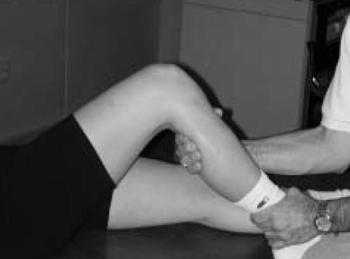
First part of Godfrey's test. Note sag of anterior tibia. The sag is more pronounced than that seen in Figure 7.
Figure 9.

Second part of Godfrey's test. Proximal tibia translates anteriorly with resisted initiation of knee extension.
DISCUSSION
The above cases represent patients with variable degrees of PCL laxity, and differing stages of injury at presentation. They each did well originally with rehabilitation, and it was felt they would be able to return to their pre-injury level of activity. It was noted that each had a grossly positive posterior functional drawer test at presentation, and that the test became negative, or nearly so, during rehabilitation in the athletes that were able to successfully resume play. One eventually came to reconstruction after she was unable to resume her training secondary to pain, and in this case the PFD remained positive despite full rehabilitation. The football players were able to resume play with use of a brace, but occasionally lost time from mild repeat injury. During these times after repeat injury, the PFD again became positive. All of these cases illustrate the usefulness of the PFD in the diagnosis and clinical follow-up of functional PCL instability.
Fowler and Messieh11 followed 13 patients with isolated PCL injuries, and reported in 1996 a 100% return to previous level of activity at an average of 2.6 years. Prior to this Parolie and Bergfeld12 reported only an 84% return to activity, and only a 68% return at the pre-injury level. They found an increase of 2mm in the side-to-side KT-1000 difference in the unsatisfied patients, versus the satisfied patients, when tested both passively, and with an active hamstring contraction. This however did not reach statistical significance. They concluded that the amount of posterior laxity did not correlate with the ability to return to sports, but felt it was more related to the patient's ability to regain quadriceps strength equal to or greater than the uninjured side. Shelbourne, et al13 reported on a prospectively followed group of patients with isolated PCL rupture and found that at an average of 5.4 years, only 50% of conservatively treated patients were able to return to their pre-injury level. They also showed this was not correlated with degree of laxity. This reflects our experience.
Our patients have all regained full quadriceps strength during rehabilitation, but those requiring reconstruction continued to exhibit a positive PFD test. This test does not quantify amount of posterior translation, but rather that it occurs, and that it causes pain and weakness of the hamstrings. We therefore believe this test is useful in identifying those patients who have completed a rehabilitation protocol and have full quadriceps strength, who are unable to return to a high level of sports activity. These patients may require use of a brace, or PCL reconstruction. The test can also be used to assess the effectiveness of a PCL brace, as the test should revert to negative if the brace is effectively resisting posterior translation of the proximal tibia. This test does not measure or directly identify posterior laxity, but rather identifies patients that have functional disability (pain and hamstring weakness) as a result of abnormal posterior translation of the proximal tibia. It also identifies those patients that have overcome this disability with rehabilitation, and ultimately do well with non-operative management.
We have also found PFD test to be very helpful in the diagnosis of acute PCL ruptures, especially in those cases when the diagnosis is not obvious. It is quickly and easily applied in the office or on the sidelines, and results are independent of the examiner. The Proximal Tibial Percussion test requires no effort or cooperation from the patient, and can be applied to the acutely injured, apprehensive patient. We feel these tests should be a useful part of the sports medicine physician's clinical diagnostic armamentarium.
In conclusion, two new clinical tests have been presented that we feel are very sensitive in diagnosing acute PCL injury and subtle cases of functional PCL instability. These tests can also be used to follow a patient's ability to return to high level activities during a rehabilitative program, and assess brace function.
References
- 1.Clancy WG, et al. Treatment of knee joint instability secondary to rupture of the posterior cruciate ligament. Report of a new procedure. JBJS. 1983;65A:310–322. [PubMed] [Google Scholar]
- 2.Daniel DM, et al. Use of the Quadriceps Active Test to diagnose posterior cruciate-ligament disruption and measure posterior laxity of the knee. JBJS. 1988;70A:386–391. [PubMed] [Google Scholar]
- 3.Muller W, et al. OAK knee evaluation: a new way to assess knee ligament injuries. CORR. 1988;232:37–50. [PubMed] [Google Scholar]
- 4.Liorzou G. Knee Ligaments, Clinical Examination. Springer-Verlag; 1990. pp. 31–33. [Google Scholar]
- 5.Trillat A, Dejour H, Bousquet G. Lere Jounies Luonnaises de Chirurgie du Genou. Lyon: Bernadet; 1971. [Google Scholar]
- 6.Shelbourne KD, et al. Dynamic posterior shift test. An adjuvant in evaluation of posterior tibial subluxation. AJSM. 17(2):275–277. doi: 10.1177/036354658901700221. [DOI] [PubMed] [Google Scholar]
- 7.Rubinstein RA, Jr, et al. The accuracy of the clinical examination in the setting of posterior cruciate ligament injuries. AJSM. 1994;22(4):550–557. doi: 10.1177/036354659402200419. [DOI] [PubMed] [Google Scholar]
- 8.Hewett TE, Noyes FR, Lee MD. Diagnosis of complete and partial posterior cruciate ligament ruptures. Stress radiography compared with KT-1000 arthrometer and posterior drawer testing. AJSM. 1997;25(5):648–655. doi: 10.1177/036354659702500510. [DOI] [PubMed] [Google Scholar]
- 9.Eakin CL, Cannon WD., Jr Arthrometric evaluation of posterior cruciate ligament injuries. AJSM. 1998;26(1):96–102. doi: 10.1177/03635465980260013401. [DOI] [PubMed] [Google Scholar]
- 10.Gross ML, et al. Magnetic resonance imaging of the posterior cruciate ligament. Clinical use to improve diagnostic accuracy. AJSM. 1992;20(6):732–737. doi: 10.1177/036354659202000615. [DOI] [PubMed] [Google Scholar]
- 11.Fowler PJ, Messieh SS. Isolated posterior cruciate ligament injuries in athletes. AJSM. 1987;15(6):553–557. doi: 10.1177/036354658701500606. [DOI] [PubMed] [Google Scholar]
- 12.Parolie JM, Bergfeld JA. Long-term results of nonoperative treatment of isolated posterior cruciate ligament injuries in the athlete. AJSM. 1986;14(1):35–38. doi: 10.1177/036354658601400107. [DOI] [PubMed] [Google Scholar]
- 13.Shelbourne KD, Davis TJ, Patel DV. The natural history of acute, isolated, nonoperatively treated posterior cruciate ligament injuries. A prospective study. AJSM. 1999;27(3):276–283. doi: 10.1177/03635465990270030201. [DOI] [PubMed] [Google Scholar]
- 14.Hughston JC, et al. Classification of Knee Ligament Instabilities Part I and Part II. JBJS. 1976;58A:159–179. [PubMed] [Google Scholar]
- 15.Committee on the Medical Aspects of Sports, American Medical Association. Standard Nomenclature of Athletic Injuries. American Medical Association; 1968. pp. 92–101. [Google Scholar]