

Abstract
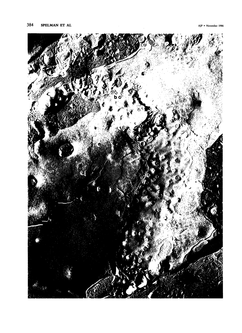
The authors have investigated early changes in liver cell gap and tight junctions that occur when rats are fed a carcinogenic diet. Animals were fed a choline-deficient diet that contained 0.1% ethionine (CDE) for periods up to 6 weeks. Short-term feeding of this diet results in the rapid proliferation of so-called "oval cells" within the liver, which is reversible upon returning the rats to a normal diet. Livers from animals fed the diet were removed at various times during feeding and during recovery from the diet and were analyzed by light and electron microscopy. The freeze-fracture technique was used to produce extended views of the internal structure of liver cell membranes at each stage under study. The characteristic junctional complex surrounding canalicular regions in normal liver disappears after only 2 weeks of the CDE regimen. Gap junctions were not found after 4 weeks of the diet, and tight junctions became increasingly disorganized. Tight junction elements were observed, however, between hepatocytes and oval cells, which indicated that these two cell types do interact directly. Changes occur in the structural complexity of tight junction elements between hepatocytes and between hepatocytes and oval cells. Recovery from the CDE diet results in a rapid increase in junctional complexity, and the large gap junction plaques characteristic of normal liver are visible within 2 weeks after cessation of the CDE regimen. These and other observations demonstrate that reversible alterations in hepatocyte gap and tight junctions occur as a result of administration of a diet that induces oval cell proliferation. The relationship of these changes to those that have been reported during other processes of cell proliferation are discussed.
Full text
PDF


Images in this article


Selected References
These references are in PubMed. This may not be the complete list of references from this article.
- ABERCROMBIE M. Localized formation of new tissue in an adult mammal. Symp Soc Exp Biol. 1957;11:235–254. [PubMed] [Google Scholar]
- Branton D., Bullivant S., Gilula N. B., Karnovsky M. J., Moor H., Mühlethaler K., Northcote D. H., Packer L., Satir B., Satir P. Freeze-etching nomenclature. Science. 1975 Oct 3;190(4209):54–56. doi: 10.1126/science.1166299. [DOI] [PubMed] [Google Scholar]
- Claude P., Goodenough D. A. Fracture faces of zonulae occludentes from "tight" and "leaky" epithelia. J Cell Biol. 1973 Aug;58(2):390–400. doi: 10.1083/jcb.58.2.390. [DOI] [PMC free article] [PubMed] [Google Scholar]
- Clement B., Guguen-Guillouzo C., Campion J. P., Glaise D., Bourel M., Guillouzo A. Long-term co-cultures of adult human hepatocytes with rat liver epithelial cells: modulation of albumin secretion and accumulation of extracellular material. Hepatology. 1984 May-Jun;4(3):373–380. doi: 10.1002/hep.1840040305. [DOI] [PubMed] [Google Scholar]
- De Vos R., Desmet V. J. Morphologic changes of the junctional complex of the hepatocytes in rat liver after bile duct ligation. Br J Exp Pathol. 1978 Apr;59(2):220–227. [PMC free article] [PubMed] [Google Scholar]
- FARBER E. Similarities in the sequence of early histological changes induced in the liver of the rat by ethionine, 2-acetylamino-fluorene, and 3'-methyl-4-dimethylaminoazobenzene. Cancer Res. 1956 Feb;16(2):142–148. [PubMed] [Google Scholar]
- Farber E., Cameron R. The sequential analysis of cancer development. Adv Cancer Res. 1980;31:125–226. doi: 10.1016/s0065-230x(08)60658-2. [DOI] [PubMed] [Google Scholar]
- GRISHAM J. W., PORTA E. A. ORIGIN AND FATE OF PROLIFERATED HEPATIC DUCTAL CELLS IN THE RAT: ELECTRON MICROSCOPIC AND AUTORADIOGRAPHIC STUDIES. Exp Mol Pathol. 1964 Jun;86:242–261. doi: 10.1016/0014-4800(64)90057-7. [DOI] [PubMed] [Google Scholar]
- Hayner N. T., Braun L., Yaswen P., Brooks M., Fausto N. Isozyme profiles of oval cells, parenchymal cells, and biliary cells isolated by centrifugal elutriation from normal and preneoplastic livers. Cancer Res. 1984 Jan;44(1):332–338. [PubMed] [Google Scholar]
- Koga A., Todo S. Morphological and functional changes in the tight junctions of the bile canaliculi induced by bile duct ligation. Cell Tissue Res. 1978 Dec 28;195(2):267–276. doi: 10.1007/BF00236724. [DOI] [PubMed] [Google Scholar]
- Larsen W. J. Biological implications of gap junction structure, distribution and composition: a review. Tissue Cell. 1983;15(5):645–671. doi: 10.1016/0040-8166(83)90041-1. [DOI] [PubMed] [Google Scholar]
- Loewenstein W. R. Junctional intercellular communication and the control of growth. Biochim Biophys Acta. 1979 Feb 4;560(1):1–65. doi: 10.1016/0304-419x(79)90002-7. [DOI] [PubMed] [Google Scholar]
- Meyer D. J., Yancey S. B., Revel J. P. Intercellular communication in normal and regenerating rat liver: a quantitative analysis. J Cell Biol. 1981 Nov;91(2 Pt 1):505–523. doi: 10.1083/jcb.91.2.505. [DOI] [PMC free article] [PubMed] [Google Scholar]
- Ogawa K., Minase T., Onhoe T. Demonstration of glucose 6-phosphatase activity in the oval cells of rat liver and the significance of the oval cells in azo dye carcinogenesis. Cancer Res. 1974 Dec;34(12):3379–3386. [PubMed] [Google Scholar]
- Petropoulos C. J., Yaswen P., Panzica M., Fausto N. Cell lineages in liver carcinogenesis: possible clues from studies of the distribution of alpha-fetoprotein RNA sequences in cell populations isolated from normal, regenerating, and preneoplastic rat livers. Cancer Res. 1985 Nov;45(11 Pt 2):5762–5768. [PubMed] [Google Scholar]
- Porvaznik M., Johnson R. G., Sheridan J. D. Intercellular junctions and other cell surface differentiations of H4-IIE hepatoma cells in vitro. J Ultrastruct Res. 1976 Jun;55(3):343–359. doi: 10.1016/s0022-5320(76)80092-5. [DOI] [PubMed] [Google Scholar]
- Rassat J., Robenek H., Themann H. Freeze-fracture elucidation of hepatocyte membrane alterations following beta-pyridylcarbinol application. Exp Pathol. 1982;22(2):103–112. doi: 10.1016/s0232-1513(82)80031-5. [DOI] [PubMed] [Google Scholar]
- Sell S., Leffert H. L. An evaluation of cellular lineages in the pathogenesis of experimental hepatocellular carcinoma. Hepatology. 1982 Jan-Feb;2(1):77–86. doi: 10.1002/hep.1840020113. [DOI] [PubMed] [Google Scholar]
- Sells M. A., Katyal S. L., Shinozuka H., Estes L. W., Sell S., Lombardi B. Isolation of oval cells and transitional cells from the livers of rats fed the carcinogen DL-ethionine. J Natl Cancer Inst. 1981 Feb;66(2):355–362. [PubMed] [Google Scholar]
- Shinozuka H., Lombardi B., Sell S., Iammarino R. M. Early histological and functional alterations of ethionine liver carcinogenesis in rats fed a choline-deficient diet. Cancer Res. 1978 Apr;38(4):1092–1098. [PubMed] [Google Scholar]
- Staehelin L. A. Structure and function of intercellular junctions. Int Rev Cytol. 1974;39:191–283. doi: 10.1016/s0074-7696(08)60940-7. [DOI] [PubMed] [Google Scholar]
- Swift J. G., Mukherjee T. M., Rowland R. Intercellular junctions in hepatocellular carcinoma. J Submicrosc Cytol. 1983 Jul;15(3):799–810. [PubMed] [Google Scholar]
- Vitkauskas G. V., Canellakis E. S. Intercellular communication and cancer chemotherapy. Biochim Biophys Acta. 1985 Nov 12;823(1):19–34. doi: 10.1016/0304-419x(85)90013-7. [DOI] [PubMed] [Google Scholar]
- Weinstein R. S., Merk F. B., Alroy J. The structure and function of intercellular junctions in cancer. Adv Cancer Res. 1976;23:23–89. doi: 10.1016/s0065-230x(08)60543-6. [DOI] [PubMed] [Google Scholar]
- Yaswen P., Hayner N. T., Fausto N. Isolation of oval cells by centrifugal elutriation and comparison with other cell types purified from normal and preneoplastic livers. Cancer Res. 1984 Jan;44(1):324–331. [PubMed] [Google Scholar]
- Yee A. G., Revel J. P. Loss and reappearance of gap junctions in regenerating liver. J Cell Biol. 1978 Aug;78(2):554–564. doi: 10.1083/jcb.78.2.554. [DOI] [PMC free article] [PubMed] [Google Scholar]