Abstract
The purpose of this study was to provide clinical and radiographic evaluation after a minimum of two years in patients who had an anterior cervical corpectomy and a fibular allograft strut. Nineteen patients returned for a follow-up visit which included independent radiographic evaluation as well as completing a Visual Analogue Scale and Oswestry and Short-Form 36 questionnaires. The categories of fusion were as follows: 1) definitely fused (84%) 2) questionably fused (11%) 3) definitely not fused (5%). The average VAS was 29 mm (range 0-85). The Oswestry Back Scores showed relatively low levels of significant pain with an average score of 29 (range 0-73). Anterior cervical corpectomy followed by an allograft fibular strut provides for relatively high rates of arthrodesis without severe loss of height or sagittal alignment at long term radiographic follow-up.
INTRODUCTION
Anterior cervical corpectomy and stabilization by arthrodesis is a reliable surgical procedure for relieving spinal cord compression.10,16 Patients with cervical spondylosis, trauma, or ossification of the posterior longitudinal ligament (OPPL) may require this procedure for decompression of the spinal cord. Strut grafts stabilize the segment after the corpectomy to prevent progressive kyphosis. Both fibular allograft and autograft have been used as struts.
For single-level anterior cervical spine surgery, there is no significant difference in fusion rates between allograft and autograft.2,20 Few studies are available with which to compare the use of allograft or autograft after corpectomy. Long-term arthodesis rates of 97-100% have been reported after corpectomy and autologous fibular grafts.3,21 However, the use of autologous bone causes an increase in donor site morbidity (19%), operative time, and wound infections.6,11 Because of these concerns, fibular allografts have been used as strut grafts.
Only a few published studies exist which provide long-term radiographic rates of union after cervical corpectomy and grafting with fibular allograft.3,12, 21 The purpose of this study was to provide clinical and radiographic evaluation after a minimum of two years in patients who had this procedure for a variety of pathological conditions.
MATERIALS AND METHODS
Twenty-five patients underwent anterior cervical corpectomy and grafting with a freeze-dried fibular allograft strut at our institutions between November 1991 and April 1997. Hospital charts and radiographs were reviewed. Nineteen patients (76% of index) were available for follow-up at a minimum of two years (mean 45 months, range 25-85 months). The operative procedure consisted of a standard Smith-Robinson anterior cervical approach. The level and number of corpectomies were determined pre-operatively from evaluation of clinical and radiographic findings. Fibular allograft struts were used in all cases. Seventeen of the eighteen patients had an anterior cervical plate applied, which spanned the fusion. No posterior procedures were performed. The standard postoperative immobilization was with a rigid collar.
At the follow-up visit, each participant had cervical spine radiographs taken consisting of a standard anterior-posterior and lateral, as well as flexion and extension lateral views. A swimmer's view was taken if there was any difficulty seeing the caudal portion of the graft. A history and physical exam were performed on each patient by two of the authors (BEM; JKW). Oswestry17 and Short Form-36 (SF-36)18 questionnaires were completed. The patient's average level of cervical pain was documented with a Visual Analogue Scale (VAS).14,15 The VAS is a 10 centimeter line with one end designated as "No Pain," and the opposite end designated as "Unbearable Pain." The patients made a mark on the line, which represented their average daily pain over the preceding month.
Immediate postoperative or intra-operative radiographs were collected for evaluation and comparison to follow-up radiographs. Both sets of films were read by a blinded board certified musculoskeletal radiologist for height and angulation of the grafted segments as described by Emery et al.4 The height of the fused segment was obtained by drawing a line across the superior endplate of the uppermost vertebra and the inferior endplate of the lowest vertebra involved in the fusion. A vertical line was drawn between the midpoint of each endplate identified. This represented the height of the fused segment measured in millimeters. The angle of the fused segments was determined by drawing lines across the superior endplate of the uppermost vertebra and the inferior endplate of the lowest involved vertebra. The Cobb Method of obtaining the angle between these lines was utilized by drawing perpendicular lines and measuring the angle at the intersection.
The grafts on the follow-up films were then graded for fusion status. Each follow-up film was categorized into one of three groups: 1) definitely fused, 2) questionable fusion, and 3) definitely not fused. Lateral and flexion-extension lateral views were evaluated to determine fusion status. Bridging bony trabeculae with one millimeter or less of motion was evidence of arthrodesis.2
RESULTS
Of the initial twenty-five patients, three patients died from causes unrelated to their surgery. One patient declined to participate, and sufficient post-operative films could not be obtained in two patients. The remaining nineteen patients (76% of index) were available for follow-up at a minimum of two years (mean 45 months, range 25-85 months).
The average age of patients was 56 years old. Eleven males and eight females participated. Five of the nineteen patients were smokers. The majority of the patients had myelopathy resulting from either spondylosis (7 patients), OPPL (2 patients), or trauma (2 patients). Seven patients were diagnosed with spondylotic cervical radiculopathy. One patient had cord compression from multiple myeloma. One patient had a single segment fusion with subtotal corpectomies above and below. Twelve patients had a two segment fusion, with a single corpectomy and fusion of the interspaces above and below. Three patients had a three segment fusion with corpectomy of two adjacent vertebral bodies. Three patients had a four segment fusion involving corpectomy at three adjacent levels.
Seventy percent of patients did not have significant neck pain defined as a VAS of greater than 30. The mean VAS was 27 mm (range 0-85 mm). Pre-morbid activities were resumed by 81.3% of participants. The SF-36 is composed of a physical and mental component score. This well validated questionnaire can be compared to means from uninjured controls. The average for both the physical and mental component scales in the United States population is 50. The average SF-36 Physical Component Score in our study population was 42.8, and the average Mental Component Score was 44.8. The Oswestry Back scores showed relatively low levels of significant pain with an average of score of 29 (range 0-73).
Surgical complications consisted of dysphagia which required plate removal (1 patient), transient unilateral upper extremity weakness (1 patient), transient hoarseness (1 patient), and persistent CSF leak (1 patient). There were no infections, postoperative hematomas, or persistent hoarseness. No obvious hardware failure occurred.
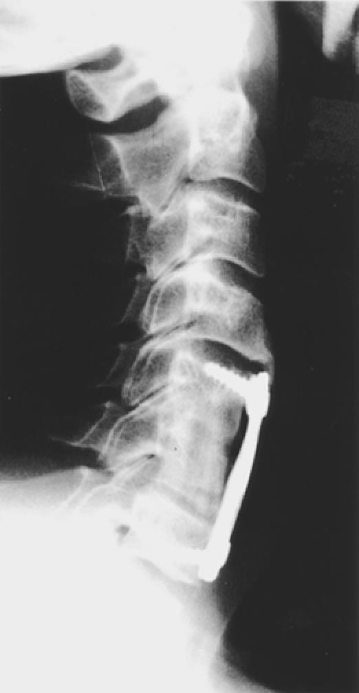
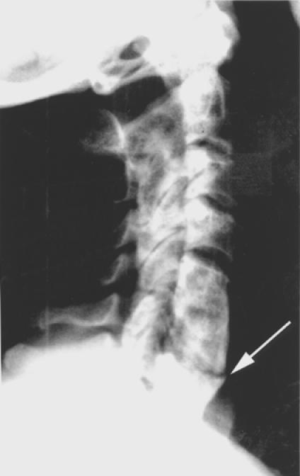
Nineteen patients were classified according to their fusion status at a minimum follow-up of two years. The categories of fusion were as follows: 1) definitely fused (16/19; 84%) (Figure 1); 2) questionable fusion (2/19; 11%); and 3) definitely not fused (1/19; 5%) (Figure 2). No evidence of instability was noted in any category. Radiographic instability was defined by >3.5 mm of sagittal plane translation (or 20%) and 20° of sagittal plane rotation on dynamic X-rays.19 The average loss of height from post-op to follow-up was 8 mm (range, 0 to 39 mm). The average angulation change was 2° of kyphosis (range , -4° to 13°). Angulations less than 0° represented a lordotic change.
Figure 1.

Lateral-extension cervical radiograph of a 40 y/o patient four years status post C6 corpectomy, fibular allograft strut (C5-C7), and anterior cervical plate (C5-C7). This graft is well united with bridging bony trabeculae and was categorized as definitely fused (Class 1).
Figure 2.

Lateral-extension cervical radiograph of a 62 y/o patient four years status post C6 corpectomy, fibular allograft strut (C5-C7), and anterior cervical plate (C5-C7). The plate was removed nine months post-operatively because of dysphagia. This was the only patient who definitely did not fuse (arrow) and was categorized as a Class 3.
DISCUSSION
When a decompressive corpectomy is used for spondylosis, tumor, kyphotic deformity, or stenosis, a fibular strut graft is well suited for surgical reconstruction. Both fibular allografts and autografts have been used. Several studies have evaluated fusion rates using autologous bone.1,8–10 However, the use of autologous bone is not without problems. Hematomas, wound infections, neuropraxia, and pain are known complications when harvesting autologous bone.6 In an effort to eliminate donor site morbidity, spinal surgeons have begun using fibular allograft. Only a few studies have documented cervical fusion rates when using fibular allograft after corpectomy.3,12,21 The purpose of this study was to provide long-term clinical and radiologic evaluation in patients who had anterior cervical corpectomy and stabilization with fibular allograft.
The majority of patients in the current study obtained a solid radiographic fusion after cervical corpectomy and grafting with a fibular allograft. Eighty-four percent of the patients were classified as definitely fused at a minimum two year follow-up. Eleven percent were classified as possibly fused because of uncertainty in radiographic views. Thus, ninety-five percent were either definitely or possibly fused. Only one patient was definitely not fused because of more than one millimeter movement on flexion-extension views. Of note, this patient was a heavy smoker and did not have any clinical symptoms or complaints at four year follow-up. Her physical exam was normal without any upper motor neuron signs. Significantly, this patient had her plate removed after nine months because of complaints of dysphagia. This may have led to the failure of fusion radiographically.
Patients in this study functioned well after surgery without significant complications. Eighty-eight percent of the patients did not have significant neck pain. Eighty-one percent regained their premorbid activities. No patient had pain or functional impairment which interfered with their activities of daily living. Oswestry Back Scores were consistent with these findings at an average score of twenty-nine.
Only a few studies have been published which evaluate fusion rates after corpectomy and grafting with a fibular allograft. Fernyhough et al.5 compared 67 patients, who had discectomy, corpectomy and fusion with fibular autograft, verses 59 patients who had the same procedure with fibula allograft. The average follow-up was 83 months. Osseous union occurred in 73% of patients with autograft fibula and in 59% of allograft fibula. In contrast to our study, this group did not use any supplemental fixation or hardware, which may account for the low percentage of osseous union with allograft fibula.
Grossman et al.7 peformed anterior decompression by a partial or complete corpectomy followed by allograft strut fusion in eight patients. These patients were followed for over one year. All grafts progressed to fusion. Clinical follow-up revealed one patient with excellent results, six patients with good results, and one patient with fair results. No patient had a plate. All patients were treated with a halo vest for 12 weeks postoperatively. The additional stability with the halo vest after surgery possibly resulted in the high fusion rate.
Zdeblick and colleagues followed fourteen patients with severe cervical kyphosis and myelopathy for over two years.21 All of their patients were treated with corpectomies and arthodesis. Only eight patients in this series had a fibular allograft strut used. However, all of these subjects had a solid radiographic union at a minimum follow-up of two years. Emery et al found similar results in their review of 108 patients with cervical spondylotic myelopathy who had been managed with anterior decompression and fusion.3 Thirty-eight patients in this study had fibular allografts placed after a corpectomy. Ninety-seven percent of the patients had radiographic union after two years.
Two studies have evaluated fusion rates following corpectomy, fibular allografts, and plate osteosynthesis. Macdonald et al.12 followed 36 patients with a pre-operative diagnosis of myleopathy. All patients received a multilevel corpectomy and fibula allograft. Only 15 patients had an anterior cervical plate placed. Patients were placed in halo immobilization for 3 months after surgery. Twenty-six patients were available for more than 24 months follow-up, and 97% of these patients had solid osseous union. Mayr et al. followed 261 patients for more than 2 years but did not document length of radiographic follow-up.13 Eighty-seven percent of this study achieved a solid union. A single surgeon performed all operations with anterior cervical plates used in each case. The results of our current study are comparable with both of these studies using supplemental plating.
The length of follow-up in this study is a significant strength. Sufficient time is necessary for strut grafts to incorporate in the surrounding vertebral bodies. All of our patients were followed for at least two years (mean 46 months) with some followed as long as 85 months. Another strength of this study is having both clinical and radiographic follow-up. All patients returned to the clinic for radiographs as well as a clinical exam. An SF-36 and Oswestry Back Scale were completed by each patient to help assess their function objectively. All radiographs were classified by a blinded board certified musculoskeletal radiologist.
This study had several strengths, but the limitations need to be mentioned as well. Because this was a retrospective study, we did not have SF-36 scores or Oswestry Score pre-operatively. In addition, six patients were not included in the study for the reasons outlined. However, at their last documented clinic visit, this group of patients was radiographically and clinically similar to the study group. None of these patients had a nonunion of their fusion. The data from our study strongly indicate that the use of a fibular allograft strut for cervical interbody fusion after corpectomy provides for relatively high rates of arthrodesis without severe loss of height or sagittal alignment at long term radiographic followup. The majority of these patients are able to regain pre-morbid activities and are free of significant pain without potential donor site morbidity.
Footnotes
Funded by a research grant from Sofamor-Danek, Inc.
References
- 1.Boni M, Cherubino P, Denaro V, Benazzo F. Multiple subtotal somatectomy. Technique and evaluation of a series of 39 cases. Spine. 1984;9(4):358–362. [PubMed] [Google Scholar]
- 2.Brown JA, Malinin TI, Daavis PB. A roentgenographic evaluation of frozen allografts versus autografts in anterior cervical spine fusions. Clin Orthop. 1976;119:231–236. [PubMed] [Google Scholar]
- 3.Emery SE, Bohlman HH, Bolesta MJ, Jones PK. Anterior cervical decompression and arthrodesis for the treatment of cervical spondylotic myelopathy. Two to seventeen-year follow-up. J Bone Joint Surg. (Am) 1998;80(7):941–951. doi: 10.2106/00004623-199807000-00002. [DOI] [PubMed] [Google Scholar]
- 4.Emery SE, Bolesta MJ, Banks MA, Jones PK. Robinson anterior cervical fusion comparison of the standard and modified techniques. Spine. 1994;19(6):660–663. [PubMed] [Google Scholar]
- 5.Fernyhough JC, White JI, LaRocca H. Fusion rates in multilevel cervical spondylosis comparing allograft fibula with autograft fibula in 126 patients. Spine. 1991;16(10 Suppl):S561–564. doi: 10.1097/00007632-199110001-00022. [DOI] [PubMed] [Google Scholar]
- 6.Gore DR, Sepic SB. Anterior cervical fusion for degenerated or protruded discs. A review of one hundred forty-six patients. Spine. 1984;9(7):667–671. doi: 10.1097/00007632-198410000-00002. [DOI] [PubMed] [Google Scholar]
- 7.Grossman W, Peppelman WC, Baum JA, Kraus DR. The use of freeze-dried fibular allograft in anterior cervical fusion. Spine. 1992;17(5):565–569. doi: 10.1097/00007632-199205000-00015. [DOI] [PubMed] [Google Scholar]
- 8.Hanai K, Fujiyoshi F, Kamei K. Subtotal vertebrectomy and spinal fusion for cervical spondylotic myelopathy. Spine. 1986;11(4):310–315. doi: 10.1097/00007632-198605000-00003. [DOI] [PubMed] [Google Scholar]
- 9.Hanai K, Inouye Y, Kawai K, Tago K, Itoh Y. Anterior decompression for myelopathy resulting from ossification of the posterior longitudinal ligament. J Bone Joint Surg. (Br) 1982;64(5):561–564. doi: 10.1302/0301-620X.64B5.6815199. [DOI] [PubMed] [Google Scholar]
- 10.Herman JM, Sonntag VK. Cervical corpectomy and plate fixation for postlaminectomy kyphosis. J Neurosurg. 1994;80(6):963–970. doi: 10.3171/jns.1994.80.6.0963. [DOI] [PubMed] [Google Scholar]
- 11.Kurz LT, Garfin SR, Booth RE., Jr Harvesting autogenous iliac bone grafts. A review of complications and techniques. Spine. 1989;14(12):1324–1331. doi: 10.1097/00007632-198912000-00009. [DOI] [PubMed] [Google Scholar]
- 12.Macdonald RL, Fehlings MG, Tator CH, Lozano A, Fleming JR, Gentili F, Bernstein M, Wallace MC, Tasker RR. Multilevel anterior cervical corpectomy and fibular allograft fusion for cervical myelopathy. J Neurosurg. 1997;86(6):990–997. doi: 10.3171/jns.1997.86.6.0990. [DOI] [PubMed] [Google Scholar]
- 13.Mayr MT, Haid RW, Comey CH, Rodts GE. Anterior Cervical Corpectomy, fibular allograft, and plate osteosynthesis: Results and Complications. Transactions North American Spine Society. 1998. pp. 13–20.
- 14.Scott J, Huskisson EC. Graphic representation of pain. Pain. 1976;2(2):175–184. [PubMed] [Google Scholar]
- 15.Scott J, Huskisson EC. Vertical or horizontal visual analogue scales. Ann Rheum Dis. 1979;38(6) doi: 10.1136/ard.38.6.560. [DOI] [PMC free article] [PubMed] [Google Scholar]
- 16.Seifert V, Stolke D. Multisegmental cervical spondylosis: treatment by spondylectomy, microsurgical decompression, and osteosynthesis. Neurosurgery. 1991;29(4):498–503. [PubMed] [Google Scholar]
- 17.Triano JJ, McGregor M, Cramer GD, Emde DL. A comparison of outcome measures for use with back pain patients: results of a feasibility study [see comments] J Manipulative Physiol Ther. 1993;16(2):67–73. [PubMed] [Google Scholar]
- 18.Ware JE, Jr, Sherbourne CD. The MOS 36-item short-form health survey (SF-36). I. Conceptual framework and item selection. Med Care. 1992;30(6):473–483. [PubMed] [Google Scholar]
- 19.White A, Panjabi M. Clinical biomechanics of the spine. 2nd. Philadelphia: JB Lippincott Co; 1990. [Google Scholar]
- 20.Young WF, Rosenwasser RH. An early comparative analysis of the use of fibular allograft versus autologous iliac crest graft for interbody fusion after anterior cervical discectomy. Spine. 1993;18(9):1123–1124. doi: 10.1097/00007632-199307000-00002. [DOI] [PubMed] [Google Scholar]
- 21.Zdeblick TA, Bohlman HH. Cervical kyphosis and myelopathy Treatment by anterior corpectomy and strut-grafting. J Bone Joint Surg. (Am) 1989;71(2):170–182. [PubMed] [Google Scholar]


