Abstract
Case report of a complete arcuate foramen in a human atlas vertebra inhibiting the placement of lateral mass screw instrumentation at C1.
Our objective is to report the presentation of the case, the operative considerations, and the management for this anatomic variation.
The groove for the vertebral artery on the posterolateral surface of the atlas (C1) varies in size and depth from a slight impression to a clear sulcus. With anomalous ossification the sulcus can be bridged which results in a posterolateral tunnel within the posterior arch of the atlas. With increasing rates of screw fixation instrumentation that include the atlas, it is of paramount importance to know the location and course of the vertebral artery in relation to the planned route of instrumentation.
The patient underwent a posterolateral fusion from C1 to C4 using autogenous iliac crest bone graft. Internal fixation from C2 to C4 was obtained using lateral mass screw instrumentation. After the vertebral artery was identified passing through the posterior arch of C1, sublaminar wires were utilized for fixation from C1 to C2. The patient responded well to surgical intervention without complications.
Abnormal vertebral artery coursing through a posterolateral tunnel in the posterior arch of C1 has been described and its incidence has a range from 1.14% to 18%. When this variant is present, lateral mass screw fixation at C1 may be contraindicated. We recommend close scrutiny of preoperative radiographs to avoid the possibility of endangering the vertebral artery when this situation exists.
PRECIS
This is a case report of a patient with rheumatoid arthritis and cervical instability requiring surgical intervention found to have a complete arcuate foramen within the posterior arch of C1 through which the vertebral artery coursed. The clinical presentation, operative considerations, and surgical management are outlined.
INTRODUCTION
The posterolateral margin of the atlas contains a sulcus or groove for the vertebral artery which can vary in size and depth.1 This groove can be bridged by anomalous ossification and a posterior ponticulus (Latin for bridge). The opening in the posterior arch of the atlas is termed the arcuate foramen, through which pass the vertebral artery and first cervical nerve. This foramen has been known by many names, but most frequently by the eponym "Kimmerle's anomaly" since Kimmerle was an early describer of this structure.2 Other terms appear in the anatomy literature to describe the same structure include: "foramen sagittale", "foramen atlantoideum", "foramen retroarticulare superior", "canalis vertebralis", "retrocondylar vertebral artery". In addition to its anatomic significance, the arcuate foramen has been postulated to play a role in clinically relevant entities such as migraines and vertebrobasilar artery stroke.3,4 The incidence of the arcuate foramen range from 1.14% to 18% depending on the study.1,4–8 Previous studies vary in study design (radiographic vs. cadaveric analysis), population studied, and grouping of the various types of arcuate foramen phenotypes.
If surgical management is performed, stabilization of the C1-C2 joint is typically accomplished through reduction and fusion of the atlantoaxial complex with internal fixation through a posterior approach. The type of internal fixation varies from wiring procedures such as the Brooks and Gallie techniques to Magerl's transarticular screw technique.9–12 More recently, Harms and Melcher have published a posterior C1-C2 fusion using polyaxial screw fixation.13 Biomechanically, screw fixation has been shown to be superior to posterior wiring.14–19 In addition, fusion rates using screw fixation are also improved over wiring.20–26 Although screw fixation has a biomechanical advantage and superior fusion rates compared to wire fixation, the technique is more demanding and carries greater risk of injuring the vertebral artery. When the vertebral artery courses above the posterior arch of C1, the placement of lateral mass screws is relatively safe27; however, the risk may increase significantly with any anomalous course of the vertebral artery. Indeed, the above-mentioned arcuate foramen would place the vertebral artery in the path of any C1 lateral mass screw.
This report describes a case of an anomalous vertebral artery coursing through the posterior arch of the atlas that precluded C1 lateral mass screw fixation in a patient with rheumatoid arthritis undergoing reduction and internal fixation of her cervical spine.
CASE
The patient is a 67-year-old female with rheumatoid arthritis who presented with complaints of neck pain with radicular symptoms into both upper extremities. Her pain was located posteriorly along the cervical spine with radiation up to the occiput and down into the shoulders bilaterally. She had been plagued by this pain for one year prior to presentation with significant worsening over the two previous months. She also complained of numbness in her hands in a glove-like distribution extending just proximal to the wrists bilaterally. She had difficulty buttoning buttons, holding coffee cups, and writing. Her pain and numbness persisted despite chiropractic treatment, physical therapy, and multiple narcotic medications. Her medical history included hypothyroidism and osteoporosis. Her surgical history included a right total hip arthroplasty in 1986, with a revision in 1997, a total abdominal hysterectomy and bilateral salpingoophrectomy in 1975, an L5-S1 decompression and fusion in 2000, appendectomy, and holecystectomy. Physical examination revealed full range of motion of the neck with pain elicited on extension and lateral bending. She had a normal gait. There was 4 out of 5 strength in all muscle groups in the left upper extremity with numbness of both hands in a glove-like distribution extending just proximal to the wrist. Examination of the hands revealed mild metacarpo-phalangeal swelling diffusely and symmetrical ulnar drift of the digits bilaterally. Deep tendon reflexes in both upper and lower extremities were normal and symmetrical with no pathologic reflexes. Radiographs revealed diffuse cervical spine degeneration with notable C1-2, C2-3 anterolisthesis. The anterior atlanto-dens interval measured 8mm and the posterior atlanto-dens interval measured 15mm in flexion with near anatomic correction in extension. There were no signs of cranial settling. Further imaging with CT and MRI scans confirmed these findings.
There were many factors that prompted the decision to offer operative management to this patient. The major concern was quality of life. In her condition at presentation, the cervical instability caused intractable pain and was associated with progressive myelopathic symptoms involving her hands.
The patient underwent Halo vest placement one day prior to surgery. Once the halo vest was placed, the patient's cervical spine was positioned so that the anterior atlanto-dens interval was less than four millimeters. Radiographs were taken to verify proper positioning. On the following day, the patient underwent C1 to C4 posterior arthrodesis utilizing autogenous iliac crest bone graft. After the patient was placed prone, the back of the halo vest was removed while leaving the front of the brace intact to maintain adequate alignment of the cervical spine. Segmental instrumentation was achieved using lateral mass screw fixation at the C2, C3, and C4 levels bilaterally. When the posterior arch of C1 was approached, the vertebral artery was found to enter the lateral aspect of C1 rather than coursing along its superior aspect. In order to avoid placing a C1 lateral mass screw through the vertebral artery, lateral mass screw fixation was abandoned and substituted with sublaminar wire placement from C1 to C2 using the Brooks technique to achieve fixation at this level. The postoperative course was uneventful, and the patient was discharged home on postoperative day nine and managed with halo vest brace immobilization. Two months after surgery, the patient is doing well. She reports mild neck pain much improved compared to her preoperative status as well as mild hand pain bilaterally. She has noticed a significant improvement in the ability to use her hands for fine motor tasks in addition to a decrease in her hand numbness. Radiographs at this time show intact instrumentation and well-maintained correction of her cervical spine.
DISCUSSION
A review of the literature revealed multiple studies with differing results regarding incidence of the arcuate foramen. These results are further confounded by the differing types of studies, populations studied, and differing groupings of foramen types. Pyo and Lowman8 found an incidence of 38 (12.67%) partial and complete foramen in 300 patients while Dugdale7 found 47 complete and 37 partial foramen in 316 patients. Kendrick and Biggs5 had a 15.8% combined incidence in 353 white children (ages 6-17) with a 5.49% and 9.15% incidence of complete and partial foramina, respectively. Stubbs6 found a complete foramen in 13.5% and a partial foramen in 5.2% of the study population (n=1000). More recently however, a cadaveric study performed by Hasan et al.1 on 350 dried macerated north Indian atlas vertebrae only reported a 3.42% incidence of complete foramen and a 1.14% incidence of a posterolateral tunnel. This study made a distinction between a complete posterior ponticulus and a more extensive posterolateral tunnel-like canal.
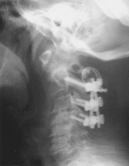
Screw fixation of C1 is evolving into newer techniques such as the one described recently by Harms and Melcher using C1-C2 polyaxial screws to achieve fixation.13 This case illustrates the clinical relevance of a posterolateral tunnel in the posterior arch of C1 carrying the vertebral artery. When this abnormality is found, screw fixation through the lateral mass of C1 is not feasible and other modalities of internal fixation must be pursued. In our case, we found the abnormal vertebral artery entering the lateral aspect of C1 intra-operatively and opted to obtain C1-C2 fixation with sublaminar wires. However, in retrospect, the pre-operative lateral radiograph did demonstrate the posterolateral tunnel. Careful analysis could have prevented this near complication.
CONCLUSION
Abnormal vertebral artery coursing through a posterolateral tunnel in the posterior arch of C1 has been described and its incidence has a range from 1.14% to 18%. When this variant is present, lateral mass screw fixation at C1 may be contra-indicated. We recommend close scrutiny of pre-operative plain films to avoid the possibility of endangering the vertebral artery when this situation exists.
Figure 1.

Postoperative lateral radiograph demonstrating a complete arcuate foramen in the posterior arch of C1 precluding facile screw placement.
References
- 1.Hasan M, et al. Posterolateral tunnels and ponticuli in human atlas vertebrae. J Anat. 2001;199(3):339–343. doi: 10.1046/j.1469-7580.2001.19930339.x. [DOI] [PMC free article] [PubMed] [Google Scholar]
- 2.Kimmerle A. Ponticulus posticus. Rontgenprax. 1930;2:479–483. [Google Scholar]
- 3.Cushing KE, et al. Tethering of the vertebral artery in the congenital arcuate foramen of the atlas vertebra: a possible cause of vertebral artery dissection in children. Dev Med Child Neurol. 2001;43(7):491–496. doi: 10.1017/s0012162201000901. [DOI] [PubMed] [Google Scholar]
- 4.Wight S, Osborne N, Breen AC. Incidence of ponticulus posterior of the atlas in migraine and cervicogenic headache. J Manipulative Physiol Ther. 1999;22(1):15–20. doi: 10.1016/s0161-4754(99)70100-4. [DOI] [PubMed] [Google Scholar]
- 5.Kendrick C, Biggs N. Incidence of the ponticulus posticus of the first cervical vertebra between ages six to seventeen. Anat Rec. 1963;145:449–451. doi: 10.1002/ar.1091450308. [DOI] [PubMed] [Google Scholar]
- 6.Stubbs DM. The arcuate foramen. Variability in distribution related to race and sex. Spine. 1992;17(12):1502–1504. [PubMed] [Google Scholar]
- 7.Dugdale L. The ponticulus posterior of the atlas. Australas Radiol. 1981;25(3):237–238. doi: 10.1111/j.1440-1673.1981.tb02254.x. [DOI] [PubMed] [Google Scholar]
- 8.Pyo J, Lowman R. The "ponticulus posticus" of the first cervical vertebra. Radiology. 1959;72:850–854. doi: 10.1148/72.6.850. [DOI] [PubMed] [Google Scholar]
- 9.Brooks AL, Jenkins EB. Atlanto-axial arthrodesis by the wedge compression method. J Bone Joint Surg. (Am) 1978;60:279–284. [PubMed] [Google Scholar]
- 10.Gallie W. Fractures and dislocations of the cervical spine. Am J Surg. 1939;46:495–459. [Google Scholar]
- 11.Holness RO, et al. Posterior stabilization with an interlaminar clamp in cervical injuries: Technical note and review of long term experience with the method. Neurosurgery. 1984;14:318–322. doi: 10.1227/00006123-198403000-00010. [DOI] [PubMed] [Google Scholar]
- 12.Magerl F, Seemann PS. Stable posterior fusion of the atlas and axis by transarticular screw fixation. In: Kehr P, Weidner A, editors. Cervical Spine. One. New York/Wein: Springer; 1986. pp. 322–327. [Google Scholar]
- 13.Harms J, Melcher RP. Posterior c1-c2 fusion with polyaxial screw and rod fixation. Spine. 2001;26(22):2467–2471. doi: 10.1097/00007632-200111150-00014. [DOI] [PubMed] [Google Scholar]
- 14.Crisco JJI, et al. Bone graft translation of four upper cervical spine fixation techniques in a cadaveric model. J Orthop Res. 1991;9:835–846. doi: 10.1002/jor.1100090609. [DOI] [PubMed] [Google Scholar]
- 15.Grob D, et al. Biomechanical evaluation of four different posterior atlantoaxial fixation techniques. Spine. 1992;17:480–490. doi: 10.1097/00007632-199205000-00003. [DOI] [PubMed] [Google Scholar]
- 16.Grob D, et al. Dorsal atlanto-axial screw fixation: A stability test in vitro and in vivo. Orthopaedics. 1991;20:154–162. [PubMed] [Google Scholar]
- 17.Henriques T, et al. Biomechanical comparison of five different atlantoaxial posterior fixation techniques. Spine. 2000;25:2877–2883. doi: 10.1097/00007632-200011150-00007. [DOI] [PubMed] [Google Scholar]
- 18.Naderi S, et al. Biomechanical comparison of C1-C2 posterior fixations. Cable, graft, and screw combinations. Spine. 1998;23(18):1946–1956. doi: 10.1097/00007632-199809150-00005. [DOI] [PubMed] [Google Scholar]
- 19.Smith MD, et al. A biomechanical analysis of atlantoaxial stabilization methods using a bovine model. C1/C2 fixation analysis. Clin Orthop. 1993;290:285–295. [PubMed] [Google Scholar]
- 20.Conye TJ, et al. C1-C2 posterior cervical fusion: Long-term evaluation of results and efficacy. Neurosurgery. 1995;37:688–692. doi: 10.1227/00006123-199510000-00012. [DOI] [PubMed] [Google Scholar]
- 21.Dickman CA, Sonntag VK. Posterior C1-C2 transarticular screw fixation for atlantoaxial arthrodesis. Neurosurgery. 1998;43:275–280. doi: 10.1097/00006123-199808000-00056. [DOI] [PubMed] [Google Scholar]
- 22.Dickman CA, Sonntag VK. Surgical management of atlantoaxial nonunions. Neurosurgery. 1995;83:248–253. doi: 10.3171/jns.1995.83.2.0248. [DOI] [PubMed] [Google Scholar]
- 23.Grob D, et al. Atlanto-axial fusion with transarticular screw fixation. J Bone Joint Surg. (Br) 1991;73:972–976. doi: 10.1302/0301-620X.73B6.1955447. [DOI] [PubMed] [Google Scholar]
- 24.Grob D, Magerl F. Surgical stabilization of C1 and C2 fractures. Orthopade. 1987;16:46–54. [PubMed] [Google Scholar]
- 25.Jeanneret B, Magerl F. Primary posterior fusion C1/2 in odontoid fractures: Indications, technique, and results of transarticular screw fixation. J Spinal Disord. 1992;5:464–475. doi: 10.1097/00002517-199212000-00012. [DOI] [PubMed] [Google Scholar]
- 26.Stillerman CB, Wilson JA. Atlanto-axial stabilization with posterior transarticular screw fixation: technical description and report of 22 cases. Neurosurgery. 1993;32(6):948–955. doi: 10.1227/00006123-199306000-00011. [DOI] [PubMed] [Google Scholar]
- 27.Gupta S, Goel A. Quantitative anatomy of the lateral masses of the atlas and axis vertebrae. Neurol India. 2000;48(2):120–125. [PubMed] [Google Scholar]


