Abstract
Tuberculosis of the bone is a well-recognized clinical condition that can be diagnosed and managed by physicians and orthopaedic surgeons, often with an excellent outcome. However, the occurrence of multifocal skeletal involvement in immunocompetent patients is rare, even in countries where tuberculosis is endemic. Patients with multifocal skeletal tuberculosis may present with multiple vague somatic symptoms. We report three cases of multifocal skeletal tuberculosis in non-immunocompromised patients. All three patients were effectively treated with antituberculous drugs.
INTRODUCTION
Tuberculosis of the bone, constituting 10 to 20 percent of all tuberculosis, is a well-recognized clinical condition that is easily diagnosed and managed by physicians and orthopaedic surgeons with an excellent outcome. However, the occurrence of multifocal skeletal involvement is exceptional, constituting less than five percent of all bony tuberculosis, even in countries where tuberculosis is commonplace. Tuberculosis with multiple-bone involvement is extremely unlikely in immunocompetent patients and in those with normal pulmonary findings. Nevertheless, since patients with multifocal skeletal tuberculosis may present with vague multiple somatic symptoms, orthopaedic surgeons should maintain a high degree of suspicion in order to diagnose this condition. Multifocal skeletal tuberculosis must be considered in the differential diagnosis in the presence of multiple destructive skeletal lesions. Furthermore, this condition may mimic malignant disease both clinically and radiologically. Radiographs should be done as a first screening test, while bone scan may help to detect multiple bone involvement at an early stage. CT scans may be useful to demonstrate the extent of the lytic lesions, while biopsy confirms the diagnosis. We report three cases of multifocal tuberculosis in non-immunocompromised patients. All three patients were effectively treated with antituberculous drugs.
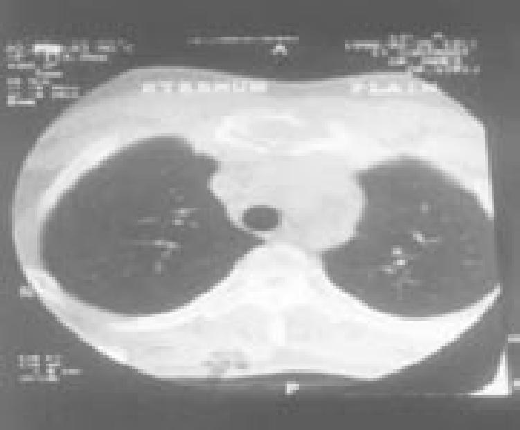
Case 1: A 19-year-old engineering student presented to us with complaints of diffuse pain over the back, chest, and right hip region of six months’ duration. He gave a history of decreased appetite and significant weight loss. There was no history of night sweats, chronic cough, or exposure to tuberculosis. On examination, he had tenderness over the upper thoracic spine, the thoracolumbar junction, the sternum, and the right sacroiliac joint. There was no paraspinal swelling or spasm. The peripheral blood count was normal. His erythrocyte sedimentation rate (ESR) was 54mm and Mantoux test was non-reactive. Radiographs of the thoracolumbar spine, chest, and pelvis were normal. Isotope scan showed an increased uptake in T1, T2, T9 through L2, the right sacroiliac joint, and the sternum (Figure 1). CT scan showed destruction of the involved vertebral bodies, the right sacroiliac joint, and the sternum (Figure 2). Biopsy of the sternal lesion showed a granulomatous lesion suggestive of tuberculosis.
CASE 1.
Figure 1.
CT scan of the spine showing lesions at T9, T10, and L2.
Figure 2.

CT scan of the chest showing a lesion in sternum.
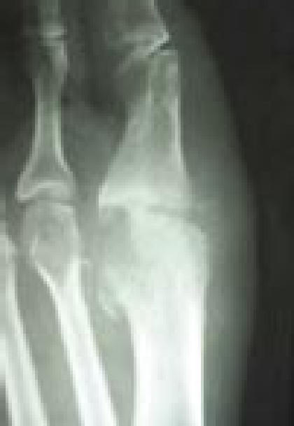
Case 2:A 41-year-old executive came to our outpatient department with complaints of pain over the mid and low back, the right side of the chest, the left hip region, and the right first metatarsophalangeal (MTP) joint of eight months’ duration. There were no constitutional symptoms of tuberculosis. On examination, he had tenderness over the mid-thoracic spine, thoracolumbar junction, and left sacroiliac joint. Paraspinal spasm was present, but there was no paraspinal swelling. His ESR was 38mm. Radiographs showed lytic lesions in the first MTP joint (Figure 3), and the T7 and T8 vertebral bodies. A bone scan showed increased uptake in T7, T8, L2, the left sacroiliac joint, the right fourth and fifth ribs, and the right first MTP joint. His CT scan showed lytic lesions in the left ilium in addition to lesions at T7, T8, and L2 (Figure 4). His Mantoux test was positive, showing a 14 x 15 mm area of induration at 48 hours. Biopsy of the first metatarsal head confirmed tuberculosis.
CASE 2.
Figure 3.

Radiograph of the right first metatarsophalangeal joint reveals a lytic lesion.
Figure 4.

CT scan of the pelvis reveals lytic lesions in the left ilium.
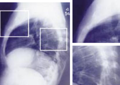
Case 3: A 55-year-old housewife presented with pain in the upper back and left hip region and a discharging sinus over the sternum of five months’ duration. On examination, there was gibbus at T6 and T7, as well as tenderness at the left sacroiliac joint and a discharging sinus over the sternum. Her ESR was 93mm and Mantoux test was strongly positive. Radiographs showed erosive lesions in the sternum and left sacroiliac joint and loss of the intervertebral space between T6 and T7 (Figure 5). Tuberculosis was confirmed by biopsy of the sternal lesion.
CASE 3. Figure 5.
Lateral radiograph of the chest and thoracic spine with lytic lesions visible at T6 and T7 as well as the sternum.
Treatment Course
Enzyme-linked immunosorbent assay (ELISA) testing for the human immunodeficiency virus (HIV) was done for all three patients and all were negative for the presence of HIV. All three patients were treated conservatively with bracing and antituberculous drugs. They had two months of intensive therapy with four drugs: isoniazid, rifampin, ethambutol, and pyrazinamide, followed by four months’ treatment with isoniazid and rifampin. Follow-up CT scan after 18 months showed resolution of all lesions in all the patients.
DISCUSSION
Tuberculosis of the bone is a relatively common clinical condition, constituting 10 to 20 percent of all tuberculosis. It can occur at any age and at almost any site of the body. In the spine, the thoracolumbar region is the most commonly affected. The multifocal skeletal form of tuberculosis is exceptional even in endemic countries, representing less than 5 percent of all bony tuberculosis.1 The multifocal skeletal form is usually seen associated with pulmonary tuberculosis. A suppressed host immune response also predisposes to multifocal tuberculosis. Tuberculosis with multiple bone involvement is exceedingly rare in non-immunocompromised patients and in those with normal pulmonary findings.
In the first patient, we initially suspected leukemia based on his history and clinical examination and because one of his first-degree relatives had died of leukemia. However, his peripheral blood count was normal. Following his isotope scan and CT scan, our differential diagnoses included bony secondary metastases, multiple myeloma, or hyperparathyroidism. However, the bone marrow aspiration, serum electrophoresis, and parathyroid hormone assay were normal. The final diagnosis was confirmed by biopsy of the sternum which showed a granulomatous lesion suggestive of tuberculosis.2
In the second patient, based on his clinical findings and bone scan, we considered multiple myeloma, multiple secondary metastases, and multifocal tuberculosis in the differential diagnosis. Bone marrow aspirate, serum electrophoresis, and urine testing for Bence-Jones proteins were negative. Biopsy of the first metatarsal head confirmed tuberculosis.
For the third patient, the diagnosis of tuberculosis was more obvious with a sternal sinus and thoracic spine lesions with deformity. Tuberculosis was confirmed by biopsy of the sternal lesion.
Approximately 50 percent of patients with bone and joint tuberculosis have negative findings on chest X-ray.3 We feel that the difficulty in diagnosing multifocal skeletal tuberculosis is due to both the generalized somatic symptoms at presentation, and the non-specific physical findings and radiologic results, all of which give rise to a large differential diagnosis. Therefore, physicians should maintain a high degree of suspicion for tuberculosis if a patient presents with multiple somatic symptoms, particularly if the patient is from an area where tuberculosis is endemic. Multifocal skeletal tuberculosis must be considered in the differential diagnosis of multiple destructive skeletal lesions. This condition may mimic malignant disease both clinically and radiographically.4
The mainstay of treatment of spinal tuberculosis is conservative management with bracing and anti-tuberculosis drugs. Surgery is needed only if there is a neurologic deficit or spinal instability. These lesions respond rapidly to anti-tuberculosis drugs and quiescence is evidenced by the return of bone density to normal, as illustrated in these three cases.
References
- 1.Moujtahid M, Essadki B, Lamine A, Bennouno D, Zryouil B. Multifocal bone tuberculosi: Apropos of a case. Rev Chir Orthop Reparatrica. Appar Mot. 1995;81(6):553–556. French. [PubMed] [Google Scholar]
- 2.Vaylet F, deMuizon H, L’Her P, Natali F, Allard P. Multifocal tuberculosis of bone. Apropos of an exceptional case. Rev Pneumol Clin. 1989;45(2):81–85. French. [PubMed] [Google Scholar]
- 3.Davidson PT, Horowity I. Skeletal tuberculosis: A review with patient presentations and discussions. Am J Med. 1970;48:77–84. doi: 10.1016/0002-9343(70)90101-4. [DOI] [PubMed] [Google Scholar]
- 4.Muadali D, Gold WL, Vellend H, Becker E. Multifocal osteoarticular tuberculosis: report of four cases and review of management. Clin Infect Dis. 1993 Aug;17(2):204–209. [PubMed] [Google Scholar]
- 5.Lachenaver CS, Consentino S, Wood RS, Yousef Zedeh DK, McCarthy CA. Multifocal skeletal tuberculosis presenting as osteomyelitis of the jaw. Pae Inf Dis J. 1991;10:940–944. doi: 10.1097/00006454-199112000-00012. [DOI] [PubMed] [Google Scholar]


