Abstract
A 20-year-old man presented to our clinic with pain and swelling in the right knee of one year's duration. Biopsy of the patella revealed an aneurysmal bone cyst secondary to a giant cell tumor. He was treated by curettage and bone cement to fill the defect. The rarity of this lesion in the patella and its treatment modalities are discussed.
INTRODUCTION
We present a case of an aneurysmal bone cyst secondary to a giant cell tumor in a 20-year-old patient, at an unusual site - the patella. He was treated by curettage of the lesion and bone cement to fill the cavity. A review of the literature reveals only one other case of a secondary aneurysmal bone cyst of the patella.
CASE REPORT
A 20-year-old college student presented to our orthopaedic outpatient department with complaints of pain in the right knee of three years' duration, and swelling of the right knee for one year. He also had fevers in the evenings and loss of appetite for three weeks. There was no history of weight loss or exposure to tuberculosis.
On examination, the patella appeared bigger and more prominent on the right side compared to the left. There was no localized warmth, but the patella was notably tender. In addition, there was a palpable bony thickening of the patella. No synovial thickening or knee joint effusion was noted. His knee range of motion was 0 to 110 degrees with pain at terminal flexion. There was no ligamentous laxity.
Laboratory studies revealed a hemoglobin of 13.5 gm/percent and white blood cell count of 13,000 cu.mm. Inflammatory markers were within the normal limits; his erythrocyte sedimentation rate was 18, C-reactive protein was negative, and alkaline phosphatase was 286 units. Mantoux test was non-reactive.
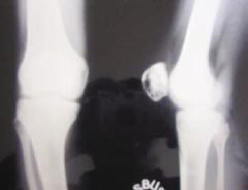
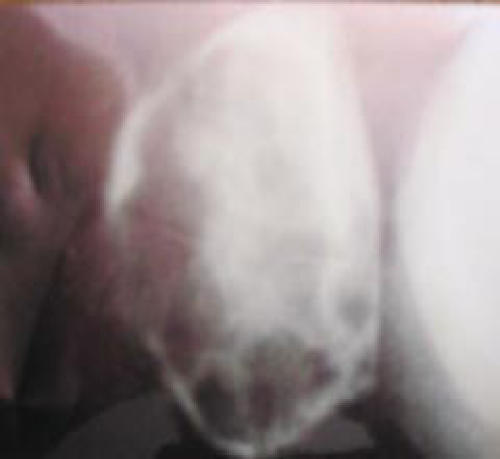
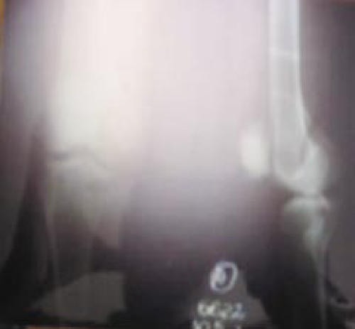
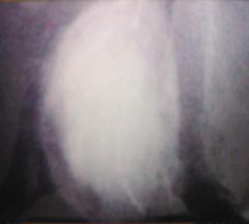
Radiographs of the right knee showed multiple lytic lesions in the patella (Figure 1). Core biopsy of the patella was done. The histology was consistent with an aneurysmal bone cyst. The patient then underwent curettage of the lesion using a high speed burr through a window made on the medial aspect of the patella. The material was sent for formal histopathological examination. The cavities in the patella were packed with bone cement (Figure 2). The biopsy showed an aneurysmal bone cyst with multiple giant cells confirming the diagnosis of aneurysmal bone cyst secondary to giant cell tumor.
Figure 1.
Radiographs of the right knee (1A) and magnified view of the patella (1B) revealing lytic lesions in the patella.
Figure 1A.
Figure 1B.
Figure 2.
Post-operative radiographs of the knee (2A) and magnified view of the patella (2B) after curettage and placement of bone cement.
Figure 2A.
Figure 2B.
DISCUSSION
Aneurysmal bone cyst (ABC) is an expansile cystic lesion, often occurring in the second decade of life,1 with a slightly increased incidence in women. It constitutes one to six percent of all primary bone tumors. Long bones are the most often affected, but the spine is involved in 30 percent of patients with ABC.
ABCs, although benign, can be locally aggressive. The etiology of this condition is not definitively known, although most believe it is a vascular malformation within the bone. The proposed theories for the origin of this malformation are that it either occurs de novo, when it is called primary ABC, or secondary to tumors or trauma, when it is called secondary ABC. Secondary ABCs can be seen associated with giant cell tumor (GCT), chondroblastoma, osteoblastoma, or osteosarcoma. Recent studies have identified chromosomal abnormalities suggesting that the tumor, once thought to be a reactive process, may actually be a neoplasm.2 This case is unusual in that it identifies an ABC occurring at a rare site (the patella) and secondary to a giant cell tumor (a lesion which also occurs rarely in the patella).
ABCs are usually painful, so late presentation as a pathological fracture is less common than with unicameral bone cysts.3 Although both conventional radiology and MRI are useful for diagnosing ABCs, biopsy is often needed to confirm the diagnosis. In this patient, initial radiographs showed multiple lytic lesions in the right patella. At the time of presentation, our differential diagnoses included tuberculosis, bone cyst, or giant cell tumor. However, the ESR was normal and the Mantoux test was not reactive. We then performed a core biopsy of the patella which showed features suggestive of an aneurysmal bone cyst.
The mainstay of treatment of ABCs is intralesional curettage with locally applied adjuvants such as liquid nitrogen or phenol.4 Other options include enbloc dissection or selective arterial embolization. The recurrence following curettage of an ABC secondary to GCT has been reported as 2 to 25 percent. Young age and open physes are associated with an increased risk of local recurrence. 5
We followed the patient for two years with radiographs of the right knee. There was no evidence of local recurrence at the end of two years, but the patient still requires longer follow-up.
References
- 1.Preiberg AA, Loder RT, Heidelberger KP, Hensinger RN. Aneurysmal bone cyst in young children. J Pediatr Orthop. 1994;14(1):86–91. doi: 10.1097/01241398-199401000-00018. [DOI] [PubMed] [Google Scholar]
- 2.Panoutsakoponlos G, Pandis N, Kysiagiglon I, Gustafson P, Mertens F, Mandahl N. Recurrent t(16;17)(q22;p13) in aneurysmal bone cysts. Genes Chromosomes Cancer. 1999 Nov;26(3):265–266. doi: 10.1002/(sici)1098-2264(199911)26:3<265::aid-gcc12>3.0.co;2-#. [DOI] [PubMed] [Google Scholar]
- 3.Bollini G, Jouve JL, Cottalorda J, Petit P, Panuel M, Jacquemier M. Aneurysmal bone cyst in children: analysis of twenty-seven patients. J Pediatr Orthop B. 1998 Oct;7(4):274–285. doi: 10.1097/01202412-199810000-00005. [DOI] [PubMed] [Google Scholar]
- 4.Marcove RC, Shelt DS, Takemoto S, Healey JH. The treatment of aneurysmal bone cyst. Clin Orthop Relat Res. 1995. pp. 157–163. [PubMed]
- 5.Gibbs CP, Jr, Hefale MC, Peabody TD, Montag AG, Aithal V, Simon MA. Aneurysmal bone cyst of the extremities: factors related to local recurrence after curettage with a high-speed burr. J Bone Joint Surg Am. 1999;81(12):1671–1678. doi: 10.2106/00004623-199912000-00003. [DOI] [PubMed] [Google Scholar]


