Abstract
Dislocation of the posterior tibial tendon has rarely been reported in the English literature. The most common mechanism is a traumatic injury. We present two patients with a traumatic dislocation. One patient was delayed in presentation to the treating physician by seven months. The second patient presented within one week. Both underwent surgical stabilization with repair of the torn retinaculum and deepening of the groove posterior to the medial malleolus. They have both returned to their pre-injury level of activity without any recurrence of dislocation.
INTRODUCTION
Traumatic dislocation of the posterior tibial tendon is a rare occurrence with only 32 cases reported in the English literature. These cases are often delayed in diagnosis, having undergone prior treatments for ankle sprains,1–3 tendinitis,4 or subtalar dislocation.5 Physical examination and a careful history remain the most reliable methods for diagnosis, but the diagnosis can be easily overlooked upon presentation following acute injury. The most common mechanism is a traumatic injury in which the foot is inverted and either dorsiflexed or plantarflexed with a violent contraction of the posterior tibial tendon. At surgical exploration, findings include a tear or avulsion of the flexor retinaculum,1,5,6,8,14,15,16,17 a shallow retromalleolar groove,1,2,7,16 elevation of the retinaculum from the tibia in a "retinacular-periosteal sleeve,"4,10 or a lax retinaculum.8,9,11,12,13 Patients typically present to the treating physician with prolonged medial ankle symptoms refractory to conservative treatment. Surgical stabilization has been successful, and most patients return to full function.
The posterior tibial tendon is the most superficial structure coursing behind the medial malleolus. It is held within the retromalleolar groove by a strong fibro-osseous tunnel and the flexor retinaculum originating from the tip of the medial malleolus and inserting into the calcaneus (Figure 1). The flexor digitorum longus, flexor hallucis longus, and the posterior tibial neurovascular bundle are deeper structures and do not appear to be involved in the dislocation. Tearing of the fibro-osseous tunnel and flexor retinaculum allows the posterior tibial tendon to dislocate anteriorly over the medial malleolus (Figure 2).
Figure 1.
Posterior tibial tendon within flexor retinaculum and fibro-osseous tunnel.
Figure 2.
Tear of flexor retinaculum and fibro-osseous tunnel allows anterior dislocation of the posterior tibial tendon.
Case 1
A 28-year-old recreational athlete presented seven months after sustaining a twisting injury to her left ankle while running downhill. She initially was unable to ambulate. Radiographs obtained following the initial injury were normal, and she was treated conservatively. She was later able to compete in a triathlon without difficulty. Several weeks after the triathlon, she developed pain along the medial ankle and was injected with a corticosteroid without relief. An MRI was obtained six months following her initial injury that demonstrated posterior tibial tendon inflammation with increased intrasubstance signal and edema in the distal medial tibia and plafond. The posterior tibial tendon was not dislocated. She presented to the treating physician (RMK) seven months following her injury. On examination, she had a clinically subluxable posterior tibial tendon. She reported mild medial ankle pain with rest and severe pain with activity. She underwent surgical exploration with partial excision of the torn portion of the posterior tibial tendon, groove deepening along the course of the posterior tibial tendon, and repair of the flexor retinaculum as illustrated in Figure 3. Her postoperative course consisted of partial weight-bearing at one week in a CAM (controlled ankle motion) boot plus range-of-motion exercises. She progressed to full weight-bearing six weeks following surgery with a removable ankle brace. Swimming and biking activities were initiated for cross training. At four months following surgery, she complained of pain only with running. On physical examination, she had slightly decreased ankle range of motion (dorsiflexion decreased 5° and plantar flexion decreased 10°) and normal strength. At final follow-up 13 months following surgery, she had resumed competing in triathlons without difficulty. Her strength and range of motion had returned to normal, and she experienced no recurrence of tendon subluxation.
Figure 3.
Repair of flexor retinaculum and fibro-osseous tunnel with relocation of the posterior tibial tendon.
Case 2
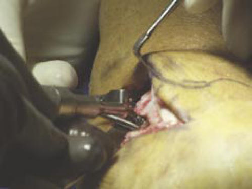
A 37-year-old male orthopaedic surgeon was waterskiing when he fell forward from his skis. He experienced some pain along the medial aspect of his ankle and could not continue waterskiing. Ten minutes later, he twisted his ankle a second time, felt a pop and noted a significant amount of medial ankle pain and swelling (Figure 4). He presented two days later to the treating physician (RMK) with left ankle pain isolated to the medial malleolar region. On examination, the posterior tibial tendon could be displaced anteriorly. An MRI was obtained which demonstrated that the posterior tibial tendon was dislocated anteriorly (Figure 5). He underwent surgical exploration one week following the injury (Figure 6). A groove-deepening procedure was performed (Figure 7) followed by repair of the flexor retinaculum (Figure 8). The tendon remained located with intra-operative range of motion of the ankle. He did not undergo physical therapy. At six months follow-up he has had no recurrent subluxation and has skied multiple times without pain. On physical examination, he has lost 5° of active dorsiflexion and has normal plantar flexion. His strength has returned to pre-injury level.
Figure 4.
Swelling and ecchymosis around the medial ankle region.
Figure 5.
Axial MRI demonstrating anterior dislocation of the posterior tibial tendon.
Figure 6.
Posterior tibial tendon dislocation.
Figure 7.
Deepening of the retromalleolar groove.
Figure 8.
Repair of the flexor retinaculum.
DISCUSSION
Ouzanian and Myerson1 have reported the largest clinical series with seven cases involving dislocation of the posterior tibial tendon. They presented six traumatic dislocations and one that occurred following multiple cortisone injections over an 18-month period. The average length of time to diagnosis was nine months, illustrating the common delay in presentation with this condition. The patients underwent various conservative treatment modalities without success, and all eventually required operative treatment. Only two patients had MRI findings consistent with a dislocated posterior tibial tendon. Upon surgical exploration, an inflamed posterior tibial tendon was encountered in three of seven patients with a torn, avulsed, or redundant retinaculum. The retro-malleolar groove was also noted to be shallow in four of the seven cases. Operative treatment consisted of retinacular repair in four cases and recreation of the retinaculum using local tissue in three cases. Two patients had a groove-deepening procedure. At final follow-up, five patients were asymptomatic, one was improved, and one had continued difficulty.
Bencardino et al.8 reported on the MRI findings of seven cases of posterior tibial tendon dislocation. The mechanism of injury was acute ankle dorsiflexion in two and major trauma in five. There were three medial malleolar fractures. In one patient, the diagnosis was made prior to MRI examination. MRI demonstrated a dislocated tendon in five patients and subluxation in two. One tendon demonstrated a partial tendon tear. The retromalleolar groove was shallow in one, slanted in one, and normal in five. The retinaculum was disrupted in two and avulsed from the tibia in five. Only two patients underwent surgery for their tendon dislocation in this primarily radiographic study. The authors concluded that MRI was a valuable tool for diagnosis and surgical planning for patients suspected of having a posterior tibial tendon dislocation.
In the English literature, 15 other series reported a total of 18 more cases of dislocation of the posterior tibial tendon. A tear in the flexor retinaculum or its avulsion from the tibia was noted in seven series for a total of eight cases.2,5,6,14,15,16,17 A lax retinaculum without tear was noted in a total of four patients, one in each of four series. 9,11,12,13 Two series reported on one patient each with an elevated "retinacular-periosteal sleeve."10,14 A shallow groove was reported at surgical exploration in three series,2,7,9 and in one case demonstrated by computed tomography.16 Three series reported a normal retromalleolar groove at the time of surgical exploration,4,6,15 while the rest did not mention the groove.
Plain radiographic examination was reported in ten series representing 11 patients.2,4–6,9,11,13,15–17 Ten of the 11 plain radiographs were reported as normal. One patient had a small fleck of bone near the medial malleolus representing an avulsion fracture.2 MRI was also used in several series to aid in evaluation of the extremity. 3–5,9,11,17 Two of the six patients who underwent MRI examination had a normal reading despite later confirmation of dislocation of the posterior tibial tendon on surgical exploration, demonstrating that MRI can miss a dynamic posterior tibial tendon dislocation that may be relocated at the time of the exam. Computed tomography was used twice in the literature.3,16 Rolf et al. reported one case of dislocation of the posterior tibial tendon that was documented by CT and ultrasound.3 In this study, there was no mention of the architecture of the retromalleolar groove on CT. A study by Soler et al.16 also mentions the use of CT. In their study, the authors report the anatomic variations of the retromalleolar groove in 25 cadaveric specimens as measured on plaster molds made of the specimens. The variation in width of the groove was large in this series, ranging from six to 15 millimeters in width and 1.5 to four millimeters in depth. Based on these cadaveric findings, the authors concluded that the patient had a sulcus that was less than normal size when measured on the CT scan images. Perlman et al. performed tenography of the posterior tibial tendon in their series and demonstrated a dislocated posterior tibial tendon in both of the cases presented in their report.2
There is no strong agreement in the literature on what is the best method of treatment. Simple flexor retinacular repair was made in six series3,5,6,10,14,15 versus a complex reconstruction with a periosteal sleeve (one series of one patient),16 an Achilles tendon flap (two patients from two different series),3,17 or suture anchor repair (two patients in two different series).4,11 There were four series in which the authors felt there was a shallow retromalleolar groove; however, groove deepening procedures were performed in two other series where no mention of the depth of the groove was made.2,7,9,12,13,16 Soler et al. reported that the retromalleolar groove was hypoplastic at the time of surgical exploration, but the authors did not perform a groove deepening procedure and instead reconstructed the flexor retinaculum with a periosteal sleeve held in place with sutures through bone tunnels. 16 In two of the remaining series in which a groove-deepening procedure was performed, a burr was used once7 and an osteotome and curette were used once.12 Perlman et al. performed a groove-deepening procedure by sliding a cortical bone slot graft posteriorly 1.5 cm to hold the relocated tendon in place.2 Sharon et al. similarly used a cortical graft from the tibia anterior to the retromalleolar groove to maintain reduction of the posterior tibial tendon. 13 Healy et al. elevated a cortical periosteal flap that was held open by a local cancellous bone graft obtained by deepening the retromalleolar groove in one patient.9 All reported good or excellent results with resumption of pre-injury level of activity.
CONCLUSION
Dislocation of the posterior tibial tendon is an uncommon injury rarely reported in the English literature with only 32 cases since 1968. The first described case was by Martius on himself when he fell from a balloon in 1874.18 Presentation to the diagnosing physician is often delayed, with the patients having undergone conservative treatment for various incorrect diagnoses. Conservative treatment is uniformly unsuccessful in the literature. Surgical stabilization by relocating the tendon and repairing or reconstructing the flexor retinaculum, with or without groove deepening, has been shown to be highly successful, with most patients returning to their pre-injury level of function. In our two patients, one presented late at seven months following injury with the initial diagnosis of a sprained ankle. The second patient was an orthopaedic surgeon; he made his own diagnosis and referral on an acute basis. They have both since returned to full function, with one competing in triathlon events. We do recognize that the two new cases presented in this report are of relatively short follow-up. Finally, MRI and other imaging modalities can help to establish the diagnosis, but have been negative in some series.4,8,9 A careful history and thorough physical examination remain the mainstay of diagnosis. Delay in diagnosis and treatment did not appear to have a deleterious effect on patient outcome in our case, nor did it adversely affect patients reported in the literature. Posterior tibial tendon dislocation should be considered in a patient with chronic medial ankle pain following injury with normal radiographic examination.
References
- 1.Ouzanian TJ, Myerson MS. Dislocation of the posterior tibial tendon. Foot and Ankle. 1992;13:215–219. doi: 10.1177/107110079201300409. [DOI] [PubMed] [Google Scholar]
- 2.Perlman MD, Wertheimer SJ, Leveille DW. Traumatic dislocation of the tibialis posterior tendon: a review of the literature and two case reports. J of Foot Surgery. 1990;29:253–259. [PubMed] [Google Scholar]
- 3.Rolf C, Guntner P, Ekenman I, Turan I. Dislocation of the tibialis posterior tendon: diagnosis and treatment. J of Foot and Ankle Surgery. 1997;36:63–65. doi: 10.1016/s1067-2516(97)80013-0. [DOI] [PubMed] [Google Scholar]
- 4.Wong YS. Recurrent dislocation of the posterior tibial tendon secondary to detachment of a retinacular- periosteal sleeve: a case report. Foot and Ankle International. 2004;25:602–604. doi: 10.1177/107110070402500815. [DOI] [PubMed] [Google Scholar]
- 5.Loncarich DP, Clapper M. Dislocation of posterior tibial tendon. Foot and Ankle International. 1998;19:821–824. doi: 10.1177/107110079801901205. [DOI] [PubMed] [Google Scholar]
- 6.Nava BE. Traumatic dislocation of the tibialis posterior tendon at the ankle. JBJS. 1968;50B:150–151. [PubMed] [Google Scholar]
- 7.Langan P, Weis CA. Subluxation of the tibialis posterior, a complication of tarsal tunnel decompression: a case report. Clin Orthop. 1980;146:226–227. [PubMed] [Google Scholar]
- 8.Bencardino J, Rosenberg ZS, Beltran J, Broker M, Cheung Y, Rosemberg LA, Scheitzer M, Hamilton W. MR imaging of dislocation of the posterior tibial tendon. Am J Roentgenol. 1997;169(4):1109–1112. doi: 10.2214/ajr.169.4.9308473. [DOI] [PubMed] [Google Scholar]
- 9.Healy WA, Starkweather KD, Gruber MA. Chronic dislocation of the posterior tibial tendon: a case report. Am J of Sports Medicine. 1992;23:776–777. doi: 10.1177/036354659502300625. [DOI] [PubMed] [Google Scholar]
- 10.Mittal RL, Jain NC. Traumatic dislocation of the tibialis posterior tendon. International Orthopaedics. 1988;12:259–260. doi: 10.1007/BF00547173. [DOI] [PubMed] [Google Scholar]
- 11.Nuccion SL, Hunter DM, Difiori J. Dislocation of the posterior tibial tendon without disruption of the flexor retinaculum: a case report and review of the literature. Am J of Sports Medicine. 2001;29:656–659. doi: 10.1177/03635465010290052101. [DOI] [PubMed] [Google Scholar]
- 12.Stanish WD, Vincent N. Recurrent dislocation of the tibialis posterior tendon: A case report with new surgical approach. Can J of Appl Sports Sci. 1984;9:220–222. [PubMed] [Google Scholar]
- 13.Sharon SM, Knudson HA, Gastwirth CM. Post-traumatic recurrent dislocation of the tibialis posterior tendon: a case report. J Am Podiatric Medicine. 1978;68:500–502. doi: 10.7547/87507315-68-7-500. [DOI] [PubMed] [Google Scholar]
- 14.Larsen E, Lauridsen F. Dislocation of the tibialis posterior tendon in two athletes. Am J of Sports Med. 1984;12:429–430. doi: 10.1177/036354658401200604. [DOI] [PubMed] [Google Scholar]
- 15.Beidert R. Dislocation of the tibialis posterior tendon. Am J of Sports Med. 1992;20:775–776. doi: 10.1177/036354659202000625. [DOI] [PubMed] [Google Scholar]
- 16.Soler RR, Gastany FJG, Ferret JR, Ramiro SG. Traumatic dislocation of the tibialis posterior tendon at the ankle level. J of Trauma. 1986;26:1049–1052. doi: 10.1097/00005373-198611000-00016. [DOI] [PubMed] [Google Scholar]
- 17.Ballasteros R, Chacon M, Cimarra A, Ramos L, Gomez-Barrena E. Traumatic dislocation of the tibialis posterior tendon: a new surgical procedure to obtain a strong reconstruction. J of Trauma. 1995;26:1198–1200. doi: 10.1097/00005373-199512000-00037. [DOI] [PubMed] [Google Scholar]
- 18.Martius C. Notes sur un cas de luxation du muscle tibial posterieur, etc. Bull R Med Belg. 1874;4:103. [Google Scholar]


