INTRODUCTION
The glomus tumor is a rare benign neoplasm that arises from the neuroarterial structure called a glomus body1, which accounts for 1 % to 4.5 % of tumors in the hand. The normal glomus body is located in the stratum reticulare throughout the body, but is more concentrated in the digits6. They are believed to function in thermal regulation. The average age at presentation is from 30 to 50 years of age, although can occur at any age1. Typical time from onset of symptoms to the correct diagnosis is seven years.
The patient with glomus tumor seeks medical attention early, but the mass is frequently too small to be identified on physical examination5. Although the classic triad of moderate pain, temperature sensitivity, and point tenderness has been described, these are nonspecific and not all may be present6. Furthermore, because the mass is usually less than 7 mm in diameter, it is very difficult to palpate. Upon biopsy, the histopathology typically reveals organoid appearance of polyhedral cells. It also can display darkly staining nuclei with fibrous stroma, and few blood cells1.
The plain radiographs are typically normal, although in longer standing lesions bony erosions can be seen. Although ultrasound has been advocated to aid in the diagnosis of glomus tumor, it is operator and techniques dependent2. Magnetic Resonance has become the imaging modality of choice when evaluating soft tissue masses. However, MRI descriptions of glomus tumors are rare in the English literature4,5.
ILLUSTRATIVE CASE REPORT
A 43 year old Korean female presented to the orthopaedic clinic with a six year history of pain at the tip of her left non-dominant thumb. She had numerous visits to different health care providers in the past, with various diagnosis documented in her medical records to include neuroma, radiculitis, Raynaud's phenomenon, and conversion reaction. There were no systemic complaints.
The patient complained of sharp pain whenever pressure was applied to the volar tip of her thumb during the activities of daily living. Although the patient denied night pain or cold sensitivity, she stated that she could feel a "grainy" mass at the tip of the digit.
The physical examination revealed no discoloration of the digit and the nail appeared normal. Although a distinct mass could not be appreciated by the examiner, the reproduction of pain was achieved by palpating the central area of the pulp. The radiographs and routine laboratory results were within normal limits.
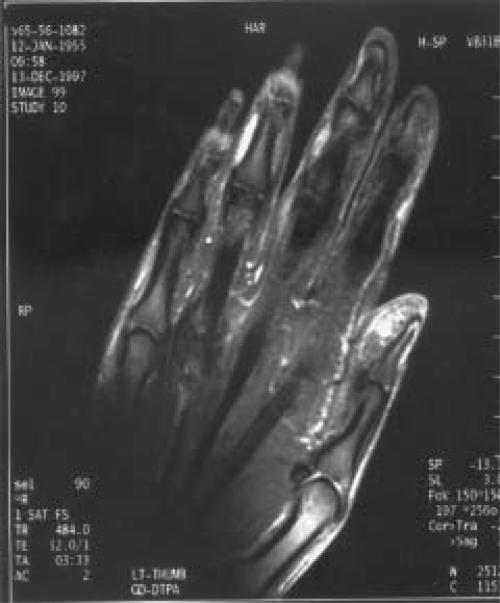
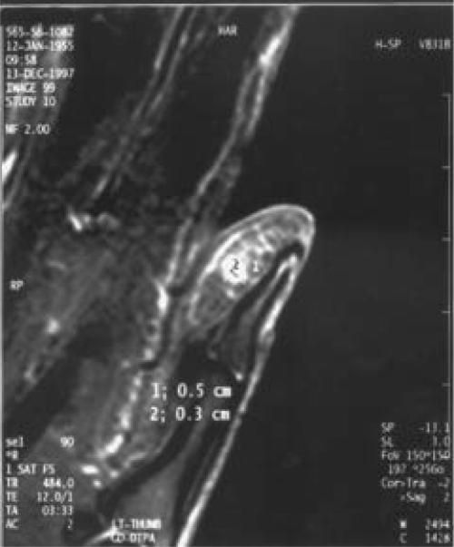
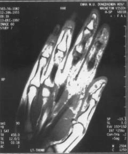
The MRI obtained revealed a spherical mass at the tip of distal phalanx. The lesion appears as a dark, well-defined mass on T1 weighted images and as a bright contrast enhancing mass on T1 post-gadolinium fat saturation images (Figures 2, 3). The small size (3 mm x 5 mm) and the spherical nature of the lesion was easily demonstrated.
Figures 2, 3.
T1 weighted, post-gadolinium, fat saturation images demonstrating the 3 mm x 5 mm mass.
Figure 2.
Figure 3.
A volar approach to the thumb was made, and excision performed. A spherical-brownish mass measuring 5 mm was identified, without gross surrounding tissue abnormality. The resulting histopathology was consistent with the pre-operative diagnosis of glomus tumor.
At follow up, the patient reported complete relief of her pre-operative symptoms.
DISCUSSION
Glomus tumor is a benign condition in which a complete excision usually leads to cure, with low incidence of recurrence. However, this benign condition has an unusually high morbidity to the patient before the correct diagnosis is made. This attests to the difficulty in correctly diagnosing this lesion initially. Although history and carefully performed physical examination significantly narrow the differential diagnosis, the plain radiographs are minimally helpful until the bony erosion occurrs at the later stages of the disease.
Glomus tumor is a vascular entity, reflecting its typically dark on T1 and bright MRI appearance on T2 weighted images. Post-gadolinium and fat saturation images further delineate the mass. Although this signal pattern can be seen with any vascular tumor, the location at the digits and its small size should lead one to suspect glomus tumor in most cases.
With increased index of suspicion, carefully performed history and physical examination, along with the findings on the MRI, the treating orthopaedist can significantly decrease the pre-operative morbidity to the patient with glomus tumor.
Figure 1.
T1 weighted image of the mass, pre-gadolinium
References
- 1.Carroll RE, Berman AT. Glomus tumors of the hand. J Bone Joint Surg. 1972;54A(4):691–703. [PubMed] [Google Scholar]
- 2.Fornage BD. Glomus tumors in the fingers: Diagnosis with US. Radiology. 1988;167(1):183–185. doi: 10.1148/radiology.167.1.2831563. [DOI] [PubMed] [Google Scholar]
- 3.Heys SD, Brittenden J, Atkinson P, Eremin O. Glomus tumor: An analysis of 43 patients and the review of the literature. Br J Surg. 1992;79:345–347. doi: 10.1002/bjs.1800790423. [DOI] [PubMed] [Google Scholar]
- 4.Matloub HS, Muoneke VN, Prevel CD, Sanger JR, Yousif NJ. Glomus tumor imaging: use of MRI for localization of occult lesions. J Hand Surg. 1992;17A:472–275. doi: 10.1016/0363-5023(92)90353-q. [DOI] [PubMed] [Google Scholar]
- 5.Mohler DG, Lim CK, Martin B. Glomus tumor of the plantar arch: A case report with magnetic resonance image findings. Foot & Ankle Int. 1997;18(10):672–674. doi: 10.1177/107110079701801014. [DOI] [PubMed] [Google Scholar]
- 6.Rettig AC, Strickland JW. Glomus tumor of the digits. J Hand Surg. 1977;2A(4):261–265. doi: 10.1016/s0363-5023(77)80121-4. [DOI] [PubMed] [Google Scholar]


