Abstract
Correction of adult scoliosis frequently involves long segmental fusions, but controversy still exists whether these fusions should include the sacrum. It has been suggested that forces associated with activities of daily living transfer the stresses to the remaining levels of the spine and to the pelvis. The case described here was a 43-year-old woman with scoliosis and chronic back pain refractory to non-surgical modalities. Radiographically, the patient had a 110 degree lumbar curve. An anterior and posterior fusion with Luque-Galveston instrumentation was performed. Six months postoperatively the patient returned with a 2-week history of right hip pain with no history of trauma. There was radiographic evidence of a displaced femoral neck fracture and pubic rami fractures. The femoral neck fracture was treated with a total hip replacement. Further surgeries were required to correct a lumbar pseudoarthrosis and hardware failure. We believe that this case provides evidence that fusion into the lumbosacral junction may distribute forces through the pelvic bones and hip resulting in stress and potential hardware complications, especially in patients at risk due to osteopenic conditions.
Correction of adult scoliosis often involves long segmental fusions into the lower lumbar spine and sacrum. However, inclusion of the lumbosacral joint in the fusion remains controversial. Following arthrodesis, forces associated with the activities of daily living transfer the stresses to the remaining unfused levels of the spine and pelvis, and may cause pseudoarthrosis, pedicle fractures, disc space narrowing, posterior facet hypertrophy and symptoms of mechanical back pain.2–5,9–12 The purpose of this paper is to present a case illustrating that spinal fusion to the sacrum may distribute forces through the pelvic bones and hip, resulting in stress fractures and hardware complications.
CASE REPORT
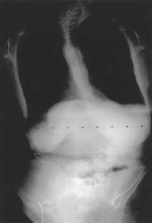
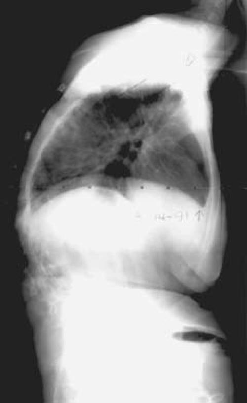
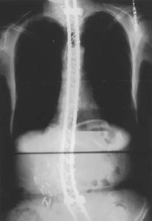
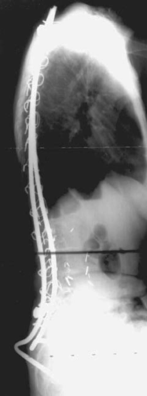
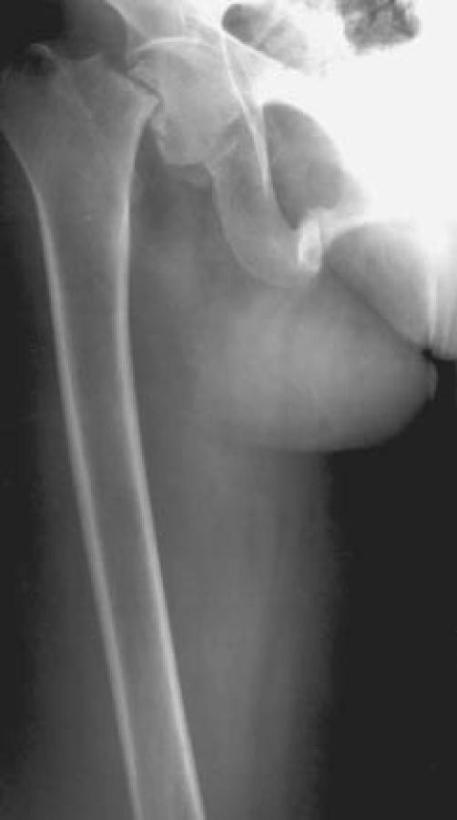
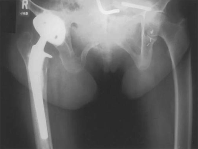
A 43-year-old woman consulted the Department of Orthopaedic Surgery at the University of Iowa in 1988 for chronic back pain secondary to scoliosis which was diagnosed at the age of 18. The patient had back pain refractory to physical therapy and analgesic medication over the past 3 years. Her past medical history was significant for hypothyroidism with replacement therapy and varicose veins. She was recently diagnosed with probable Marfan's syndrome with elements of Ehlers-Danlos syndrome. The neurological exam was normal. Radiographically, the patient had an 18 degree left thoracic curve from T5 to T10 and an 88 degree right curve extending from T11 to L4. A kyphotic deformity at the cervico-thoracic junction was also noted. The lumbar curve measured 110 degrees, which corrected only to 90 degrees on lateral bending (Fig. 1). Due to progression and back pain, she was indicated for spinal arthrodesis and instrumentation. In August 1991, an anterior release and fusion, and posterior fusion with Luque-Galveston segmental instrumentation was performed concurrently (Fig. 2). There were no surgical complications, and postoperatively the patient was ambulating with a TLSO. The patient had significant pain relief over the next eight months, at which time the TLSO was discontinued. However, the patient began having discomfort at the iliac region. She was treated with nonsteroidal anti-inflammatories and instructed to decrease her activity level. Six months later, the patient returned with a 2-week history of right hip pain with no history of trauma. There was radiographic evidence of a displaced femoral neck fracture and superior and inferior pubic rami fractures (Fig. 3-A). The femoral neck fracture was treated with a total hip replacement. The surgery was complicated by a transient femoral nerve palsy. (Fig. 3-B). The pubic rami fracture was treated non-operatively.
Figure 1.
Preoperative standing postero-anterior and lateral radiographs of the spine.
Figure 2.
Postoperative standing postero-anterior and lateral radiographs after corrective surgery and fusion with Luque-Galveston segmental instrumentation up to the sacrum.
Figure 3A.
Anteroposterior radiograph of the right hip demonstrating displaced femoral neck fracture.
Figure 3B.
Anteroposterior radiograph of the pelvis after total hip replacement. Note left side Luque rod fracture.
Lack of lumbar lordosis was causing the patient difficulty with ambulation and a feeling of being "thrown forward" while walking. Therefore, spine revision surgery was performed to correct the position of the lumbar fusion. Several osteotomies through the fusion mass, which was found to be solid and without signs of pseudoarthrosis, followed by further bending of the Luque rods, achieved a significant increase in lumbar lordosis and a better gait pattern for the patient. The pubic rami fractures were healed by that time.
The patient underwent further spine revision surgery in July 1994 for a broken Luque rod related to pseudoarthrosis at the lumbosacral junction area. This was addressed with removal of the distal end of the Luque rod, insertion of VSP screws and TSRH hooks connected to short Luque rods and linking to the previous rods. Additional lordosis was obtained through the pseudoarthrosis. Extreme osteopenia was noted at that time. The patient was treated with a corset for 7 months. Skin breakdown over a prominent TSRH hook resolved with conservative treatment. When last seen in July 1995, all wounds were healed, the patient had no pain related to the spine, but still complained of a "pitching forward" sensation.
DISCUSSION
The management of the adult patient with progressive and painful scoliosis remains one of the most challenging problems for the spinal surgeon. When proper patient selection is made, adult scoliosis corrective surgery has been shown to improve patient self-reported health assessment and function.1 However, specific considerations of adult scoliosis such as rigidity, associated degenerative disc and facet disease, and osteopenia make the surgery more complex. In addition, there is a higher incidence of complications when compared to similar surgery in adolescents.4,6,7,8,11,12 Mortality is in the range of 0.7 to 5.6%, and the incidence of both minor and major complications ranges between 41 and 86%.4,7,9,11
Anterior and posterior combined procedures, as well as long segmental fusions into the lower lumbar spine when degenerative changes or instability are present, are frequently necessary.2 When considering whether or not to extend a long fusion to the sacrum, the surgeon must take into account both the risks of complications resulting from arthrodesis or fixation of L5-S1 (12-70%) and the chances of iatrogenic instability or lumbosacral pain (75-90%).2 If the fusion does extend to the sacrum, supplemental anterior fusion is recommended in most patients to minimize nonunion and to preserve the lordosis.8 However, when a solid fusion mass is obtained, a stress riser may develop in the lower mobile segments. Kostuik4 noted a 49% loss of lordosis and 22% pseudoarthrosis rate in 45 patients fused to the sacrum. Knight3 reported a case in which the dissipation of forces through the remainder of the spinal column resulted in a stress fracture of the pedicle of the lower fused vertebra.
In this case, although spinal fusion was initially achieved, there was a definite loss of lumbar lordosis. We believe that the lack of spinal flexibility, failure to restore the normal sagittal spinal contours and altered gait apparently concentrated the biomechanical stresses on the next mobile segment, in this case the pubic bones and the hip. These abnormal stresses, when coupled with the patient's osteopenia, caused stress fractures to occur in these structures. Subsequent hardware failure associated with pseudoarthrosis of the fusion mass required further surgeries. Although secondary procedures were successful in alleviating the stresses in the pelvis and hip, we believe this case provides evidence of the progression of stresses within an abnormal lumbar spine to more mobile segments.
References
- 1.Albert TJ, Purtill J, Mesa J, McIntosh T, Balderston A. Health outcome assessment before and after adult deformity surgery. A prospective study. Spine. 1995;20:2002–2004. doi: 10.1097/00007632-199509150-00009. [DOI] [PubMed] [Google Scholar]
- 2.Horton WC, Holt RT, Muldowny DS. Fusion of L5-S1 in adult scoliosis. Spine. 1996;21:2520–2521. doi: 10.1097/00007632-199611010-00024. [DOI] [PubMed] [Google Scholar]
- 3.Knight RQ, Chan DPK. Idiopathic scoliosis with unusual stress fracture of the pedicle within solid fusion mass. A case report. Spine. 1992;17:849–851. doi: 10.1097/00007632-199207000-00023. [DOI] [PubMed] [Google Scholar]
- 4.Kostuik JP, Israel J, Hall JE. Scoliosis surgery in adults. Clin Orthop. 1973;93:225–234. doi: 10.1097/00003086-197306000-00022. [DOI] [PubMed] [Google Scholar]
- 5.McDonnell MF, Glassman SD, Dimar JR, Rolando MP, Johnson JR. Perioperative complications of anterior procedures on the spine. J Bone Joint Surg. (A) 1996;78:839–847. doi: 10.2106/00004623-199606000-00006. [DOI] [PubMed] [Google Scholar]
- 6.Moskowitz A, Moe JH, Winter RB, Binner H. Long-term follow-up of scoliosis fusion. J Bone Joint Surg. (A) 1980;62:364–376. [PubMed] [Google Scholar]
- 7.Ponder RC, Dickson JH, Harrington PR, Erwin WD. Results of Harrington instrumentation and fusion in the adult idiopathic scoliosis patient. J Bone Joint Surg. (A) 1975;57:797–801. [PubMed] [Google Scholar]
- 8.Saer EH, Winter RB, Lonstein JE. Long scoliosis fusion to the sacrum in adults with nonparalytic scoliosis. An improved method. Spine. 1990;15:650–653. doi: 10.1097/00007632-199007000-00007. [DOI] [PubMed] [Google Scholar]
- 9.Simmons ED, Kowalski JM, Simmons EH. The results of surgical treatment for adult scoliosis. Spine. 1993;18:718–724. doi: 10.1097/00007632-199305000-00008. [DOI] [PubMed] [Google Scholar]
- 10.Sponseller PD, Cohen MS, Nachemson AL, Hall JE, Wohl MEB. Results of surgical treatment of adults with idiopathic scoliosis. J Bone Joint Surg. (A) 1987;69:667–675. [PubMed] [Google Scholar]
- 11.Swank S, Lonstein JE, Moe JH, Winte RB, Bradford DS. Surgical treatment of adult scoliosis. J Bone Joint Surg. (A) 1981;63:268–287. [PubMed] [Google Scholar]
- 12.Van Dam B, Bradford D, Lonstein J, et al. Adult idiopathic scoliosis treated by posterior spinal fusion and Harrington instrumentation. Spine. 1987;12:32. doi: 10.1097/00007632-198701000-00006. [DOI] [PubMed] [Google Scholar]


