Abstract
Degenerative change involving the acromioclavicular (AC) is frequently seen as part of a normal aging process. Occasionally, this results in a painful clinical condition. Although AC joint symptoms commonly occur in conjunction with other shoulder pathology, they may occur in isolation. Treatment of isolated AC joint osteoarthritis is initially non-surgical. When such treatment fails to provide lasting relief, surgical treatment is warranted. Direct (superior) arthroscopic resection of the distal (lateral) end of the clavicle is a successful method of treating the condition, as well as other isolated conditions of the AC joint. The following article reviews appropriate patient evaluation, surgical indications and technique.
INTRODUCTION
Symptomatic osteoarthritis (OA) of the AC joint can be treated effectively with both non-surgical and surgical means. Historically, patients that have failed nonoperative management have been treated with resection of the distal clavicle, typically through an open superior incision.14,19,31,33 With the acceptance of the arthroscopic treatment of shoulder pathology, many surgeons now prefer arthroscopic resection of the distal clavicle. Technically, arthroscopic resection of the distal clavicle is feasible either from a direct (superior) approach, in which the instrumentation and dissection remains entirely intra-articular, or from a bursal approach, which is usually performed concomitantly with acromioplasty, subacromial bursectomy, and/or rotator cuff repair. Occasionally, AC joint arthrosis occurs in isolation; in these situations, it may be preferred to avoid trauma to the subacromial region, thereby minimizing postoperative inflammation and scarring, and potentially reducing recovery time. The following review article discusses appropriate patient selection, technique, and expected results when performing a direct arthroscopic distal clavicle resection.
ANATOMY
The AC joint is a diarthrodial joint formed by the medial facet of the acromion and the lateral, or distal, end of the clavicle, both of which are covered with hyaline cartilage. The joint has a variable degree of inclination in both sagittal and coronal planes. There is an intra-articular fibrocartilaginous disk of variable size and shape; a complete disk is noted in less than 10% of the population.4,15 Developmentally, there is no physeal plate at the distal clavicle. The articular cartilage of the distal clavicle may function in the longitudinal growth of the clavicle.12
Stability of the AC joint is primarily provided by the capsular ligaments that are located anterior, posterior, superior, and inferior.15,20,21 The superior AC ligament is the strongest and blends with the fascial attachments of the deltoid and trapezius muscles. The capsular ligaments provide anteroposterior (horizontal) stability of the distal clavicle.7,22,30 Slight upward and downward movement between the clavicle and acromion results in approximately 20 degrees of rotational movement between the two structures; however, this is likely variable on an individual level.12 The capsular insertion is approximately 1.5 cm medial from the end of the distal clavicle; therefore, resections of greater than 2 cm can compromise horizontal stability. The vertical stability of the distal clavicle is provided by the coracoclavicular (CC) ligaments, which are injured in moderate to high grade (i.e. II-V) AC joint separations.
NATURAL HISTORY
It is well documented that the fibrocartilaginous disk of the AC joint deteriorates with age. This natural aging process begins in the second decade of life.4 Several studies have demonstrated such changes in cadaveric studies or radiographic or magnetic resonance imaging of asymptomatic patients.9,17–18,23,25–28 Despite the abundant evidence that degenerative changes at the AC joint are common with aging, little is understood about the likelihood that such changes will eventually result in clinical symptoms.
In a study of 100 patients with osteoarthritis of other joints, there was a 70% incidence of AC joint tenderness. Treatment was provided for 30 consecutive patients in the series with intra-articular corticosteroid injections with successful results.32 The long-term efficacy of injection therapy for AC arthritis is unclear. While the previous study demonstrated success, others have found such injections provide only short-term relief, with a majority of patients ultimately requiring surgical treatment.10
EVALUATION
History
A detailed history is critical to making an accurate diagnosis of AC joint pathology. Pain is universally the chief complaint. The patient should be questioned to determine the onset, location, and character of pain. In addition, a history of prior trauma or surgery should be excluded. Finally, previous treatment, and the response to that treatment, should be known.
Pain associated with isolated AC joint arthrosis or synovitis is typically anterosuperior in location, in direct proximity to the joint itself. Not infrequently, the pain may radiate anterior, posterior, proximal, or distal, and may mimic pain experienced with other shoulder conditions, such as rotator cuff tears, subacromial or subcoracoid impingement, bicipital tendonitis, or labral or other intra-articular pathology. Most commonly, pain is described as a dull ache, but may be sharp or burning. Symptoms are usually activity related but may be present either at rest or during the night, and patients may experience symptoms when sleeping on the affected shoulder.
The patient should be asked about typical activities that produce symptoms. Using the affected arm across the body (in shoulder adduction) or behind the back (such as reaching a wallet or tucking in a shirt) may be particularly bothersome. As well, reaching with the arm elevated or overhead can produce pain, not unlike that associated with impingement pathology. The patient may also describe crepitation that localizes to the AC joint with certain maneuvers. However, such mechanical sensations can be difficult to localize and should be correlated during the physical examination.
Physical Examination
The examination begins with a thorough inspection of the entire shoulder girdle. Male patients should have the shirt removed, while female patients should be appropriately gowned to leave the shoulder girdle exposed. The resting posture of the shoulder is assessed, including the sternoclavicular (SC) joint, clavicle, AC joint, scapula, and surrounding musculature. Prior incisions should be documented, particularly in the region of the AC joint. Occasionally, the AC joint may demonstrate swelling, hypertrophic change, or resting deformity consistent with prior AC joint ligamentous injury.
Anatomic landmarks should be palpated for tenderness. This includes the SC joint, AC joint, acromion, proximal humerus, and periscapular region. Palpation tenderness at the AC joint is the hallmark of the condition. However, this should be compared to the unaffected side. As well, the patient may not have direct tenderness at the joint on examination, but should be asked whether the joint is the typical location of pain.
Next, shoulder range of motion is assessed. Movement in forward elevation, abduction, external rotation (both adducted and abducted) and internal rotation are documented. In patients with isolated AC joint pathology, there typically is no primary limitation to shoulder movement. As well, scapular control and scapulohumeral rhythm are evaluated to rule out underlying neurologic injuries or dysfunction. Additionally, strength and function of the rotator cuff, deltoid, trapezius and distal extremity are assessed.
Finally, specific tests to elicit isolated AC joint symptoms are performed. There are three tests which are utilized. First, the cross-body adduction test is performed with the arm in 90° elevation and maximal adduction across the body. The maneuver increases contact pressure at the AC joint and often reproduces symptoms. Second, the active compression test is performed with the arm in 90° elevation and 10-15° adduction, with the arm in maximal internal rotation with the thumb pointed down. Likewise, this maneuver loads the AC joint and often reproduces pain. It must be determined the pain is located mainly at the AC joint, as this test is also used to demonstrate superior labral pathology.16 Finally, pain reproduced at the AC joint with the arm in maximal internal rotation is a common finding, and may be the most sensitive test. Ranging the shoulder through full circumduction may also reproduce pain and crepitation at the AC joint.
Imaging
A standard radiographic series is obtained on all patients with a chief complaint of shoulder pain. In general, a series of four views are recommended that will evaluate for most degenerative and inflammatory conditions of the shoulder. Typically, true anteroposterior (AP) views of the glenohumeral joint with the humerus in both internal and external rotation, supraspinatus outlet, and axillary radiographs are obtained. In addition, when the clinician desires a specific evaluation of the AC joint, a Zanca radiograph is preferred, which is taken in the AP plane of the joint with a 10 degree cephalic tilt.34 This view is also particularly helpful in the postoperative evaluation of a distal clavicle resection.
Further imaging is often performed prior to considering surgical treatment. Magnetic resonance imaging (MRI) is preferred due to its ability to evaluate both bone and soft tissue. MRI of the shoulder allows detailed visualization of the rotator cuff, subacromial and subdeltoid bursae, acromion, greater tuberosity, coracoid process, subcoracoid interval, and medial clavicle. Intra-articular contrast may be used when further detail of the glenohumeral chondral surfaces or glenoid labrum is desired. It is important to include T2 weighted sequences that will demonstrate edema of the distal clavicle and medial acromion. Such findings are more suggestive of symptomatic AC joint arthrosis than are hypertrophic changes (i.e., osteophyte formation) alone.25 In addition, MRI allows evaluation of the coracoclavicular (CC) ligaments in situations where the patient has AC joint symptoms following a prior type I or II AC separation. Such an evaluation may be helpful in determining treatment in these instances. It is recommended that patients with prior AC joint injuries, even in the remote past, be thoroughly examined for residual distal clavicular instability. Caution should be exercised in selecting such patients for the procedure, as it has been demonstrated that such patients are at higher risk for postoperative failure.2,6
NONSURGICAL TREATMENT
Nonsurgical modalities include medical management with nonsteroidal anti-inflammatories (NSAIDs) and analgesics, local modalities such as moist heat, ice, or ultrasound, and physical rehabilitation to correct underlying rotator cuff or intra-articular pathology, or periscapular dysfunction. When associated conditions have been either ruled out or corrected, the ideal form of treatment of isolated AC joint arthrosis or synovitis is an intra-articular corticosteroid injection. When symptoms have persisted beyond 6 months despite transiently effective injections, surgical treatment of the problem is reasonable.
SURGICAL INDICATIONS
The indication for surgical treatment of AC joint arthrosis is the failure of nonsurgical management to return the patient to the desired level of daily work and recreational functioning. With regard to isolated treatment of the AC joint (i.e., with open or arthroscopic resection of the distal clavicle), it cannot be overemphasized that the key to an appropriate surgical indication is making an accurate preoperative diagnosis. This is again done with a specific and detailed history, physical examination, evaluation of pertinent radiographic and MRI images, and a documented clinical response to diagnostic and therapeutic injections.
TECHNIQUE
The author prefers patient positioning in the beach chair position; however, the procedure may be alternatively be performed in the lateral decubitus position. A regional interscalene block for shoulder surgery is utilized. Care is taken to position the head and neck in neutral, and all potential peripheral pressure areas are thoroughly padded. The patient is given appropriate preoperative antibiotics. The shoulder is draped sterile using a standard arthroscopic shoulder drape that provides excellent exposure and collects excess fluid during the procedure. Bony landmarks are then marked, which is critical to creating the precise portals necessary to perform the procedure. The scapular spine, coracoid process, acromion, and clavicle are all accurately drawn.
The standard portals for direct arthroscopic resection of the distal clavicle are typically 1.5 centimeters directly anterior and posterior to the AC joint itself. In larger patients with thicker subcutaneous tissue, the portals must be referenced further from the joint, such that the instruments will track beneath the skin for a distance, thus entering the joint at the appropriate location.
The joint initially is localized using two 22 gauge needles and a 10 cc syringe with normal saline. The needles are directed into the joint from anterior and posterior, and fluid injected to distend the joint. With appropriate placement, the anterior needle should allow outflow from the joint, suggesting accurate portal location. The skin is then incised in the desired locations with a #11 blade. The joint is initially inspected with a 2.7mm arthroscope (i.e., used for ankle or wrist arthroscopy) through the posterior portal. A needle is then utilized to create an anterior portal using an outside in technique. The portal is created with a #11 blade and widened at the level of the joint capsule with a blunt obturator. A 3.5 mm full radius resector is used to remove any inflammatory tissue or remaining meniscal remnants which may be present. A tissue ablation device (TurboVac 90, Arthrocare, USA) is used to subperiosteally expose the lateral end of the clavicle in its entirety from the level of the anterior capsule to the posterior capsule. As well, the bone must be exposed from inferior to superior, while keeping the respective capsular attachments intact. Maintaining the integrity of the capsular attachments minimizes the amount of bleeding and trauma into the subacromial space, thereby decreasing postoperative pain and shortening the recovery period. As well, preserving the posterior and superior aspects of the capsule avoids creating iatrogenic distal clavicular horizontal instability. This is particularly important in the unusual situation where the procedure is being performed following a previous type I or II AC separation (see previous text).
Once the lateral clavicle is exposed and any impinging soft tissue removed, the surgeon may switch to visualization with a standard 4.0 mm arthroscope, if the patient's anatomy allows. Resection of the distal clavicle is then performed with a 6.5 mm oval bur, beginning anterior and working toward the posterior aspect of the joint. If the joint space is excessively narrow to begin with, resection may be started with a smaller bur (i.e. 3.5mm round), before progressing to the larger bur. Care is taken to create an even resection from superior to inferior; typically, 6-7 mm of bone is removed, which has been shown to be adequate to prevent bone to bone contact with rotation of the scapula.30
The arthroscope is then moved to the anterior AC joint portal, to allow complete visualization of the posterior aspect of the distal clavicle and posterior capsule. If necessary, the ablation device is used to further expose the posterior clavicle. The bur is then used through the posterior portal to complete an even resection of bone, and confirm adequate decompression of the posterior portion of the joint. This step is critical, as a common technical error is inadequate visualization and resection of the posterior clavicle when performing the procedure arthroscopically.
Finally, in situations where it is desirable to inspect the glenohumeral joint as part of the same procedure, this can be done through a standard posterior portal established in the soft spot just medial and inferior to the posterolateral corner of the acromion. The anterior AC joint portal may be used for outflow and instrumentation, or a more standard anterior glenohumeral portal can be established. Such an inspection may or may not be preferred, depending upon the index of suspicion of intra-articular pathology based on clinical exam or by imaging modalities.
The portals are closed with nylon suture and standard dressings applied. The patient is placed in a Cryocuff (Aircast, USA) for postoperative comfort and to minimize swelling, as well as protect the arm while the regional block remains effective. It is typically discontinued 3-4 days after surgery. The patient begins gentle postoperative therapy 3-5 days after surgery, performing active assisted range of motion exercises within their tolerance level. The level of therapy is progressed to full active motion and isometrics in the ensuing 7-10 days. Typically, the patient progresses to a strengthening protocol 3 to 4 weeks post surgery. Most patients can expect full recovery in 8 to 10 weeks post surgery.
Postoperative radiographs, consisting of a Zanca and axillary lateral view, are obtained to confirm the clavicular resection has been adequate. Alternatively, these can be obtained immediately postoperatively or intraoperatively, depending on the confidence level of the surgeon. The author recommends the routine use of intra-operative fluoroscopy or plain radiography when the technique is being used for resection of distal clavicle nonunions or excision of ectopic bone. This avoids the unwanted complication of inadequate resection, which may lead to continued symptoms postoperatively.
RESULTS
The results of direct arthroscopic distal clavicle resection have been published previously. In a series of 29 patients with isolated AC joint arthrosis, 93% had excellent or good results at minimum 2 year follow up.6 This is consistent with the extensive reported literature evaluating either open distal clavicle resection,14,19,31,33 or arthroscopic resections performed using the more common bursal approach.1,5,8,11,13,24,29 A higher incidence of failure has been demonstrated in patients with prior AC joint instability (i.e., previous type II AC separation).2,6
CASE EXAMPLES
Patient One
A 39-year-old female was referred for evaluation of persistent anterosuperior shoulder pain 6 months following prior arthroscopic subacromial decompression. The patient was active in racquet sports and bowling, and reported discomfort with rotational and overhead movements of the shoulder. In particular, overhead weighted presses, pushups, and reaching behind the back were inciting activities.
Physical examination demonstrated healed prior arthroscopic portals and no resting deformity of the shoulder. Range of motion was full in all planes, although maximal internal rotation behind the back produced pain specifically at the AC joint. Neer and Hawkins impingement signs were negative. Cross body adduction and active compression tests reproduced symptoms at the AC joint. No signs of generalized laxity or pathologic glenohumeral instability were evident.
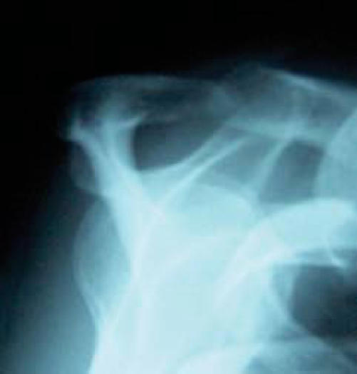
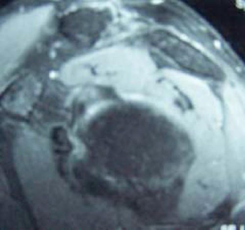
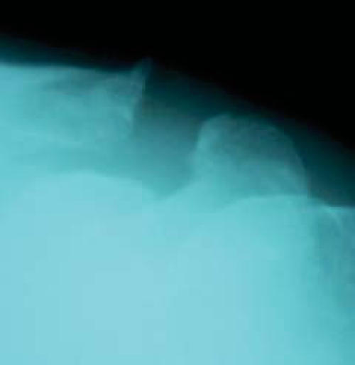
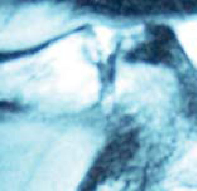
Standard radiographs demonstrated prior acromioplasty and relatively minimal degenerative change of the AC joint (Figure 1). An MRI was obtained which revealed brightly enhancing edema pattern of the distal clavicle and medial acromion, with associated subchondral cystic change (Figures 2A-C). No rotator cuff, bicipital, or labral pathology was identified.
Figure 1.
Supraspinatus outlet view in patient one demonstrating a flat acromial morphology consistent with successful prior arthroscopic acromioplasty. Patient continued to complain of anterosuperior shoulder pain.
Figure 2.
T2-weighted coronal (A), sagittal (B), and axial (C) MRI sequences of patient one. Note the edema pattern of the distal clavicle and medial acromion, with subchondral cyst formation.
Figure 2A.
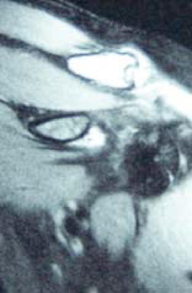
Figure 2B.
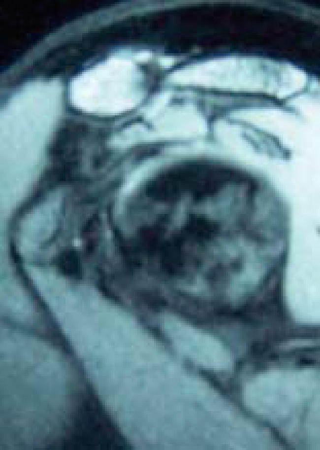
Figure 2C.
The patient underwent a diagnostic and therapeutic AC injection of 4cc 1% plain Lidocaine and 1cc of depomedrol, which demonstrated complete resolution of symptoms. Other nonsurgical treatment included scheduled NSAID use, modification of activity, and rotator cuff and periscapular strengthening. She experienced lasting relief for roughly 5 to 6 weeks, with gradual return of pain. A subsequent injection produced similar results in terms of duration and clinical efficacy.
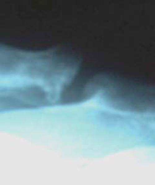
When her symptoms again recurred, the patient then elected to proceed with arthroscopic distal clavicle resection. Postoperative radiographs demonstrated adequate clavicular resection (Figure 3). The patient underwent standard postoperative protection and physiotherapy, and returned to full activity without restriction at 8 weeks following surgery. At 6 months, she continues to be pain free and pleased with the outcome.
Figure 3.
Postoperative Zanca radiograph of patient one after arthroscopic distal clavicle resection. Note the even resection of bone.
Patient Two
A 61-year-old male presented with an 8 month history of left anterosuperior shoulder pain of gradual onset. He reported an AC joint separation suffered roughly 23 years prior that was treated nonsurgically without complication. He denied subjective instability to the distal clavicle with functional activities, which included weight training, swimming, hunting, and fishing. Once again, pain was particularly noted when reaching across the body or behind the back.
Physical examination demonstrated moderate hypertrophic change at the AC joint, but no superior translation of the distal clavicle relative to the acromion. There were no previous incisions. Range of motion and strength of the shoulder were symmetric to the contralateral side, and impingement signs were negative. There was no gross manual translational instability of the distal clavicle, although slight crepitation in this region was produced with circumduction of the shoulder. Positive tests included cross-body adduction and active compression, with pain located specifically at the AC joint.
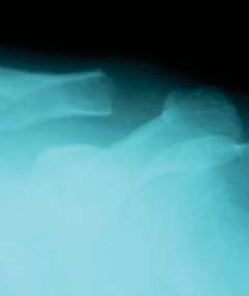
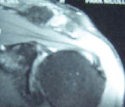
Standard radiographs demonstrated maintenance of the AC joint space, but marked hypertrophic degenerative change (Figure 4). MRI demonstrated an effusion of the AC joint with a moderate edema pattern on T2 weighted sequences (Figure 5). The coracoclavicular ligaments were demonstrated and intact on sagittal oblique sequences (Figure 6). The remaining bone, cartilage, and rotator cuff were normal in appearance.
Figure 4.
Preoperative AP radiograph of patient two demonstrating hypertrophic degenerative change of the AC joint.
Figure 5.
T2-weighted coronal MRI sequence of patient two demonstrating edema pattern of the medial acromion and fluid within the AC joint space.
Figure 6.
T2-weighted sagittal MRI sequence. Note the intact coracoclavicular ligaments. The patient had suffered a prior AC joint separation.
This patient likewise underwent a diagnostic and therapeutic AC injection of 4cc 1% plain Lidocaine and 1cc of depomedrol, which demonstrated complete resolution of symptoms. He experienced lasting relief for roughly 7 to 8 months, with gradual return of pain. A subsequent injection produced similar improvement for 2 months. He also elected to proceed with arthroscopic distal clavicle resection (Figure 7A-C).
Figure 7.
A.
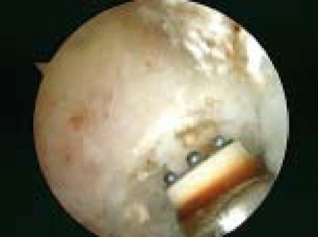
A tissue ablation device is used to denude the distal end of the clavicle of residual soft tissue, in preparation for resection. The device is also used to maintain hemostasis.
B.
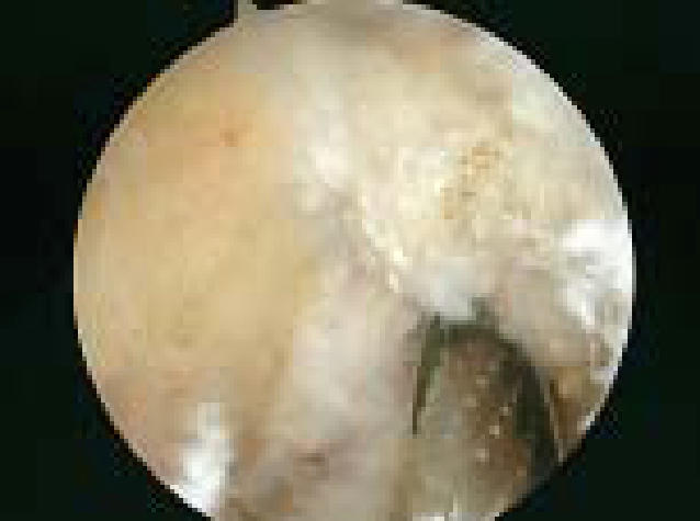
View from anterior portal of the left AC joint. A 6.5mm bur is used to complete the posterior resection of the clavicle
C.
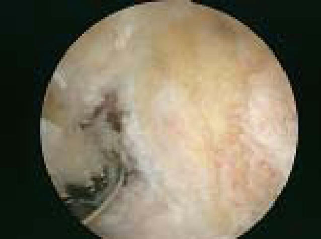
View from posterior of the completed left distal clavicle resection
Postoperative radiographs demonstrated adequate bone resection and no obvious translation of the clavicle relative to the acromion (Figure 8). He underwent a standard postoperative protocol and returned to swimming and other functional activities at 8 weeks following surgery. He remains pain free at one year following surgery, with no complaints of mechanical instability of the distal clavicle.
Figure 8.
Postoperative Zanca radiograph of same patient demonstrating distal clavicle resection. Again, note even bone resection and avoidance of excessive bone removal, particularly in the setting of a prior AC joint injury.
SUMMARY
Direct arthroscopic resection of the distal clavicle is an acceptable method of treating osteoarthrosis or synovitis of the AC joint that has failed to respond to a reasonable course of nonoperative treatment. Other AC joint disorders, such as distal clavicle osteolysis or nonunion of the distal clavicle, may also be treated with this technique. The procedure should only be performed when it is determined preoperatively that the AC joint is the isolated area of pathology. The only exception to this is the rare situation when it is preferred to diagnostically inspect the glenohumeral joint, but not perform extensive work either there, or in the subacromial or subcoracoid space. In comparison to other techniques, it allows precise resection of bone with minimal trauma to the surrounding soft tissue structures, which may decrease postoperative discomfort and shorten the recovery time. In addition, the procedure is quite pleasing from a cosmetic standpoint, and minimizes scar irritation from clothing or luggage straps that may occur with a larger superior incision.
References
- 1.Auge WK, Fisher RA. Arthroscopic distal clavicle resection for isolated atraumatic osteolysis in weight lifters. AJSM. 1998;26:189–192. doi: 10.1177/03635465980260020701. [DOI] [PubMed] [Google Scholar]
- 2.Bigliani LU, Nicholson GP, Flatow EL. Arthroscopic resection of the distal clavicle. Orth Clin N America. 1993;24:133–141. [PubMed] [Google Scholar]
- 3.Branch TP, Burdette HL, Shatiriari AS, et al. The role of the acromioclavicular ligaments and the effect of distal clavicle excision. AJSM. 1996;24:293–297. doi: 10.1177/036354659602400308. [DOI] [PubMed] [Google Scholar]
- 4.DePalma AF. The role of the disks of the sternoclavicular and the acromioclavicular joints. CORR. 1959;13:222–233. [Google Scholar]
- 5.Flatow EL, Cordasco FA, Bigliani LU. Arthroscopic resection of the outer end of the clavicle from a superior approach: A critical, quantitative radiographic assessment of bone removal. Arthroscopy. 1992;8:55–64. doi: 10.1016/0749-8063(92)90136-y. [DOI] [PubMed] [Google Scholar]
- 6.Flatow EL, Duralde XA, Nicholson GP, Pollock RG, Bigliani LU. Arthroscopic resection of the distal clavicle with a superior approach. J Shoulder Elbow Surg. 1995;4:41–50. doi: 10.1016/s1058-2746(10)80007-2. [DOI] [PubMed] [Google Scholar]
- 7.Fukuda K, Craig EV, An K, Cofield RH, Chao EYS. Biomechanical study of the ligamentous system of the acromioclavicular joint. JBJS. 1986;68A:434–440. [PubMed] [Google Scholar]
- 8.Gartsman GM. Arthroscopic resection of the acromioclavicular joint. AJSM. 1993;21:71–77. doi: 10.1177/036354659302100113. [DOI] [PubMed] [Google Scholar]
- 9.Horvath F, Ke·ry L. Degenerative deformations of the acromioclavicular joint in the elderly. Arch Gerontol Geriatric. 1984;3:259–265. doi: 10.1016/0167-4943(84)90027-x. [DOI] [PubMed] [Google Scholar]
- 10.Jacob AK, Sallay PI. Therapeutic efficacy of corticosteroid injections in the acromioclavicular joint. Biomed Sci Instrum. 1997;34:380–385. [PubMed] [Google Scholar]
- 11.Jerosch J, Steinbeck J, Schroder M, Castro WHM. Arthroscopic resection of the acromioclavicular joint. Sports Traum Arthroscopy. 1993;1:209–215. doi: 10.1007/BF01560209. [DOI] [PubMed] [Google Scholar]
- 12.Jobe CM, Coen MJ. Rockwood, et al., editors. Gross anatomy of the shoulder. The Shoulder. 3rd edition 2004.
- 13.Kay SP, Ellman H, Harris E. Arthroscopic distal clavicle excision. CORR. 1994;301:181–184. [PubMed] [Google Scholar]
- 14.Mumford EB. Acromioclavicular dislocation. JBJS. 1941;23:799–802. [Google Scholar]
- 15.Nuber FW, Bowen MK. Iannotti, Williams, editors. Disorders of the Acromioclavicular Joint: Pathophysiology, diagnosis, and management. Disorders of the Shoulder: Diagnosis and Management. 1999.
- 16.O'Brien SJ, Pagnani MJ, Fealy S, McGlynn SR, Wilson JB. The active compression test: A new and effective test for diagnosing labral tears and acromioclavicular joint abnormality. AJSM. 1998;26:610–613. doi: 10.1177/03635465980260050201. [DOI] [PubMed] [Google Scholar]
- 17.Petersson CJ. Degeneration of the acromioclavicular joint: A morphological study. Acta Orth Scand. 1983;54:434–438. doi: 10.3109/17453678308996597. [DOI] [PubMed] [Google Scholar]
- 18.Petersson CJ, Redlund-Johnell I. Radiographic joint space in normal acromioclavicular joints. Acta Orth Scan. 1983;54:490–491. doi: 10.3109/17453678308996596. [DOI] [PubMed] [Google Scholar]
- 19.Petersson CJ. Resection of the lateral end of the clavicle: a 3- to 30-year follow-up. Acta Orth Scand. 1983;54:904–907. doi: 10.3109/17453678308992931. [DOI] [PubMed] [Google Scholar]
- 20.Richards RR. Acromioclavicular joint injuries. Inst Course Lect. 1993;42:259–269. [PubMed] [Google Scholar]
- 21.Rockwood CA, Jr, Williams GR, Jr, Young DC. Rockwood, et al., editors. Disorders of the Acromioclavicular Joint. The Shoulder. 3rd edition 2004.
- 22.Salter EG, Jr, Nasca RJ, Shelley BS. Anatomical observations on the acromioclavicular joint and supporting ligaments. AJSM. 1987;15:199–206. doi: 10.1177/036354658701500301. [DOI] [PubMed] [Google Scholar]
- 23.Schweitzer ME, Magbalon MJ, Frieman BG, et al. Acromioclavicular joint fluid: Determination of clinical significance with MR imaging. Radiology. 1994;192:205–207. doi: 10.1148/radiology.192.1.8208939. [DOI] [PubMed] [Google Scholar]
- 24.Snyder SJ, Banas MP, Karzel RP. The arthroscopic Mumford procedure. Arthroscopy. 1995;11:157–164. doi: 10.1016/0749-8063(95)90061-6. [DOI] [PubMed] [Google Scholar]
- 25.Stein BE, Wiater JM, Pfaff HL, Bigliani LU, Levine WN. Detection of acromioclavicular joint pathology in asymptomatic shoulders with magnetic resonance imaging. J Shoulder Elbow Surg. 2001;10:204–208. doi: 10.1067/mse.2001.113498. [DOI] [PubMed] [Google Scholar]
- 26.Stenlund B, Marions O, Engstrom KF, Goldie I. Correlation of macroscopic osteoarthrotic changes and radiographic findings in the acromioclavicular joint>. Acta Radiol. 1988;29:571–576. [PubMed] [Google Scholar]
- 27.Stenlund B, Goldie I, Hagberg M, et al. Radiographic osteoarthritis in the acromioclavicular joint resulting from manual work or exposure to vibration. Br J Sports Med. 1992;49:588–593. doi: 10.1136/oem.49.8.588. [DOI] [PMC free article] [PubMed] [Google Scholar]
- 28.Stenlund B. Shoulder tendonitis and osteoarthrosis of the acromioclavicular joint and their relation to sports. Br J Sports Med. 1993;27:125–130. doi: 10.1136/bjsm.27.2.125. [DOI] [PMC free article] [PubMed] [Google Scholar]
- 29.Tolin BS, Snyder SJ. Our technique for the arthroscopic Mumford procedure. Orth Clinic N America. 1993;24:143–151. [PubMed] [Google Scholar]
- 30.Urist MR. Complete dislocations of the acromioclavicular joint: The nature of the traumatic lesion and effective methods of treatment with analysis of 41 cases. JBJS. 1946;28:813–837. [PubMed] [Google Scholar]
- 31.Wagner C. Partial claviculectomy. Am J Surg. 1953;85:259–265. doi: 10.1016/0002-9610(53)90607-2. [DOI] [PubMed] [Google Scholar]
- 32.Waxman J. Acromioclavicular disease in rheumatologic practice—the forgotten joint. J La State Med Soc. 1977;129:1–3. [PubMed] [Google Scholar]
- 33.Worchester JN, Green DP. Osteoarthritis of the acromioclavicular joint. CORR. 1968;58:69–73. [PubMed] [Google Scholar]
- 34.Zanca P. Shoulder pain: Involvement of the acromioclavicular joint: Analysis of 1000 cases. Am J Roent. 1971;112:493–506. doi: 10.2214/ajr.112.3.493. [DOI] [PubMed] [Google Scholar]


