Abstract
A 67-year-old woman with chronic lumbosacral and hip symptoms involving gluteus medius tendon rupture and strain injury is presented here. We report her work-up and management. Although this is an uncommonly reported pathology, many patients with back, buttock and leg pain see physicians who often focus on lumbar spinal stenosis, lumbar radiculopathy or hip/knee osteoarthritis. Careful physical examination guided us to this patient's diagnosis.
INTRODUCTION
The gluteus medius muscle is important in stabilizing the ipsilateral hip in the stance phase of gait. When patients are unable to maintain pelvic neutrality, the energy cost of ambulation increases. We speculate that patients with weakness in the gluteus medius will subsequently develop low back pain, buttock pain or trochanteric bursitis pain.
We present a woman whose weakness in the gluteus medius was discerned through physical examination for back pain. Her weakness was treated with an appropriate rehabilitation program. Her pain then subsequently subsided.
CASE REPORT
The patient is a 67-year-old retired woman with a chief complaint of low back pain, right buttock pain and lateral thigh pain down to the knee. Her symptoms started four years previous with no specific precipitating traumatic event. Her symptoms worsened recently. They have now limited her walking and stair climbing. She stopped many of her outside activities like shopping, exercising at the health club or babysitting her grandchildren. Her pain was aggravated the most with any weight bearing on the right side. Standing and walking were painful, as was prolonged sitting. She could not lie on her right side in bed.
She had an extensive work-up. She initially saw her family physician locally, where x-rays of the lumbar spine were obtained which indicated spondylosis. She was referred for physical therapy that included lumbar stabilization exercises. She did not appreciate much relief and was sent for a lumbar spine magnetic resonance imaging (MRI) that indicated moderate spondylosis without significant canal stenosis. She was referred to a pain management facility locally for an interlaminar epidural steroid injection followed by lumbar facet injections at L4-5 bilaterally, both only producing temporary relief. Lumbar facet radiofrequency denervation was done at the same level without significant relief. A CT-myelogram of the lumbar spine as well as hip x-rays were negative. She also had electromyography and nerve conduction studies which were normal. Two more interlaminar epidural steroid injections at the L4-5 level were done with minimal benefit. She also had a trochanteric bursal injection which afforded only temporary relief.
Since she was continuing to have worsening pain and inability to ambulate, further treatment involved consideration of a spinal cord stimulator. She requested a second opinion regarding this procedure and came to our physiatry and physical therapy clinic.
Her physical exam revealed a positive Trendelenburg sign in stance and with gait. Hip rotational movements were normal without groin pain, but painful in the buttocks on the right. Neurologic examination was normal with no neural tension signs or weakness in toe extensors or ankle dorsiflexor muscles. Trunk flexion and extension movements in standing were limited 25% with pain in the gluteal area. She had adequate abdominal muscle control. Most notably, manual muscle testing for the gluteus medius muscle (side-lying, hip in extension, with the leg in abduction) on the right was 2/5 and reproduced her pain symptoms. Testing on the left was 5/5 and pain free. There was considerable tenderness over the right trochanter and in the gluteal muscles lateral to the posterior superior iliac spine (PSIS).
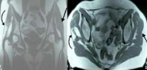
We obtained MRI of the pelvis to rule out a stress fracture or other hip joint pathology, as well as examine the integrity of the gluteal muscles. The MRI revealed a strain injury as well as a large tear of the right gluteus medius muscle at the musclotendinous junction. (Figure 1).
Figure 1.
Coronal and axial T2 MRI views of the pelvis indicating strain injury and a large tear (curved arrow) of the right gluteus medius muscle. The opposite side is normal (straight arrow).
Treatment involved protection of the strain injury during ambulation with the use of a straight cane and frequent use of cold packs over the gluteal muscles and trochanteric bursa. She also began a very low-level exercise program for strengthening the gluteus medius muscle. She was seen for follow-up in one month and reported a significant reduction in her painful symptoms and was making progress with strength. Manual muscle testing indicated a grade 3/5 that was still painful. Her exercise program was progressed and she continued use of the cane whenever she was out of the house. At four months, she was essentially asymptomatic and had returned to her regular activities. She no longer used a cane but continued to do her strengthening exercises. Manual muscle testing was a 4/5 and was not painful. A follow-up MRI was obtained which indicated healing of the strain injury, but the rupture remained (Figure 2).
Figure 2.
Coronal and axial T2 MRI views of the pelvis indicating healed muscle belly and persisting tear (curved arrow) after 4 months. The opposite side is normal (straight arrow).
DISCUSSION
In the past several years, there has been limited literature describing MRI and ultrasound diagnosis of gluteus medius strains or tears. Bird et al. found 46% of 24 patients with chronic buttock pain and symptoms ranging between 1-5 years, to have a gluteus medius tendon tear by MRI.2 The Trendelenburg test was the most sensitive and specific physical exam finding for this condition. Chung looked specifically at elderly women with buttock pain and found six out of six women age 62-79 with full- or partial- thickness tendon tears by MRI findings. Three had surgical intervention for repair.4 Connell et al. reported on ultrasound imaging in 75 patients, and found 63 had tendinopathy with 25 showing full-thickness or partial-thickness tears.5 MRI parameters for diagnosing hip abductor strain and tendinopathy have been described as yielding an accuracy of 91%. Sensitivity and specificity of T2 hyperintensity superior to the greater trochanter are 73% and 91% respectively.10
Gluteus medius tendon tears have been described mostly in relation to findings during hip surgeries or gamma nailing procedures for proximal femur fractures in which up to 27% of the gluteus medius tendon insertion is disrupted during correct placement of this hardware. 8 Kagan found seven patients who had a partial tear of the gluteus medius tendon during treatment for refractory trochanteric bursitis, and reattached the tendons. 9 At median follow-up of 45 months, patients were pain free. Other studies report the surgical repair of some tears, but some authors have concluded that not all of them need to be surgically repaired.14 Little is written regarding non-surgical treatment for gluteus medius tears. Bewyer and Bewyer reported a recommended treatment protocol and algorithm.1
The authors feel this problem is not as rare as most commonly thought. In a pilot study through our spine clinic, 70% of people presenting with lumbosacral and/or leg pain were found to have painful, weak gluteus medius muscles on examination. Not all of these people had tendon ruptures, but muscle strain versus tendinopathy versus tendon tear would seem to be a consideration. We are in the process of a more formal prevalence study of 150 consecutive patients. We hope to be able to describe this problem more completely in terms of diagnostic work-up as well as treatment.
References
- 1.Bewyer D, Bewyer K. Rationale for treatment of hip abductor pain syndrome. Iowa J Ortho. 23:57–60. [PMC free article] [PubMed] [Google Scholar]
- 2.Bird P, Oakley S, Shnier R, Kirkham B. Prospective evaluation of magnetic resonance imaging and physical examination inpatients with greater trochanteric pain syndrome. Arth and Rheum. 2001 Sep;44(9):2138–2145. doi: 10.1002/1529-0131(200109)44:9<2138::AID-ART367>3.0.CO;2-M. [DOI] [PubMed] [Google Scholar]
- 3.Bunker T. Rotator cuff tear of the hip. J Bone Joint Surg. 1997 Jul;79-B(4):618–620. doi: 10.1302/0301-620x.79b4.7033. [DOI] [PubMed] [Google Scholar]
- 4.Chung C, Robertson J, Cho G, Vaughan L, Copp S, Resnick D. Gluteus medius tendon tears and avulsive injuries in elderly women: imaging findings in six patients. Am J Roent. 1999 Aug;173(2):351–353. doi: 10.2214/ajr.173.2.10430134. [DOI] [PubMed] [Google Scholar]
- 5.Connell D, Bass C, Sykes C, Young D, Edwards E. Sonographic evaluation of gluteus medius and minimus tendinopathy. Eur Radiol. 2003 Jun;13(6):1339–1347. doi: 10.1007/s00330-002-1740-4. [DOI] [PubMed] [Google Scholar]
- 6.Gabrion A, Vernois J, Havet E, Mertl P, de Lestang M. Gluteus medius tendon tear and degenerative hip disease. Revue de Chirurgie Orthopedique et Reparatrice de l Appareil Moteur. 2003 Nov;89(7):640–642. [PubMed] [Google Scholar]
- 7.Hertel J, Sloss BR, Earl JE. Effect of foot orthotics on quadriceps and gluteus medius electromyographic activity during selected exercises. Archives of Physical Medicine & Rehabilitation. 2005 Jan;86(1):26–30. doi: 10.1016/j.apmr.2004.03.029. [DOI] [PubMed] [Google Scholar]
- 8.Honl M, Dierk O, Gauck C, Carrero V, Lampe F, Dries S, Quante M, Schwieger K, Hille E, Morlock M. Comparison of robotic-assisted and manual implantation of a primary total hip replacement. A prospective study. J Bone Joint Surg. 2003 Aug;85-A(8):1470–1478. doi: 10.2106/00004623-200308000-00007. [DOI] [PubMed] [Google Scholar]
- 9.Kagan A. Rotator cuff tears of the hip. Clin Ortho and Related Research. 1999;368:135–140. [PubMed] [Google Scholar]
- 10.Kumagai M, et al. Functional evaluation of hip abductor muscles with the use of magnetic resonance imaging. J of Clin Research. 15:888–893. doi: 10.1002/jor.1100150615. [DOI] [PubMed] [Google Scholar]
- 11.LaBan MM, Weir SK, Taylor RS. 'Bald trochanter' spontaneous rupture of the conjoined tendons of the gluteus medius and minimus presenting as a trochanteric bursitis. American Journal of Physical Medicine & Rehabilitation. 2004 Oct;83(10):806–809. doi: 10.1097/01.phm.0000140792.48248.49. [DOI] [PubMed] [Google Scholar]
- 12.Lehman G, Lennon D, Tresidder B, Rayfield B, Poschar M. Muscle recruitment patterns during the prone leg extension. BMC Musculoskeletal Disorders. 2004 Feb;5(1):3. doi: 10.1186/1471-2474-5-3. [DOI] [PMC free article] [PubMed] [Google Scholar]
- 13.McConnell T, Tornetta P, Benson E, Manuel J. Gluteus medius tendon injury during reaming for gamma nail insertion. Clin Orthop Res. 2003. Feb, pp. 199–202. [DOI] [PubMed]
- 14.Ozcakar L, Erol O, Kaymak B, Aydemir N. An underdiagnosed hip pathology: apropos of two cases with gluteus medius tendon tears. Clinical Rheumatology. 2004 Oct;23(5):464–466. doi: 10.1007/s10067-004-0917-4. [DOI] [PubMed] [Google Scholar]


