Summary
Four cases of oro-facial infection leading to life-threatening complications are reported. Although all had been treated with antibiotics prior to consultation, lack of surgical intervention had allowed the infection to progress. These cases are a reminder that acute spreading odontogenic infection can be life-threatening. Definitive treatment includes airway management, adequate resuscitation and optimization of pre-existing medical conditions prior to removal of the source of infection and drainage of pus. High dose intravenous antibiotics should be administered, with the initial choice of antibiotics modified in the light of subsequent bacteriological reports. The treatment of all odontogenic infections must include removal of the focus of infection, and drainage of pus.
Keywords: Oro-facial infection, odontogenic infection, resuscitation, supportive therapy, tooth extraction
Introduction
In the United States of America fatality involving oro-facial infection is low due to the proper use of antibiotics and prompt interventions1,2,3.
Without proper management of odontogenic infections complications such as facial cellulites, mediastinitis, brain abscess, septicaemia and thromboembolism could result1.
The importance of supportive treatment, vigorous antibiotic therapy, release of pus by extracting the involved tooth and avoidance of tracheostomy, if at all possible, has been stressed in a report, where fatalities were reported in a series of ten patients with acute spreading oro-facio-cervical infections4,5,6,7.
At the Tarkwa Gorvernment Hospital, dental infection was second only to malaria in hospital attendance for the period of 1998 to 20018.
This paper reports on life-threatening complications associated with oro-facial infections and their management in four patients seen at the Tarkwa Government Hospital during the period 1998–2001.
Case Reports
Case One
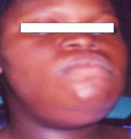
A nineteen-year-old girl (Figure 1) was rushed to the Dental Department of Tarkwa Government Hospital after collapsing with rigors at home. A pharmacist or chemical seller had previously prescribed amoxycillin for her toothache and a swelling of the lower jaw. She had no significant medical history.
Figure 1.

Patient with bilateral submandibular swelling-Ludwig's angina
On arrival she was in respiratory distress, had a pulse of 180 beats per minute and a blood pressure of 110/40mmHg. Her axillary temperature was 40.5 degrees Celsius and her Glasgow Coma Score (GCS) was 10/15. There was an obvious right submandibular and sub-mental swelling, with minor trismus. The tongue was elevated and was in contact with the palate making breathing, swallowing and feeding difficult. Full blood count (FBC) showed an Hb of 9.5g/dl and biochemical profile was within normal range. A presumptive diagnosis of septic shock secondary to dental infection i.e. Ludwig's angina 5 was made.
She was admitted and treated with high flow oxygen, intravenous fluids, ceftriazone and metronidazole. Extraction of the involved tooth, together with an incision of submental region to drain the abscess under general anaesthesia was undertaken three days after admission. Intraoperatively 20mls of pus was obtained. Staphylococcus aureus was subsequently isolated from the pus and blood culture. The patient was discharged from hospital seven days after surgery in satisfactory condition.
Case Two
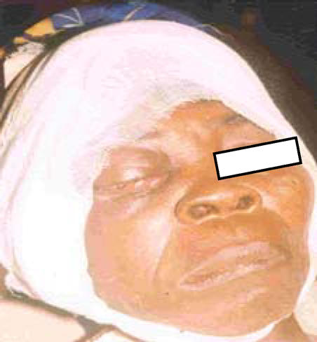
A 60 year old female (Figure 2) was admitted because of a right periorbital abscess. She was a farmer, who looked poorly nourished and dehydrated. She denied ever having had a toothache. Her full blood count showed haemoglobin of 8.5g/dL and elevated white cell count of 13.2 x 10⁹.
Figure 2.

Patient with right periorbital cellulitis
A diagnosis of right periorbital cellulitis was made.
She was rehydrated with intravenous fluids and given antibiotics prior to having the abscess drained under general anaesthesia through incisions of both eyelids and the insertion of drainage tubes.
The following day her general condition had deteriorated and she was now unresponsive with a GCS of 10/15, and in shock (pulse, 120bpm; BP, 70/40mmHg). Upon thorough intra-oral examination the source of the infection was determined as a carious upper right third molar tooth. The offending tooth was extracted under general anaesthesia during which 35mls of pus was drained from the socket and periorbital region, and this continued to discharge pus till the 4th day after the extraction.
High flow oxygen was administered postoperatively and intravenous fluids and intravenous amoxicillin and metronidazole were continued. The day after the extraction, her condition improved dramatically and she was discharged home 9 days after admission.
Case Three
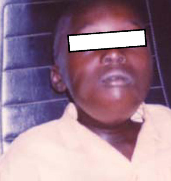
A 10 year old school boy was rushed to our unit with a day's history of swelling of the submental region. He had received antibiotics from his father who had been reluctant to take him to the hospital whenever he complained of toothache. He had no relevant medical history, but looked severely malnourished. His temperature was 39.4 degrees Celsius. He had severe trismus with interincisal opening of only 6mm. There was no sign of respiratory distress. He had a neglected dentition, with several carious teeth. The result of the full blood count demonstrated a leucocytosis (WBC 21.5 x 10⁹), and Hb of 9.0g/dl but his remaining haematological and biochemical test results were within the normal range.
A presumptive diagnosis of Ludwig's Angina was made. As it was anticipated that intubation would be difficult, the abscess was incised and drained extra orally by a median incision at the submental region under local anaesthesia releasing 35mls of pus. Intravenous antibiotics (benzyl-penicillin 1g qid and metronidazole 500mg tid) were started. The following day his condition had further deteriorated, with significant respiratory distress evident. The swelling now involved both sub-lingual spaces, and it was considered appropriate to extract all the eight carious teeth under general anaesthesia using the awake intubation method. This meant that the patient was awake and in full control of his airway while the endotracheal tube was being inserted. Though uncomfortable, this was considered the safest method of inducing anaesthesia due to the restricted mouth opening. About 30mls more pus was drained from the sublingual spaces. A drainage tube was inserted, and this continued to discharge large volumes of pus until the fifth day postoperatively. Streptococcus faecalis was subsequently isolated from the pus. He was treated as an in-patient for 15 days before being discharged home. After the first week, when his mouth opening improved the intravenous antibiotics were changed to oral axoxycillin and metronidazole.
Case Four
A fifty-seven year old male farmer was referred by a medical practitioner with swelling of his left submandibular region, left check, left eye and the left temporal region of 14 days duration. It had not responded to a course of oral amoxicillin prescribed by the doctor who referred him. He had mixed cardiac valve disease as a consequence of previous rheumatic fever. He had been unable to take neither any of his usual medication including that for his cardiac condition nor the one the doctor prescribed for him due to trismus and dysphagia for five days prior to seeing the first author (EKA)
On admission, his respiration was normal and his temperature was 38.3 degrees Celsius. His pulse was 110bpm but regular, and his BP was 135/85mmHg. There was no evidence of cardiac failure. His left eye was closed completely due to the swelling. Haematological investigation showed WBC of 13.5 x 10⁹. The cause of his facial swelling was identified as a carious lower left second molar. A diagnosis of left facial cellulitis secondary to odontogenic infection was established.
He received intravenous fluids and intravenous amoxicillin and metronidazole and, his previously prescribed lisinopril, frusemide and flecainide were administered.
The following morning he was taken to theatre and had the offending tooth extracted under general anaesthesia with additional extraoral incisions at the left submandibular, left cheek and left temporal regions which released about 70mls of pus. Drains were inserted into each of the incisions and a head bandage was applied. The drains continued to discharge large volumes of pus so 2% Hydrogen peroxide and Eusol solutions were used to irrigate the wound until the pus ceased discharging one week postoperatively. The patient was discharged from the hospital ten days after the operation.
Discussion
In the developed world, fatal dental infections are rare in patients with intact immune response. In the third world where people are generally poor and malnourished such fatal oro-facial infections are more common. It is only prompt specialised assistance that can prevent fatalities. Isolated cases have been reported of mediastinitis secondary to dental infection and bacterial endocarditis.
Occasionally however, death can result from sequelae as diverse as necrotizing fasciitis, brain abscess and disseminated intravascular coagulation (DIC). The number of “near misses” is difficult to estimate.
The absence of a universal health insurance scheme deters patients from seeking regular dental care, and may even encourage patients with acute dental infection to present directly to pharmacy shops or “quack doctors” since these consultations are considered cheaper.
Most infections of odontogenic origin are mixed with gram positive cocci and gram negative rods predominating. Such infections are responsive to the pecillins and cepholosporins.9 The isolation of Streptococcus faecalis in one patient could have been an indication of the unsanitary conditions to which the patient was exposed
The definitive treatment of serious dental infections often requires, in addition to appropriate antibiotic therapy, the extraction of the offending tooth to allow for proper drainage of pus and removal of the source of infection as have been described in this paper.
Figure 3.

A 10 year old Patient with Ludwig's angina
References
- 1.McCurdy JA, Jr, MacInnis EL, Hays LL. Fatal mediastenitis after a dental infection. J Oral Surg. 1977;35:726–729. [PubMed] [Google Scholar]
- 2.Zeitoun IM, Dhanarajani PJ. Cervical cellulites and mediastenitis caused by odontogenic infections. J Oral Maxillofac Surg. 1995;53:203–208. doi: 10.1016/0278-2391(95)90404-2. [DOI] [PubMed] [Google Scholar]
- 3.Bonapart IE, Stevens HP, Kerver AJ, Reitveld AP. Rare complications of an odontogenic abscess: mediastenitis, thoracic empyema and cardiac tamponade. J Oral Maxillofac Surg. 1995;53:610–613. doi: 10.1016/0278-2391(95)90078-0. [DOI] [PubMed] [Google Scholar]
- 4.Steiner M, Grau MJ, Wilson DL, Snow NJ. Odontogenic infection leading to cervical emphysema and fatal mediastenitis. J Oral Maxillofac Surg. 1990;28:189–193. doi: 10.1016/0278-2391(82)90293-2. [DOI] [PubMed] [Google Scholar]
- 5.Iwu CO. Ludwig's angina: report of seven cases and review of current concepts in management. Br J Oral Maxillofac Surg. 1990;28:189–193. doi: 10.1016/0266-4356(90)90087-2. [DOI] [PubMed] [Google Scholar]
- 6.Currie WJ, Ho V. An unexpected death associated with an acute dentoaveolar abscess - report of a case. Br J Oral Maxillofac Surg. 1993;31:296–298. doi: 10.1016/0266-4356(93)90063-3. [DOI] [PubMed] [Google Scholar]
- 7.Marks PV, Patel KS, Mee EW. Multiple brain abscess secondary to dental caries and severe periodontal disease. Br J Oral Maxillofac Surg. 1988;26:244–247. doi: 10.1016/0266-4356(88)90171-4. [DOI] [PubMed] [Google Scholar]
- 8.Annual Report. Ghana: Tarkwa Government Hospital; 2001. [Google Scholar]
- 9.Reza AJ, Aziz SR, Ziccardi VB. Microbiology and antibiotic sentitivities of head and neck space infections of odontogenic origin. J Oral Maxillofac J. 2006;64:1377–1380. doi: 10.1016/j.joms.2006.05.023. [DOI] [PubMed] [Google Scholar]


