Abstract
The anterior heart field (AHF), which contributes to the outflow tract and right ventricle of the heart, is defined in part by expression of the LIM homeobox transcription factor Isl-1. The importance of Isl-1–positive cells in cardiac development and homeostasis is underscored by the finding that these cells are required for cardiac development and act as cardiac stem/progenitor cells within the postnatal heart. However, the molecular pathways regulating these cells’ expansion and differentiation are poorly understood. We show that Isl-1–positive AHF progenitor cells in mice were responsive to Wnt/β-catenin signaling, and these responsive cells contributed to the outflow tract and right ventricle of the heart. Loss of Wnt/β-catenin signaling in the AHF caused defective outflow tract and right ventricular development with a decrease in Isl-1–positive progenitors and loss of FGF signaling. Conversely, Wnt gain of function in these cells led to expansion of Isl-1–positive progenitors with a concomitant increase in FGF signaling through activation of a specific set of FGF ligands including FGF3, FGF10, FGF16, and FGF20. These data reveal what we believe to be a novel Wnt-FGF signaling axis required for expansion of Isl-1–positive AHF progenitors and suggest future therapies to increase the number and function of these cells for cardiac regeneration.
Introduction
Recent evidence has demonstrated that there are 2 distinct mesodermal cell pools that contribute to the vertebrate heart: the primary heart field and the anterior heart field (AHF), which resides in the developing pharyngeal mesoderm and is located anterior to the primary cardiac crescent in early development. Previous work has established that the AHF contributes to the outflow tract, the right ventricle, portions of the atria, and the interventricular septum of the heart (reviewed in ref. 1). The LIM homeobox transcription factor Isl-1 is a marker of the AHF, and Isl-1 fate-mapped cells can contribute to the right ventricle and outflow tract of the developing heart (2). Moreover, these cells can contribute to cardiomyocyte, vascular smooth muscle, and endothelial cell lineages when clonally isolated from differentiating ES cells (3, 4). These findings suggest that Isl-1 marks an early multipotential mesodermal precursor. Isl-1–positive cells within the postnatal heart also have demonstrated cardiomyocyte progenitor capabilities including self renewal and differentiation into mature myocytes after clonal selection (5). These findings correlate with the contribution of Isl-1–positive cells to smooth muscle of the outflow tract and cardiomyocytes of the right ventricle. In addition to Isl-1, FGF10 is also a marker of the AHF, and FGF10-expressing cells can contribute to the outflow tract and right ventricle of the heart (6). However, the role for FGF signaling in AHF Isl-1–expressing cells is unclear. Given the potential therapeutic value in such reparative cells within the heart, identification of pathways and factors that enhance their proliferative and self-renewing capabilities is a high priority.
Wnt/β-catenin signaling plays critical roles in the development of multiple tissues through the regulation of cell proliferation, differentiation, and movement (reviewed in ref. 7). Wnts are secreted cysteine-rich ligands that bind to a coreceptor complex composed of the G protein receptor–like frizzled proteins and LRP5/6. Wnts can signal either through the β-catenin–dependent canonical pathway or through noncanonical pathways that involve JNK and PKC. Upon activation of the canonical pathway, GSK-3β kinase activity is inhibited, leading to a reduction in β-catenin phosphorylation, cytosolic accumulation, and nuclear translocation. In addition to acting as a coactivator for the lymphoid enhancer factor/T cell factor (LEF/TCF) family of transcription factors, β-catenin activates transcription of a poorly understood set of target genes. The role for Wnt/β-catenin signaling in cardiac development has been a source of controversy. Recent evidence has shown that Wnt/β-catenin signaling is required for mesoderm development in differentiating ES cells (8). Early in the development of the vertebrate embryo, Wnt/β-catenin signaling appears to inhibit cardiac specification by opposing bone morphogenetic protein signaling (9, 10). In contrast, noncanonical Wnt signaling mediated by Wnt11, acting through JNK and PKC, is a critical inducer of cardiac specification (11). These data point to an important positive role for Wnt/β-catenin signaling in mesoderm formation but suggest an inhibitory role for cardiac specification. However, these studies do not address the role of this pathway in later morphological events mediated by interactions between different cell lineages within the developing heart.
In the current study, we used both gain and loss of Wnt/β-catenin function and showed that this pathway is critical for expansion of Isl-1–positive AHF cells through FGF signaling. Loss of β-catenin led to decreased numbers of Isl-1–positive cells within the outflow tract and decreased FGF10 expression and FGF signaling as indicated by a loss in ERK1/2 phosphorylation. Activation of Wnt signaling increased the number of Isl-1–positive cells within the AHF and caused an increase in FGF3, FGF10, FGF16, and FGF20 expression. Conversely, loss of FGF signaling abrogated the ability of Wnt signaling to expand Isl-1–positive AHF progenitors, while activation of Wnt plus FGF signaling cooperatively increased the number of these progenitors. Thus, Wnt/β-catenin acts cooperatively with FGF signaling to promote expansion of Isl-1–positive progenitors in the AHF.
Results
Wnt/β-catenin signaling is active during development of the AHF.
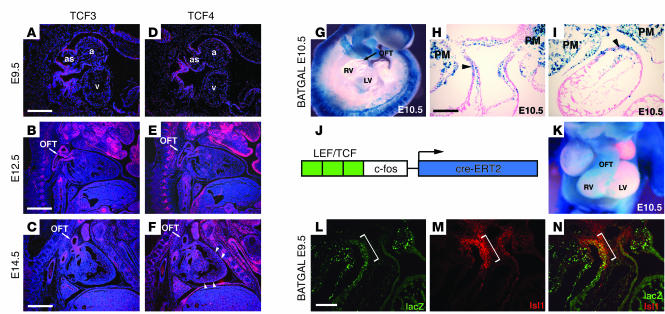
Little is known about the expression and activity of Wnt/β-catenin pathway components during cardiovascular morphogenesis. Therefore, we examined expression of the LEF/TCF family, members of which are critical effectors of Wnt/β-catenin signaling. TCF3 and TCF4 were expressed early in the developing heart in the aortic sac and developing atria (Figure 1, A and D). Later in development, TCF3 and to a lesser extent TCF4 were expressed in the developing outflow tract (Figure 1, B, C, E, and F). Low-level expression of TCF4 was also observed in the outer compact layer of the myocardium at E14.5 (Figure 1F).
Figure 1. Expression of Wnt/β-catenin signaling components during cardiac development.
Expression of TCF3 was observed in the aortic sac (as) and atria (a) at E9.5 (A) and in the developing outflow tract (OFT) at E12.5 (B) and E14.5 (C). v, ventricle. TCF4 expression was also observed in the aortic sac and atria at E9.5 (D) and at much lower levels in the developing outflow tract at E12.5 (E) and E14.5 (F). Arrowheads indicate compact zone of the myocardium. (G) BATGAL lacZ expression was not observed in the heart proper at E10.5 or any other time tested. (H and I) However, extensive lacZ expression was observed in the outflow tract and pharyngeal mesoderm (PM) at E10.5 (arrowheads). (J) Wnt signaling was fate-mapped using the TOP–cre-ERT2 transgenic line, which consists of 3 reiterated LEF/TCF DNA binding sites upstream of a minimal c-fos promoter driving the tamoxifen-inducible cre-ERT2 cDNA (14). (K) Strong lacZ expression was observed in the outflow tract and right ventricle of the heart but little contribution was observed in the left ventricle. (L–N) LacZ expression driven from the BATGAL transgene colocalized with Isl-1 expression in the outflow tract of the developing heart (brackets) using immunostaining with anti–β-galactosidase and anti–Isl-1 antibodies. Scale bars: 125 μm (A, D, H, I, and L–N); 500 μm (B, C, E, and F).
Several transgenic reporters have been generated that use reiterated LEF/TCF DNA binding sites upstream of minimal promoters to express β-galactosidase in cells where Wnt/β-catenin signaling is active. We used one of these lines, BATGAL (12), to map Wnt/β-catenin signaling during cardiovascular morphogenesis. BATGAL mice showed extensive β-galactosidase expression within the aortic sac region at E10.5 and in the dorsal aspect of the developing outflow tract and right ventricle (Figure 1, G–I). BATGAL expression was also observed in the pharyngeal mesoderm in the region harboring Isl-1–positive AHF progenitors (Figure 1, H and I). In contrast, we observed little β-galactosidase expression from either BATGAL or another Wnt reporter, TOPGAL (13), in the heart proper (Figure 1G and data not shown).
The presence of active Wnt/β-catenin signaling in the distal outflow tract, aortic sac, and pharyngeal mesoderm suggested that this pathway is important for cells that give rise to the AHF. These cells, which can be identified by Isl-1 expression, are thought to be important for development of the right ventricle and outflow tract of the heart (2). However, because there is little Wnt reporter activity in the heart proper, Wnt signaling must be active early in the AHF but turned off as cardiac differentiation progresses. To address this hypothesis, we used a fate-mapping approach to determine whether Wnt/β-catenin–positive cells are capable of generating AHF-derived structures. Using a Wnt transgenic reporter linked to a tamoxifen-inducible cre (Figure 1J) (14), we mapped Wnt-responsive cells from E8.5 through E10.5. Our results showed that cells with active Wnt signaling contributed to the right ventricle, the outflow tract, and portions of the atria, correlating closely with the regions of the heart derived from the AHF (Figure 1K). To determine whether the BATGAL-positive cells corresponded to Isl-1–positive AHF cells, we performed double immunostaining for expression of lacZ — driven from the BATGAL transgenic line — and Isl-1. These data revealed extensive overlap between cells with active Wnt/β-catenin signaling and Isl-1–positive cells within the AHF, especially as Isl-1–positive cells migrated into the outflow tract region (Figure 1, L–N). Together, these data suggest that active Wnt/β-catenin activity occurs in portions of the developing cardiovascular system analogous to the AHF.
Deletion of β-catenin in the cardiovascular system results in defective development of the AHF.
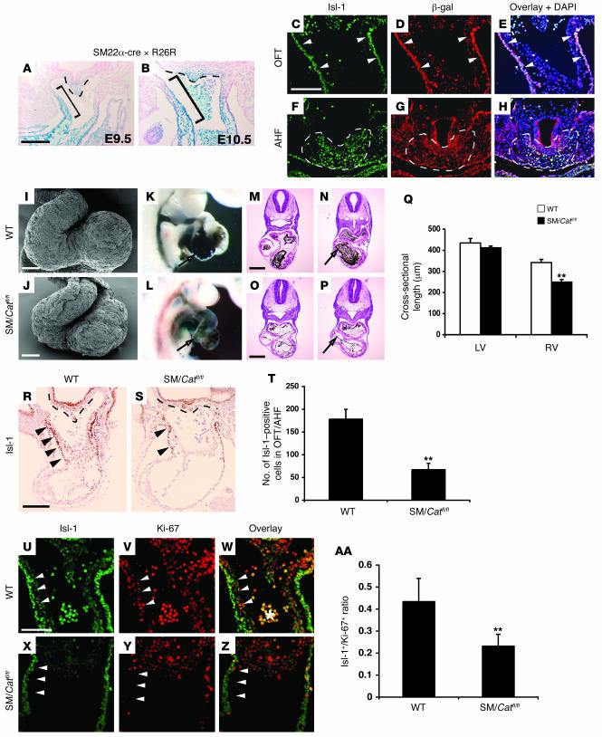
To assess the role of Wnt/β-catenin signaling in the heart, β-catenin was deleted in myocardial precursors using the previously described SM22α-cre transgenic mouse line (15). The SM22α-cre line was active within the primary heart field as well as the AHF, as evidenced by fate-mapping studies using the R26R mouse line, which showed lacZ expression in the distal outflow tract as well as the pharyngeal mesoderm (Figure 2, A and B, and ref. 15). Of note, this expression correlated closely with that observed in the BATGAL Wnt reporter line (Figure 1H), and extensive costaining was observed for Isl-1 and β-galactosidase expression in the AHF and outflow tracts of SM22α-cre × R26R embryos (Figure 2, C–H). SM22cre/Catnbflox/flox embryos die between E10.5 and E11.5 as a result of severe defects in cardiac development. Scanning electron microscopy of the hearts from E9.5 SM22cre/Catnbflox/flox embryos revealed substantial and specific reduction in the size of the right ventricle of the heart (Figure 2, I and J). Ink injections to outline the cardiovascular system showed that the right ventricle was severely hypoplastic (Figure 2, K and L). Histological examination and quantitation of right ventricular diameter showed that it was reduced by 29% in SM22cre/Catnbflox/flox embryos (P < 0.005; Figure 2, M–Q).
Figure 2. Loss of Wnt/β-catenin signaling in the AHF leads to decreased right heart development and loss of Isl-1 progenitors.
(A and B) SM22α-cre is active in the AHF, demonstrated by lacZ expression throughout the outflow tract (brackets) and in the pharyngeal mesodermal apex (dotted lines) of SM22α-cre × R26R mice at E9.5 and E10.5. (C–H) Immunofluorescent staining for Isl-1 and β-galactosidase expression shows extensive overlap within the outflow tract (arrowheads) and AHF (dotted lines) at E9.5. Loss of β-catenin using the SM22α-cre transgenic line caused hypoplastic right ventricle as assessed by scanning electron microscopy (I and J) and ink injections of wild-type (K) and SM22cre/Catnbflox/flox (SM/Catfl/fl) embryos (L) at E9.5. Histological sectioning at multiple levels showed the reduction in right ventricle size (arrows) at E9.5 in SM22cre/Catnbflox/flox (O and P) compared with wild-type embryos (M and N). (Q) Right ventricular diameter in SM22cre/Catnbflox/flox compared with wild-type embryos. (R–T) To assess changes in Isl-1 AHF progenitors, wild-type and SM22cre/Catnbflox/flox embryos were immunostained for Isl-1 protein expression. SM22cre/Catnbflox/flox mutants have severely reduced numbers of Isl-1 AHF progenitors in the outflow tract at E9.5. (U–AA) Isl-1 and Ki-67 double immunofluorescence was performed to determine changes in proliferation in AHF progenitors. Reduced Ki-67 staining in Isl-1–positive cells within the outflow tract was observed in SM22cre/Catnbflox/flox mutant embryos (arrowheads). (AA) Quantitation showed an almost 50% reduction in Isl-1 AHF progenitor proliferation. **P < 0.005. Scale bars: 100 μm (A–J); 500 μm (M–P); 125 μm (R and S); 75 μm (U–Z).
To determine whether this loss of right ventricular size was caused by loss of Isl-1–positive AHF progenitors, we assessed expression of Isl-1 in wild-type and SM22cre/Catnbflox/flox embryos by immunohistochemistry. Loss of Wnt/β-catenin signaling caused a significant loss in Isl-1–positive precursors in the outflow tract and pharyngeal mesodermal apex of SM22cre/Catnbflox/flox mutants (Figure 2, R–T). Moreover, loss of Wnt/β-catenin signaling decreased proliferation in the Isl-1–positive cell population migrating into the outflow tract of the heart (Figure 2, U–AA). These data indicate that Wnt/β-catenin signaling regulates the number and proliferation of Isl-1–positive cells within the AHF.
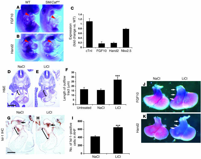
The loss of right heart development suggested that pathways important for AHF development might be preferentially compromised in SM22cre/Catnbflox/flox embryos. FGF10 and Hand2 are 2 markers known to delineate the AHF and structures within the developing heart derived from the AHF (16, 17). In situ hybridization revealed decreased expression of FGF10 and Hand2 in SM22cre/Catnbflox/flox embryos at E9.5 (Figure 3, A and B). Quantitative real-time RT-PCR (Q-PCR) revealed a preferential loss of FGF10 and Hand2 versus cTnI and Nkx2.5, suggesting a specific effect on right heart development (Figure 3C). Together, these data indicate that loss of Wnt/β-catenin signaling in the AHF leads to defective right ventricular development coincident with a loss of FGF10 and Hand2 expression.
Figure 3. Regulation of AHF marker genes and progenitor number by Wnt signaling.
(A and B) FGF10 and Hand2 expression was reduced specifically in the hearts of SM22cre/Catnbflox/flox mutants (arrowheads). (C) Q-PCR was used to quantitate expression changes in the hearts of E9.5 SM22cre/Catnbflox/flox mutants. Expression of FGF10 and Hand2 was significantly downregulated, while expression of cTnI and Nkx2.5 was not appreciably affected by loss of β-catenin. (D–F) To activate canonical Wnt signaling in vivo, developing embryos were treated with LiCl as described in Methods. LiCl treatment of embryos increased outflow tract (brackets) length. (G and H) This increased outflow tract length was associated with an increase in the number of Isl-1–positive AHF progenitors migrating into the outflow tract (brackets) and increased Isl-1 staining in the pharyngeal mesoderm harboring the Isl-1–positive AHF progenitor pool (arrows). (I) The number of Isl-1–positive cells in the outflow tract/right ventricle increased approximately 50% in Isl-1–positive AHF progenitors after LiCl treatment. (J and K) FGF10 and Hand2 expression was upregulated in the outflow tract and right ventricle after LiCl treatment (arrows). ***P < 0.001. Scale bars: 250 μm (D and E); 125 μm (G and H).
Increased Wnt/β-catenin activity leads to expansion of Isl-1–positive AHF precursors and increased FGF10 and Hand2 expression.
Next, we sought to determine whether activating Wnt/β-catenin signaling would lead to an increase in Isl-1–positive cells and markers of AHF-derived structures. To activate Wnt signaling in the developing heart, pregnant mice were injected with LiCl, which inhibits GSK-3β and in turn activates Wnt signaling (18). Embryos were injected daily starting at E8.5 and analyzed at E10.5. Activation of Wnt signaling via LiCl resulted in a significant lengthening of the outflow tract at E10.5, indicating increased development of this region of the heart (Figure 3, D–F). Remarkably, LiCl treatment also led to a dramatic increase in the number of Isl-1–positive cells within the outflow tract and pharyngeal mesoderm of the AHF (Figure 3, G–I). These results suggest that Wnt signaling activates AHF development through increased expansion of Isl-1–positive progenitors. To assess the effect of increased Wnt signaling on other components of the AHF pathway, in situ hybridization was performed to determine FGF10 and Hand2 expression. We found a dramatic increase in the expression of both FGF10 and Hand2 in the hearts of LiCl-treated embryos (Figure 3, J and K).
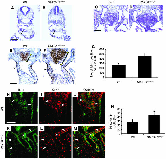
We performed 2 additional studies to verify that the LiCl gain-of-function results were caused by specific activation of canonical Wnt signaling. First, SM22α-cre mice were crossed to Catnbflox(ex3)/+ mice to specifically activate canonical Wnt/β-catenin signaling by expression of a highly stable form of β-catenin (19). SM22cre/Catnbflox(ex3)/+ embryos had enlarged right ventricles at E10.5 (Figure 4, A and B). The mesoderm surrounding the developing anterior region of the foregut analogous to the AHF was markedly enlarged, indicating expansion of this tissue (Figure 4, C and D). Moreover, the number of Isl-1–positive AHF progenitors within this region and the outflow tract was dramatically increased in SM22cre/Catnbflox(ex3)/+ embryos (Figure 4, E–G). Second, in order to determine whether cell proliferation was affected in the AHF of the SM22cre/Catnbflox(ex3)/+ embryo, Isl-1 and Ki-67 double immunofluorescent staining was performed. We found increased proliferation in Isl-1–positive cells within the AHF and outflow tracts of SM22cre/Catnbflox(ex3)/+ embryos (Figure 4, H–N). Together, these data suggest that stabilization of β-catenin in AHF progenitors increases cell proliferation, leading to expansion of Isl-1–positive cells.
Figure 4. Activation of canonical Wnt signaling by β-catenin stabilization increases AHF and right heart development and proliferation.
(A and B) SM22α-cre mice were crossed to Catnbflox(ex3)/+ mice to generate SM22cre/Catnbflox(ex3)/+ mutant embryos. SM22cre/Catnbflox(ex3)/+ mutants had enlarged hearts, in particular the right ventricles (arrows), at E9.5. (C and D) Increased cell mass was also observed in the anterior foregut mesoderm surrounding the trachea, where a pool of Isl-1–positive AHF progenitors resides, at E10.5. (E and F) Isl-1 immunostaining was increased in the AHF (brackets) and outflow tracts (arrowheads) of SM22cre/Catnbflox(ex3)/+ mutants at E10.5. (G) Quantitation of the increase in Isl-1 immunostaining revealed a greater than 50% increase in Catnbflox(ex3)/+ mutants. (H–M) Isl-1 and Ki-67 double immunofluorescent staining reveal increased proliferation in Isl-1–positive cells within the outflow tracts of SM22cre/Catnbflox(ex3)/+ mutant embryos at E10.5 (arrowheads). (N) Ki-67 staining in Isl-1–positive cells increased approximately 40% within SM22cre/Catnbflox(ex3)/+ embryos. *P < 0.02. Scale bars: 500 μm (A and B); 75 μm (C–F); 100 μm (H–M).
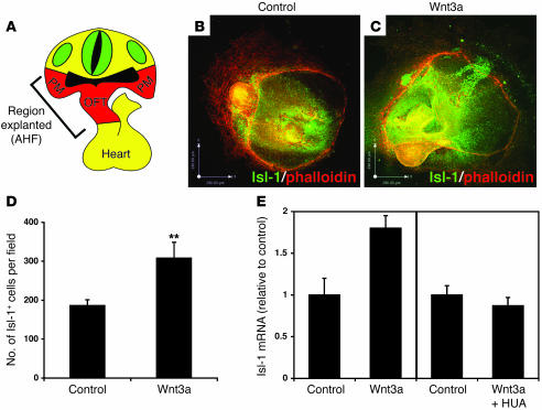
To determine whether Wnts can activate expansion of Isl-1 AHF progenitors directly, explant studies were performed by culturing the AHF (the outflow tract and part of the pharyngeal mesoderm) in the presence or absence of Wnt3a, a ligand known to activate the β-catenin–dependent canonical pathway (20). Under previously described conditions (21), these explants were capable of differentiating into beating cardiac myocytes after 2 days in culture (see Supplemental Videos 1 and 2; supplemental material available online with this article; doi:10.1172/JCI31731DS1) (21). Culturing these explants in the presence of Wnt3a led to a significant increase in Isl-1–positive cells (Figure 5, A–D). By treating explants with hydroxyurea and aphidicolin (HUA), known to impede cell proliferation (22), in addition to Wnt3a, we determined that the increase in Isl-1 expression was the result of an increase in the number of Isl-1–positive cells and not the level of Isl-1 per cell (Figure 5E). Together, these data indicate that Wnt/β-catenin signaling directs expansion of Isl-1–positive cells in the AHF both in vivo and in ex vivo explants.
Figure 5. Wnt signaling expands the number of Isl-1–positive progenitors in AHF explants.
(A–C) The AHF was explanted as shown (A) and cultured for 2 days in the absence (B) or presence (C) of Wnt3a. Isl-1 immunostaining increased upon treatment with Wnt3a. (D) Isl-1–positive cells increased approximately 50% in Wnt3a-treated explants. (E) Isl-1 mRNA expression also increased, as determined by Q-PCR. To determine whether this increase was due to an increase in Isl-1 expression per cell or due primarily to an increase in Isl-1–positive cells, explants were treated with HUA to inhibit cell proliferation. HUA treatment inhibited the increase in Isl-1 mRNA expression, which suggests that the majority of increased Isl-1 immunostaining and mRNA expression was the result of expansion of Isl-1–positive cells after Wnt3a treatment. **P < 0.005.
Wnt/β-catenin signaling acts through multiple FGFs to regulate expansion of Isl-1–positive AHF progenitors.
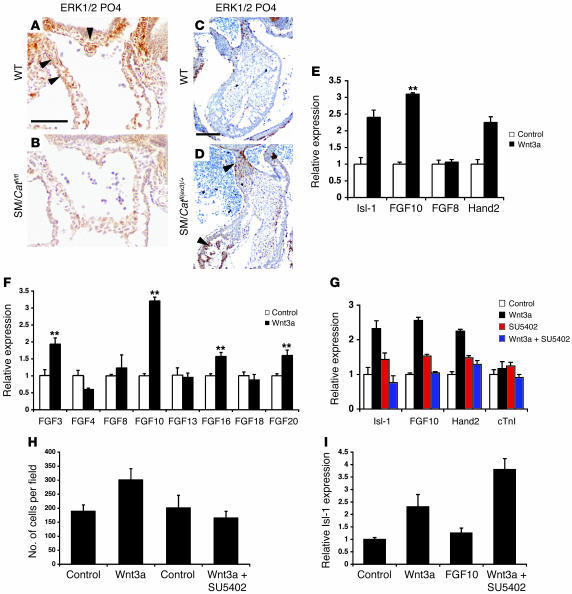
Given the increase in FGF10 expression, we sought to determine whether FGF signaling acts cooperatively with Wnt signaling to regulate AHF precursor expansion. As a first step toward testing this hypothesis, we examined phosphorylation of ERK1/2, which is activated by FGF signaling, in wild-type and SM22cre/Catnbflox/flox embryos. There was a substantial reduction in ERK1/2 phosphorylation in the outflow tract and AHF of SM22cre/Catnbflox/flox embryos at E9.5, indicating a loss of FGF signaling corresponding to a loss in FGF10 expression (Figure 6, A and B). Conversely, an increase in ERK1/2 phosphorylation was observed in SM22cre/Catnbflox(ex3)/+ embryos (Figure 6, C and D). These findings indicate that Wnt/β-catenin signaling regulates levels of overall FGF signaling activity in the AHF. However, loss of FGF10 did not lead to as dramatic phenotype as observed in SM22cre/Catnbflox/flox embryos (23). Thus, we determined whether expression of FGF8, a ligand expressed in the AHF and known to play an important part in its development (24), was altered. Surprisingly, FGF8 expression levels were unaffected by treatment of AHF explants with Wnt3a (Figure 6E). This suggested a degree of FGF ligand specificity in regulation of AHF precursors. Therefore, we performed a larger screen of FGF ligands to assess which, if any, were regulated by Wnt signaling in the AHF. These studies revealed that FGF3, FGF10, FGF16, and FGF20 were positively regulated by Wnt3a in the AHF, while FGF4 was repressed and FGF8, FGF13, and FGF18 were unaffected (Figure 6F). These data suggest a broader FGF signaling network downstream of Wnt signaling in the AHF.
Figure 6. FGF signaling is activated by Wnt signaling in the AHF.
(A and B) ERK1/2 phosphorylation was used to assess the activity of FGF signaling in the AHF. SM22cre/Catnbflox/flox mutants expressed less phosphorylated ERK1/2 in the AHF and outflow tract than did wild-type littermates at E9.5 (arrowheads). PO4, phosphorylation. (C and D) ERK1/2 phosphorylation increased in the outflow tract and right ventricular myocardium in SM22cre/Catnbflox(ex3)/+ embryos at E10.5 (arrowheads). (E) FGF10 and FGF8 expression was assessed by Q-PCR in AHF explants treated with Wnt3a. Surprisingly, FGF10 expression was significantly upregulated, while expression of FGF8, which is also expressed in the AHF, was unchanged. Expression of both Isl-1 and Hand2 was upregulated as expected. (F) Expression of additional FGF ligands was determined by Q-PCR, and FGF3, FGF10, FGF16, and FGF20 were all significantly upregulated by Wnt3a treatment, whereas FGF4 was downregulated. (G and H) Activation of AHF gene expression (G) and Isl-1–positive AHF progenitor number (H) by Wnt3a was attenuated by the FGF receptor inhibitor SU5402, indicating that these pathways act cooperatively in regulating AHF development. (I) Conversely, Wnt3a and FGF10 cooperatively increased Isl-1 expression in AHF explants, further supporting interaction between Wnt and FGF signaling in AHF development. **P < 0.005. Scale bars: 100 μm.
To test whether Wnt-mediated expansion of Isl-1–positive AHF progenitors was dependent on FGF signaling, AHF explants were treated with Wnt3a, SU5402 (a potent FGF receptor inhibitor), or both. As observed above, Wnt3a significantly increased expression of FGF10 as well as Isl-1 and Hand2 (Figure 6G). Addition of SU5402 to Wnt3a attenuated this response compared with explants treated with Wnt3a alone (Figure 6G). SU5402 alone caused a slight increase in expression of Isl-1 and FGF10, possibly by inhibiting a negative feedback loop regulating Wnt activity. Such a negative feedback loop has been proposed to involve sprouty proteins: inhibitors of FGF signaling, some of which are positively regulated by both Wnt and FGF signaling (25, 26). Thus, SU5402 inhibition may result in a low-level threshold increase in FGF signaling by inhibiting an FGF inhibitor such as sprouty. The number of Isl-1–positive cells was assessed after Wnt3a treatment alone or with Wnt3a plus SU5402. Inhibition of FGF signaling by SU5402 abrogated the increase in Isl-1–positive cells in AHF explants (Figure 6H), showing that FGF signaling acts in the same pathway as Wnt signaling to expand Isl-1 AHF progenitors. Conversely, the combination of Wnt3a and FGF10 led to a cooperative increase in Isl-1–positive cells in AHF explants (Figure 6I). The finding that treatment of explants with FGF10 did not lead to a maximal increase in the number of Isl-1–positive cells further supports the requirement of multiple FGF ligands in AHF progenitor expansion. Together, these data suggest a direct link between Wnt and FGF signaling in the expansion of Isl-1–positive progenitors in the AHF and imply that FGF signaling acts downstream of Wnt in this process.
FGF10 is a direct target of Wnt signaling in the AHF.
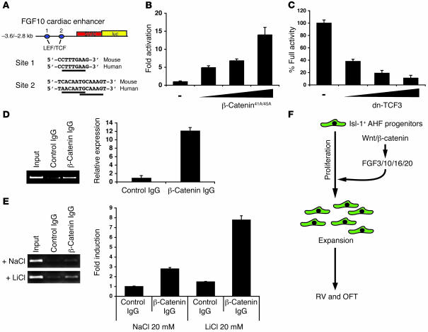
Promoter/enhancer elements from the FGF10 locus have been shown to direct expression in the AHF/outflow tract of transgenic mice, and FGF10 expression has been used to define the AHF (6, 17). Examination of this FGF10 enhancer sequence, hereafter referred to as the FGF10-AHF enhancer, revealed the presence of 2 LEF/TCF DNA binding sites conserved in both mice and humans (Figure 7A). Using the FGF10-AHF enhancer linked to a luciferase reporter, we performed transactivation assays to determine its sensitivity to Wnt signaling through expression of a constitutively active form of β-catenin (27). Remarkably, the FGF10-AHF enhancer was activated to high levels in a dose-dependent manner by an activated form of β-catenin (Figure 7B). Conversely, the FGF10-AHF enhancer was repressed by a dominant-negative TCF3 lacking the β-catenin binding site, further indicating that the enhancer is Wnt responsive (Figure 7C). To determine whether β-catenin could form a complex on the conserved LEF/TCF DNA binding sites in the FGF10-AHF enhancer in vivo, we performed chromatin immunoprecipitation (ChIP) assays using chromatin from the AHF and outflow tracts of E10.5 embryos and found that β-catenin associated with the FGF10-AHF enhancer element in vivo (Figure 7D). In order to determine whether activation of Wnt signaling could increase the association of β-catenin with the FGF10-AHF enhancer, 293 T cells were transfected with the FGF10-AHF enhancer reporter plasmid and treated with either NaCl or LiCl, after which ChIP assays were performed. LiCl increased the level of β-catenin and FGF10-AHF enhancer association by almost 3-fold (Figure 7E). Together, these data indicate that FGF10 is a direct target of Wnt/β-catenin signaling and identify a signaling axis in which Wnt/β-catenin acts upstream of FGF10, in addition to FGF3, FGF16, and FGF20, to regulate the expansion of Isl-1–positive AHF progenitors (Figure 7F).
Figure 7. FGF10 is a direct target of Wnt/β-catenin signaling in the AHF.
(A) The FGF10-AHF enhancer from the FGF10 gene contains 2 cross-species–conserved LEF/TCF DNA binding sites. (B and C) The FGF10-AHF enhancer is activated and repressed by an activated form of β-catenin (β-catenin41A/45A) and dominant-negative TCF3 (dnTCF2), respectively. (D and E) ChIP assays showed that β-catenin formed a complex on the FGF10-AHF enhancer in vivo (D), and LiCl treatment of 293 T cells increased the association of β-catenin with this enhancer element (E). (F) Model of Wnt/FGF signaling promoting expansion of Isl-1–positive AHF progenitors through activation of specific FGF ligands, leading to proliferation and proper development of the outflow tract and right ventricle of the heart.
Discussion
Accumulating evidence indicates that Isl-1–positive AHF progenitors play an important role in regulation of both cardiac development and postnatal cardiac homeostasis (2–4, 6, 21, 24). The results of our present study suggest that Wnt/β-catenin signaling directs the expansion of these cells through activation of multiple FGF ligands, in particular FGF10. Delineation of this signaling pathway provides critical insight into how these cells develop into right-sided cardiac structures as well as important clues as to how Isl-1–positive progenitors can be expanded for possible postnatal cardiac regenerative therapies.
Although Wnt signaling is a critical regulator of cardiac specification, to the best of our knowledge this is the first evidence of Wnt/β-catenin signaling playing an important role in a cardiac progenitor/stem cell population in vivo. Cardiac development occurs through differentiation of a specific population of early mesodermal precursors. The earliest events driving this differentiation process are still poorly understood, but there is evidence that Wnt signaling plays a critical role through regulation of bone morphogenetic protein signaling (9, 10). However, evidence as to whether canonical Wnt signaling promotes or inhibits cardiogenesis is conflicting. Previous studies have shown that inhibition of canonical Wnt signaling leads to cardiac differentiation and that noncanonical Wnt signaling via Wnt11 also promotes cardiac differentiation (9, 11). More recent evidence in ES cells shows that the role of canonical Wnt signaling in cardiogenesis is likely more complex and may include temporal restrictions in its requirement (28), possibly due to the complexity of Wnt ligands and receptors expressed during early mesoderm and cardiac development. However, it is unknown which of these are responsible for regulating early cardiac development. Previous studies demonstrated that a potential Wnt7b-directed paracrine signaling mechanism regulates vascular smooth muscle development in the lung (29, 30). In the mouse, Wnt2a and Wnt2b are expressed in an overlapping pattern in the early cardiac crescent, while Wnt8a is expressed in the myocardium through E12.5 (31–33). A previous report on the generation and characterization of Wnt2a mouse mutants did not describe any cardiac defects (32), and Wnt2b and Wnt8a null animals have not previously been reported. Thus, the lack of a phenotype in Wnt2a null animals could be due to redundancy with Wnt2b, Wnt8a, or other as-yet-unidentified Wnt ligands.
Our findings demonstrate that Wnt signaling acts upstream of several FGF ligands, including FGF3, FGF10, FGF16, and FGF20, leading to increased expansion of Isl-1–positive AHF progenitors. This corresponds to earlier studies showing that FGF10 and Isl-1 expression mark the same or a similar cell population in the AHF (2, 6). Our finding that FGF3, FGF10, FGF16, and FGF20 were upregulated by Wnt signaling underscores important redundancy in this pathway in AHF development and progenitor expansion. We demonstrated a direct link between these pathways by showing that FGF10 was a direct target of Wnt signaling. Moreover, a previous report has demonstrated that FGF20 is a direct target of Wnt signaling (34). Thus, Wnt signaling may be a focal point of an integrated signaling network required for stem/progenitor cell expansion and self renewal in the heart.
The ability of Wnt-FGF signaling to control expansion of Isl-1–positive progenitors in the AHF has several important implications. These cells have previously been shown to be critical in the development of right-sided structures in the heart including the right ventricle and the outflow tract (2). Given the significance of congenital heart defects in humans that involve these structures, including persistent truncus arteriosus, double outlet right ventricle, and hypoplastic right ventricle, it will be important to ascertain whether defective Wnt-FGF signaling in the AHF underlies some of these syndromes. Recently, Isl-1–positive cells isolated from differentiating embryoid bodies were shown to have clonal cardiac progenitor potential, including the ability to differentiate into cardiomyocytes, vascular smooth muscle, and endothelial cells (3). Isl-1–positive cells are also present in the early postnatal heart, albeit at low levels, but disappear with progressing age (5). Given the extreme rarity of these cells and their ability to act as cardiac progenitors, the capacity of Wnt-FGF signaling to expand this population may prove to be useful: in future work, the ability of Isl-1–positive cells may be harnessed to regenerate damaged cardiac tissue. Thus, controlling Wnt and FGF signaling in Isl-1–positive cells both in vivo and ex vivo may allow their controlled expansion for future reparative therapies.
Methods
Generation of SM22α-restricted excision of β-catenin.
Catnbflox/flox mice were mated to homozygosity and crossed to Catnbflox/+ × SM22α-cre mice to generate SM22cre/Catnbflox/flox embryos. Catnbflox(ex3)/+ mice were mated to SM22α-cre mice to generate SM22cre/Catnbflox(ex3)/+ embryos. Genotyping of Catnbflox/flox, Catnbflox(ex3)/+, and SM22α-cre mice was performed as previously described (15, 19, 35). Generation and genotyping of the BATGAL and TOP-creERT2 mice has been previously reported (3, 14). All animal protocols were approved by the University of Pennsylvania Institutional Animal Care and Use Committee.
Detection of Wnt/β-catenin activity through reporter mice.
Embryos at the ages indicated in the figure legends were collected and stained for lacZ expression as previously described (29). For fate mapping of canonical Wnt activity in the cardiovascular system, pregnant TOP-creERT2 mice crossed to R26R lacZ reporter mice were injected with tamoxifen at E8.5, and embryos were harvested at E10.5 and stained for lacZ expression as above.
Histology.
Embryos were isolated from pregnant dams and fixed in 4% paraformaldehyde for 24–48 hours. They were then dehydrated through a series of washes in increasing concentrations of ethanol. After paraffin embedding, embryos were sectioned and slides were stained as previously described (29, 36–38). In situ hybridization probes for TCF1, TCF3, and TCF4 were generated using RT-PCR of E10.5 mouse embryos’ cDNA. Immunohistochemistry was performed using the following antibodies: rabbit anti–β-galactosidase (catalog no. CR7001RP; Cortex Biochem), mouse anti-rat Isl-1 (University of Iowa Hybridoma Bank), rat anti-Ki67 (clone TEC-3; Dako), and mouse anti-ERK1/2 phosphorylation (E-4, catalog no. sc-7383; Santa Cruz Biotechnology Inc.). For scanning electron microscopy, hearts from E9.5 embryos were fixed in 2% gluteraldehyde and processed as described previously (29).
LiCl treatment of developing embryos.
Pregnant dams were injected with 10 μl of a 600-mM LiCl or NaCl solution each day starting at E8.5 through E10.5. Survival of LiCl-treated embryos was not significantly different from that of NaCl-treated embryos up to E12.5. Embryos were fixed and processed as described above.
Cotransfections and ChIP assays.
The –3.6- to –2.8-kb mouse FGF10 enhancer has been previously described (17). This enhancer region was subcloned into the pGL3 promoter luciferase reporter vector to generate the pGL3FGF10.luc vector. The dominant-negative TCF3 and β-catenin41A/45A expression vectors have been previously described (27, 39). We transfected 293 T cells with the increasing amounts of the above plasmids using Fugene 6 as previously described (40). Luciferase assays were performed using a Dual Luciferase Reporter Assay System (Promega), and reported values are the mean ± SEM of 3 assays performed in duplicate.
Chromatin was isolated from the AHF regions of E10.5 embryos or 293 T cells transfected with the pGL3FGF10.luc plasmid using a ChIP Assay kit (Upstate). Chromatin was then immunoprecipitated with either the β-catenin mAb (BD Biosciences — Pharmingen) or a nonimmune control antibody, washed, and subjected to PCR using the oligonucleotides listed in Supplemental Table 1. Amplified fragments were analyzed on a 1.5% agarose gel or Q-PCR was performed on an ABI 7900 cycler, and quantitative differences were measured. Each ChIP assay was performed at least 3 times, and Q-PCR results represent the mean ± SEM of 3 assays.
AHF explant culture system.
The outflow tracts and pharyngeal arches of E8.5 embryos were isolated with tungsten needles and cultured for 48 hours on collagen/fibronectin-coated tissue culture plastic multiwell chamber slides as previously described (21) in MEM containing 5% horse serum and antibiotics. Where indicated in Figures 5 and 6, medium was supplemented with HUA (20 mM hydroxyurea; 150 μM aphidicolin), 100 ng/ml Wnt3a (R&D Systems), 500 ng/ml FGF10 (R&D Systems), and/or 15 μM SU5402. Explants were stained for Isl-1 expression using the monoclonal Isl-1 antibody and counterstained with phalloidin-TRITC and DAPI. Imaging was performed on a Leica DM6000B confocal microscope using Velocity 4.02 software (Improvision).
Q-PCR.
Total RNA was isolated from E9.5 wild-type or SM22cre/Catnbflox/flox hearts or cultured AHF explants and reverse transcribed using a Reverse Transcription System (Promega). Oligonucleotides listed in Supplemental Table 1 were used to measure differences in gene expression on an ABI 7900 cycler. Reported values are the mean ± SEM of 3 assays from at least 3 hearts of each genotype or at least 20 explants.
Statistics.
Two-tailed Student’s t test was used.
Supplementary Material
Acknowledgments
The authors appreciate helpful comments during the course of this work and on the manuscript from Jon Epstein and Mark Kahn (University of Pennsylvania School of Medicine). The BATGAL mice were kindly provided by Stefano Piccolo (University of Padua, Padua, Italy). These studies were supported by NIH grants HL075215 (to E.E. Morrisey) and DC006254 (D.J. Epstein) and by a postdoctoral fellowship from the American Heart Association (to E.D. Cohen). E.E. Morrisey is an Established Investigator of the American Heart Association.
Footnotes
Nonstandard abbreviations used: AHF, anterior heart field; ChIP, chromatin immunoprecipitation; HUA, hydroxyurea and aphidicolin; LEF, lymphoid enhancer factor; TCF, T cell factor.
Conflict of interest: The authors have declared that no conflict of interest exists.
Citation for this article: J. Clin. Invest. 117:1794–1804 (2007). doi:10.1172/JCI31731
References
- 1.Kelly R.G. Molecular inroads into the anterior heart field. Trends Cardiovasc. Med. 2005;15:51–56. doi: 10.1016/j.tcm.2005.02.001. [DOI] [PubMed] [Google Scholar]
- 2.Cai C.L., et al. Isl1 identifies a cardiac progenitor population that proliferates prior to differentiation and contributes a majority of cells to the heart. Dev. Cell. 2003;5:877–889. doi: 10.1016/s1534-5807(03)00363-0. [DOI] [PMC free article] [PubMed] [Google Scholar]
- 3.Moretti A., et al. Multipotent embryonic isl1+ progenitor cells lead to cardiac, smooth muscle, and endothelial cell diversification. Cell. 2006;127:1151–1165. doi: 10.1016/j.cell.2006.10.029. [DOI] [PubMed] [Google Scholar]
- 4.Wu S.M., et al. Developmental origin of a bipotential myocardial and smooth muscle cell precursor in the mammalian heart. Cell. 2006;127:1137–1150. doi: 10.1016/j.cell.2006.10.028. [DOI] [PubMed] [Google Scholar]
- 5.Laugwitz K.L., et al. Postnatal isl1+ cardioblasts enter fully differentiated cardiomyocyte lineages. Nature. 2005;433:647–653. doi: 10.1038/nature03215. [DOI] [PMC free article] [PubMed] [Google Scholar]
- 6.Kelly R.G., Brown N.A., Buckingham M.E. The arterial pole of the mouse heart forms from Fgf10-expressing cells in pharyngeal mesoderm. Dev. Cell. 2001;1:435–440. doi: 10.1016/s1534-5807(01)00040-5. [DOI] [PubMed] [Google Scholar]
- 7.Logan C.Y., Nusse R. The Wnt signaling pathway in development and disease. Annu. Rev. Cell Dev. Biol. 2004;20:781–810. doi: 10.1146/annurev.cellbio.20.010403.113126. [DOI] [PubMed] [Google Scholar]
- 8.Lindsley R.C., Gill J.G., Kyba M., Murphy T.L., Murphy K.M. Canonical Wnt signaling is required for development of embryonic stem cell-derived mesoderm. Development. 2006;133:3787–3796. doi: 10.1242/dev.02551. [DOI] [PubMed] [Google Scholar]
- 9.Marvin M.J., Di Rocco G., Gardiner A., Bush S.M., Lassar A.B. Inhibition of Wnt activity induces heart formation from posterior mesoderm. Genes Dev. 2001;15:316–327. doi: 10.1101/gad.855501. [DOI] [PMC free article] [PubMed] [Google Scholar]
- 10.Tzahor E., Lassar A.B. Wnt signals from the neural tube block ectopic cardiogenesis. Genes Dev. 2001;15:255–260. doi: 10.1101/gad.871501. [DOI] [PMC free article] [PubMed] [Google Scholar]
- 11.Pandur P., Lasche M., Eisenberg L.M., Kuhl M. Wnt-11 activation of a non-canonical Wnt signalling pathway is required for cardiogenesis. Nature. 2002;418:636–641. doi: 10.1038/nature00921. [DOI] [PubMed] [Google Scholar]
- 12.Maretto S., et al. Mapping Wnt/beta-catenin signaling during mouse development and in colorectal tumors. Proc. Natl. Acad. Sci. U. S. A. 2003;100:3299–3304. doi: 10.1073/pnas.0434590100. [DOI] [PMC free article] [PubMed] [Google Scholar]
- 13.DasGupta R., Fuchs E. Multiple roles for activated LEF/TCF transcription complexes during hair follicle development and differentiation. Development. 1999;126:4557–4568. doi: 10.1242/dev.126.20.4557. [DOI] [PubMed] [Google Scholar]
- 14.Riccomagno M.M., Takada S., Epstein D.J. Wnt-dependent regulation of inner ear morphogenesis is balanced by the opposing and supporting roles of Shh. Genes Dev. 2005;19:1612–1623. doi: 10.1101/gad.1303905. [DOI] [PMC free article] [PubMed] [Google Scholar]
- 15.Lepore J.J., et al. High-efficiency somatic mutagenesis in smooth muscle cells and cardiac myocytes in SM22alpha-Cre transgenic mice. Genesis. 2005;41:179–184. doi: 10.1002/gene.20112. [DOI] [PubMed] [Google Scholar]
- 16.McFadden D.G., et al. A GATA-dependent right ventricular enhancer controls dHAND transcription in the developing heart. Development. 2000;127:5331–5341. doi: 10.1242/dev.127.24.5331. [DOI] [PubMed] [Google Scholar]
- 17.Ohuchi H., et al. Identification of cis-element regulating expression of the mouse Fgf10 gene during inner ear development. Dev. Dyn. 2005;233:177–187. doi: 10.1002/dvdy.20319. [DOI] [PubMed] [Google Scholar]
- 18.Klein P.S., Melton D.A. A molecular mechanism for the effect of lithium on development. Proc. Natl. Acad. Sci. U. S. A. 1996;93:8455–8459. doi: 10.1073/pnas.93.16.8455. [DOI] [PMC free article] [PubMed] [Google Scholar]
- 19.Harada N., et al. Intestinal polyposis in mice with a dominant stable mutation of the beta-catenin gene. EMBO J. 1999;18:5931–5942. doi: 10.1093/emboj/18.21.5931. [DOI] [PMC free article] [PubMed] [Google Scholar]
- 20.Nakamura T., Sano M., Songyang Z., Schneider M.D. A Wnt- and beta -catenin-dependent pathway for mammalian cardiac myogenesis. Proc. Natl. Acad. Sci. U. S. A. 2003;100:5834–5839. doi: 10.1073/pnas.0935626100. [DOI] [PMC free article] [PubMed] [Google Scholar]
- 21.Zaffran S., Kelly R.G., Meilhac S.M., Buckingham M.E., Brown N.A. Right ventricular myocardium derives from the anterior heart field. Circ. Res. 2004;95:261–268. doi: 10.1161/01.RES.0000136815.73623.BE. [DOI] [PubMed] [Google Scholar]
- 22.Harris W.A., Hartenstein V. Neuronal determination without cell division in Xenopus embryos. Neuron. 1991;6:499–515. doi: 10.1016/0896-6273(91)90053-3. [DOI] [PubMed] [Google Scholar]
- 23.Marguerie A., et al. Congenital heart defects in Fgfr2-IIIb and Fgf10 mutant mice. Cardiovasc. Res. 2006;71:50–60. doi: 10.1016/j.cardiores.2006.03.021. [DOI] [PubMed] [Google Scholar]
- 24.Ilagan R., et al. Fgf8 is required for anterior heart field development. Development. 2006;133:2435–2445. doi: 10.1242/dev.02408. [DOI] [PubMed] [Google Scholar]
- 25.Katoh Y., Katoh M. FGF signaling inhibitor, SPRY4, is evolutionarily conserved target of WNT signaling pathway in progenitor cells. Int. J. Mol. Med. 2006;17:529–532. [PubMed] [Google Scholar]
- 26.Nadeau R.J., Toher J.L., Yang X., Kovalenko D., Friesel R. Regulation of Sprouty2 stability by mammalian Seven-in-Absentia homolog 2. J. Cell. Biochem. 2007;100:151–160. doi: 10.1002/jcb.21040. [DOI] [PubMed] [Google Scholar]
- 27.Xu L., Corcoran R.B., Welsh J.W., Pennica D., Levine A.J. WISP-1 is a Wnt-1- and beta-catenin-responsive oncogene. Genes Dev. 2000;14:585–595. [PMC free article] [PubMed] [Google Scholar]
- 28.Naito A.T., et al. Developmental stage-specific biphasic roles of Wnt/beta-catenin signaling in cardiomyogenesis and hematopoiesis. Proc. Natl. Acad. Sci. U. S. A. 2006;103:19812–19817. doi: 10.1073/pnas.0605768103. [DOI] [PMC free article] [PubMed] [Google Scholar]
- 29.Shu W., Jiang Y.Q., Lu M.M., Morrisey E.E. Wnt7b regulates mesenchymal proliferation and vascular development in the lung. Development. 2002;129:4831–4842. doi: 10.1242/dev.129.20.4831. [DOI] [PubMed] [Google Scholar]
- 30.Wang Z., Shu W., Lu M.M., Morrisey E.E. Wnt7b activates canonical signaling in epithelial and vascular smooth muscle cells through interactions with Fzd1, Fzd10, and LRP5. Mol. Cell. Biol. 2005;25:5022–5030. doi: 10.1128/MCB.25.12.5022-5030.2005. [DOI] [PMC free article] [PubMed] [Google Scholar]
- 31.Jaspard B., Couffinhal T., Dufourcq P., Moreau C., Duplaa C. Expression pattern of mouse sFRP-1 and mWnt-8 gene during heart morphogenesis. Mech. Dev. 2000;90:263–267. doi: 10.1016/s0925-4773(99)00236-1. [DOI] [PubMed] [Google Scholar]
- 32.Monkley S.J., Delaney S.J., Pennisi D.J., Christiansen J.H., Wainwright B.J. Targeted disruption of the Wnt2 gene results in placentation defects. Development. 1996;122:3343–3353. doi: 10.1242/dev.122.11.3343. [DOI] [PubMed] [Google Scholar]
- 33.Zakin L.D., et al. Structure and expression of Wnt13, a novel mouse Wnt2 related gene. Mech. Dev. 1998;73:107–116. doi: 10.1016/s0925-4773(98)00040-9. [DOI] [PubMed] [Google Scholar]
- 34.Chamorro M.N., et al. FGF-20 and DKK1 are transcriptional targets of beta-catenin and FGF-20 is implicated in cancer and development. EMBO J. 2005;24:73–84. doi: 10.1038/sj.emboj.7600460. [DOI] [PMC free article] [PubMed] [Google Scholar]
- 35.Brault V., et al. Inactivation of the beta-catenin gene by Wnt1-Cre-mediated deletion results in dramatic brain malformation and failure of craniofacial development. Development. 2001;128:1253–1264. doi: 10.1242/dev.128.8.1253. [DOI] [PubMed] [Google Scholar]
- 36.Morrisey E.E., Ip H.S., Lu M.M., Parmacek M.S. GATA-6: a zinc finger transcription factor that is expressed in multiple cell lineages derived from lateral mesoderm. Dev. Biol. 1996;177:309–322. doi: 10.1006/dbio.1996.0165. [DOI] [PubMed] [Google Scholar]
- 37.Morrisey E.E., Ip H.S., Tang Z., Lu M.M., Parmacek M.S. GATA-5: a transcriptional activator expressed in a novel temporally and spatially-restricted pattern during embryonic development. Dev. Biol. 1997;183:21–36. doi: 10.1006/dbio.1996.8485. [DOI] [PubMed] [Google Scholar]
- 38.Shu W., et al. Wnt/beta-catenin signaling acts upstream of N-myc, BMP4, and FGF signaling to regulate proximal-distal patterning in the lung. Dev. Biol. 2005;283:226–239. doi: 10.1016/j.ydbio.2005.04.014. [DOI] [PubMed] [Google Scholar]
- 39.Vonica A., Weng W., Gumbiner B.M., Venuti J.M. TCF is the nuclear effector of the beta-catenin signal that patterns the sea urchin animal-vegetal axis. Dev. Biol. 2000;217:230–243. doi: 10.1006/dbio.1999.9551. [DOI] [PubMed] [Google Scholar]
- 40.Weidenfeld J., Shu W., Zhang L., Millar S.E., Morrisey E.E. The WNT7b promoter is regulated by TTF-1, GATA6, and Foxa2 in lung epithelium. J. Biol. Chem. 2002;277:21061–21070. doi: 10.1074/jbc.M111702200. [DOI] [PubMed] [Google Scholar]
Associated Data
This section collects any data citations, data availability statements, or supplementary materials included in this article.


